International Journal of
Environmental Research and Public Health Article
Dendritic Architecture of Principal Basolateral Amygdala Neurons Changes Congruently with Endocrine Response to Stress Akshaya Hegde, Poh Soh Yee and Rupshi Mitra * School of Biological Sciences, Nanyang Technological University, 60 Nanyang Drive, Singapore 637551, Singapore;
[email protected] (A.H.);
[email protected] (P.S.Y.) * Correspondence:
[email protected]; Tel.: +65-6513-8043; Fax: +65-6791-1604 Received: 12 June 2017; Accepted: 4 July 2017; Published: 14 July 2017
Abstract: Animals cope with changing environments through changes in behavior. Such plasticity is, however, marked by substantial inter-individual variability. Neuroendocrine reactivity to challenging environments can be an important predictor of resilience. Both basolateral amygdala (BLA) neurons and adrenal glucocorticoid signaling are integral parts of the stress neuroendocrine response. In this report, we test if individual variation in hormonal response to stress is associated with individual variation in the dendritic complexity of BLA neurons. We report a positive correlation between inter-individual variability in glucocorticoid response and neuronal plasticity in the BLA subsequent to a stressor. This suggests that stressful experiences in the past act as significant sculptors of BLA neuronal plasticity and congruent neuroendocrine response. Keywords: anxiety; corticosterone; inter-individual variation; amygdala; neuroendocrine; predator stress
1. Introduction Animals frequently respond to changes in the environment by changing their endocrine milieu. For example, episodic environmental challenges lead to a rise in adrenal hormones, including glucocorticoids [1]. This rise leads to the realignment of physiological resources towards more immediate needs of coping and survival, commonly known as a flight-or-fight response [2,3]. Different species show large variation in reactivity of endocrine change, or correspondence between environmental challenge and endocrine change [4,5]. Species endemic to predictable environments exhibit less reactivity, and species with more variable ecologies exhibit tighter coupling of environmental conditions with endocrine change [6–8]. Interestingly, individuals within the same population also exhibit remarkable variation in endocrine reactivity [4,5,9,10]. The coefficient of variation for glucocorticoid response to a threatening environment lies at 98% for the data presented in this report. Such large variation is consistent with previously published reports and meta-analyses conducted in a variety of animals. For example, meta-analytic estimates for the coefficient of variation at 30 to 60 min after restraint stress range from 50% to 60% in a variety of animals [4]. These numbers suggest that, generally, a large number of individuals exhibit significant departures from mean glucocorticoid response of the group. Biological underpinnings of such large inter-individual variation remain understudied at the present. Exposure to traumatic and/or chronic stress often leads to increased anxiety-like behavior in rodents. The anxiogenic effect of prior stress has been confirmed in a variety of paradigms, including chronic restraint [11–13], acute immobilization [14], and exposure to predators [15,16]. Increased anxiety-like behavior in these experiments is long-lasting for weeks after termination of the stress. Stress also causes long-lasting neuroendocrine changes in parallel to sustained anxiogenesis. On one hand, historically stressful episodes potentiate the endocrine response to subsequent stressors [9,17,18]; while Int. J. Environ. Res. Public Health 2017, 14, 779; doi:10.3390/ijerph14070779
www.mdpi.com/journal/ijerph
Int. J. Environ. Res. Public Health 2017, 14, 779
2 of 13
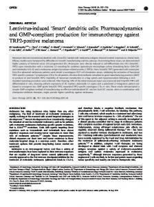
on the other hand, stress initiates structural plasticity in the basolateral amygdala (BLA). This plasticity is characterized by dendritic hypertrophy [19] and increase in spine density [20] of principal projection neurons within the BLA. Several strands of evidence suggest that the effects of stress on anxiety-like behavior, BLA dendrites, and adrenal hormones are not isolated biological entities. Rather, these effects likely represent nodes of a reciprocally interacting loop. For example, exogenous corticosterone treatment after cat exposure leads to greater anxiety in rats when measured after several days [9]. This anxiogenesis manifests as a lower exploration of open spaces in elevated plus mazes and greater acoustic startle. This is congruent with the ability of exogenous corticosterone to increase anxiety-like behavior by itself, in parallel to causing dendritic hypertrophy in the BLA [21]. Similarly, pharmacological blockade of steroidogenesis in rats prevents a rise in anxiety-like behavior after exposure to cat odor [22]. Molecular studies also support the notion that the BLA is crucial for the relationship between glucocorticoids and stress-induced anxiogenesis. For example, diversion of glucocorticoid signaling within the BLA from glucocorticoid receptors toward mineralocorticoid receptors [23] or towards estrogen receptors [24] reduces anxiety-like behavior. Moreover, experimental manipulations that reduce BLA hypertrophy also show concomitantly reduced anxiety-like behavior and reduced glucocorticoid levels [25–27]. These observations suggest that BLA hypertrophy, anxiety and hypercortisolism are part of an interacting triad that is facilitated by historical stress. This hypothesis predicts a positive relationship of BLA dendrites with anxiety-like behavior and/or glucocorticoid levels. An alternative interpretation is also possible. The observations above could be similarly explained if BLA dendritic hypertrophy were a compensatory response to mediators of anxiety-like behavior and/or endocrine activation. This hypothesis predicts a negative relationship between BLA dendrites and anxiety-like behavior and/or hormonal measures. We test these competing predictions in the present report by quantifying correlations between endpoints related to anxiety-like behavior, BLA plasticity and glucocorticoids within the same cohort of individual rats. This experimental design also encapsulates inter-individual variation for glucocorticoid response to novel stressful episodes subsequent to historical stressors, and the relationship of this inter-individual variation with that of the BLA dendritic structure. 2. Materials and Methods 2.1. Animals and Experimental Groups Adult male Wistar rats (7 weeks old, weight: 220–250 g, housed as 2 rats/cage; ad-libitum food and water; 12:12 light-dark cycles with lights on at 700 h) were procured from National University of Singapore. This outbred strain was chosen because of its greater phenotypic variability for later correlational analysis. Rats were habituated for one week before the start of the experiments. All experimental procedures were reviewed and approved by the institutional animal use and care committee of Nanyang Technological University (IACUC: A-0195). The experimental sequence is depicted in Figure 1.
Int. J. Environ. Res. Public Health 2017, 14, 779
3 of 13
Int. J. Environ. Res. Public Health 2017, 14, 779
3 of 13
Figure Schematic experimental flow. (A) Animals were sequentially tested anxiety Figure 1. 1. Schematic ofof experimental flow. (A) Animals were sequentially tested forfor anxiety inin thethe open‐field arena (OFT), exposed to predator stress and subsequently exposed to an elevated plus open-field arena (OFT), exposed to predator stress and subsequently exposed to an elevated plus maze maze (EPM). Blood for corticosterone estimates was drawn thirty minutes after OFT, EPM, and the (EPM). Blood for corticosterone estimates was drawn thirty minutes after OFT, EPM, and the animals animals were eventually sacrificed for morphological endpoints. Days elapsed from the start of the were eventually sacrificed for morphological endpoints. Days elapsed from the start of the experiment areexperiment are denoted on top. (B) A separate cohort was exposed to open field and sacrificed the denoted on top. (B) A separate cohort was exposed to open field and sacrificed the next day for next day for morphological endpoints. 30 min measurement. after OFT for hormone morphological endpoints. Blood was collectedBlood 30 minwas aftercollected OFT for hormone measurement.
2.2. Open Field Test (OFT) 2.3. Predator Odor Exposure Animals were tested for anxiety-like behavior in an open field. Exposure to open field Rats were exposed to 2 mL bobcat urine on day 5 of the experimental sequence (Figure 1A). The occurred before exposure to the stressor, i.e., before exposure to cat odor (Figure 1A). In addition, predator odor served as a stressor in our experiment [28–31]. Effect of predator odor stress on the a separate cohort of animals was exposed to OFT without subsequent stress exposures (Figure 1B). subsequent reactivity of endocrine response and anxiety‐like behavior was later measured in the A square-shaped open field was constructed from Plexiglas (100 cm × 100 cm, 30 cm wall) and elevated plus maze on day 17 Figure 1A. Exposure to predator odor occurred in a rectangular arena illuminated at the center (10 lux at center and 3–4 lux at the periphery). All tests were conducted (two 76 cm × 9 cm bisects separated by a 9 cm × 9 cm central connector, wall = 15 cm; duration = 10 between 900 and 1200 h (duration = 300 s). Time spent in the central part of the open field min). Individuals were habituated to the arena for 10 min on three successive days before actual (33 cm × 33 cm) was measured as a proxy for lower anxiety-like behavior. exposure to the predator odor (bobcat urine). Aversion to the bobcat urine was quantified as occupancy of the bisect containing odor (76 cm × 9 cm) relative to the total area of the arena (chance 2.3. Predator Odor Exposure = 47.2%, based on the area of the bisect containing predator odor vis‐à‐vis total area of the arena). Rats were exposed to 2 mL bobcat urine on day 5 of the experimental sequence (Figure 1A). The predator odor served as a stressor in our experiment [28–31]. Effect of predator odor stress on 2.4. Elevated Plus Maze (EPM) the subsequent reactivity of endocrine response and anxiety-like behavior was later measured in the Animals were exposed to an EPM on day 17 of the experimental sequence (Figure 1A), between elevated plus maze on day 17 Figure 1A. Exposure to predator odor occurred in a rectangular arena (two 900 and 1200 h, trial duration = 300 s). Relative open‐arm exploration in the EPM was measured as a 76 cm × 9 cm bisects separated by a 9 cm × 9 cm central connector, wall = 15 cm; duration = 10 min). proxy for lower anxiety. The EPM consisted of two open (75 cm × 11 cm, illuminated