Design and evaluation of a continuous-wave diffuse optical tomography system A. M. Siegel Electro-Optics Technology Center, Tufts University, Medford, MA 02155
[email protected]
J. J. A. Marota* and D. A. Boas NMR Center and the Dept. of Anesthesia and Critical Care*, Massachusetts General Hospital, Harvard Medical School, Massachusetts General Hospital East, Bldg #149, Charlestown, MA 02129
[email protected]
Abstract: Diffuse optical tomography (DOT) can image spatial variations in highly scattering optical media. We have built an inexpensive and portable continuous-wave DOT system containing 18 laser diode sources (9 at 780nm and 9 at 830nm) and 16 silicon detectors, which can acquire 288 independent measurements in less than 4 seconds. These data can then be processed using a variety of imaging algorithms. We first discuss the design of diffuse imaging equipment in general, and then describe our instrument, along with the technical issues that influenced its design. The technical challenges involved in performing DOT over large optode areas are discussed. We also present rat brain measurements following electrical forepaw stimulation using DOT. These results clearly demonstrate the capabilities of DOT and set the stage for advancement to quantitative functional brain imaging. 1999 Optical Society of America
OCIS codes: (170.6960) Tomography; (170.5280) Photon migration
_____________________________________________________________________ References and links 1. A. Yodh and B. Chance, "Spectroscopy and imaging with diffusing light," Phys. Today 48, 34-40 (1995). 2. S. R. Arridge and J. C. Hebden, "Optical Imaging in Medicine: II. Modelling and reconstruction," Phys. Med. Bio. 42, 841-854 (1997). 3. J. C. Hebden, S. R. Arridge and D. T. Delpy, "Optical imaging in medicine: I. Experimental techniques," Phys. Med. Bio. 42, 825-840 (1997). 4. P. T. Fox and M. E. Raichle, "Focal physiological uncoupling of cerebral blood flow and oxidative metabolism during somatosensory stimulation in human subjects," Proc. Natl. Acad. Sci. USA 83, 1140-4 (1986). 5. K. K. Kwong, J. W. Belliveau, D. A. Chesler, I. E. Goldberg, R. M. Weisskoff, B. P. Poncelet, D. N. Kennedy, B. E. Hoppel, M. S. Cohen, R. Turner, H.-M. Cheng, T. J. Brady and B. R. Rosen, "Dynamic magnetic resonance imaging of human brain activity during primary sensory stimulation," Proc. Natl. Acad. Sci. USA 89, 5675-9 (1992). 6. S. Ogawa, T. M. Lee, A. R. Kay and D. W. Tank, "Brain magnetic resonance imaging with contrast dependent on blood oxygenation," Proc. Natl. Acad. Sci. USA 87, 9868-72 (1990). 7. S. Ogawa, D. Tank, R. Menon, J. Ellermann, S.-G. Kim, H. Merkel and K. Ugurbil, "Intrinsic signal changes accompanying sensory stimulation: Functional brain mapping with magnetic resonance imaging," Proc. Natl. Acad. Sci. USA 89, 5951-5955 (1992). 8. R. L. Barbour, H. L. Graber, J. Chang, S. S. Barbour, P. C. Koo and R. Aronson, "MRI-guided optical tomography:prospects and computation for a new imaging method," IEEE Computation Science and Engineering 2, 63-77 (1995). 9. B. W. Pogue and K. D. Paulsen, "High-resolution near-infrared tomographic imaging simulations of the rat cranium by use of a priori magnetic resonance imaging structural information," Opt. Lett. 23, 1716-1718 (1998). 10. B. W. Pogue, M. Testorf, T. McBride, U. Osterberg and K. Paulsen, "Instrumentation and design of a frequencydomain diffuse optical tomography imager for breast cancer detection," Opt. Express 1, 391-403 (1997). http://epubs.osa.org/oearchive/source/2827.htm 11. N. Ramanujam, C. Du, Y. Ma and B. Chance, "Sources of phase noise in homodyne and heterodyne phase modulation devices used for tissue oximetry studies," Rev. Sci. Instru. 69, 3042-3054 (1998). 12. B. Chance, M. Cope, E. Gratton, N. Ramanujam and B. Tromberg, "Phase measurement of light absorption and scattering in human tissues," Rev. Sci. Instru. 689, 3457-3481 (1998).
#9224 - $15.00 US
(C) 1999 OSA
Received March 08, 1999; Revised March 30, 1999
12 April 1999 / Vol. 4, No. 8 / OPTICS EXPRESS 287
13. P. N. den Outer, T. M. Nieuwenhuizen and A. Lagendijk, "Location of objects in multiple-scattering media," J. Opt. Soc. Am. A 10, 1209-1218 (1993). 14. D. A. Boas, M. A. O'Leary, B. Chance and A. G. Yodh, "Scattering of diffuse photon density waves by spherical inhomogeneties within turbid media: analytic solution and applications," Proc. Natl. Acad. Sci. USA 91, 48874891 (1994). 15. M. S. Patterson, B. Chance and B. C. Wilson, "Time resolved reflectance and transmittance for the non-invasive measurement of tissue optical properties," Appl. Opt. 28, 2331-2336 (1989). 16. M. A. O'Leary, D. A. Boas, B. Chance and A. G. Yodh, "Refraction of diffuse photon density waves," Phys. Rev. Lett. 69, 2658-2661 (1992). 17. J. B. Fishkin and E. Gratton, "Propagation of photon density waves in strongly scattering media containing an absorbing 'semi-infinite' plane bounded by a straight edge," J. Opt. Soc. Am. A 10, 127-140 (1993). 18. A. Villringer and B. Chance, "Non-invasive optical spectroscopy and imaging of human brain function," Trends Neurosci 20, 435-442 (1997). 19. G. Gratton, M. Fabiani, P. M. Corballis, D. C. Hood, M. R. Goodman-Wood, J. Hirsch, K. Kim, D. Friedman and E. Gratton, "Fast and localized event-related optical signals (EROS) in the human occipital cortex: comparisons with the visual evoked potential and fMRI.," NeuroImage 6, 168-180 (1997). 20. B. Chance, A. Endla, N. Shoko, Z. Shuoming, H. Long, K. Worden, C. Li, T. Murray, Y. Ovetsky, D. Pidikiti and R. Thomas, "A novel method for fast imaging of brain function, non-invasively, with light," Opt. Express 2, 411423 (1998). http://epubs.osa.org/oearchive/source/4445.htm 21. M. Kohl, U. Lindauer, U. Dirnagl and A. Villringer, "Separation of changes in light scattering and chromophore concentrations during cortical spreading depression in rats," Opt. Lett. 23, 555-557 (1998). 22. J. B. Mandeville, J. J. A. Marota, B. E. Kosofsky, J. R. Keltner, R. Weissleder, B. R. Rosen and R. M. Weisskoff, "Dynamic functional imaging of relative cerebral blood volume during rat forepaw stimulation," MRM 39, 615624 (1998). 23. J. B. Mandeville, J. J. A. Marota, C. Ayata, M. A. Moskowitz, R. M. Weisskoff and B. R. Rosen, "An MRI Measurement of the Temporal Evolution of Relative CMRO2 During Rat Forepaw Stimulation," Magn. Reson. Med. In Review, (1999). 24. J. J. A. Marota, C. Ayata, M. A. Moskowitz, R. M. Weisskoff, B. R. Rosen and J. B. Mandeville, "Investigation of the Early Response to Rat Forepaw Stimulation," Magn. Reson. Med. In Press, (1999). 25. A. C. Kak and M. Slaney, Principles of Computerized Tomographic Imaging (IEEE Press, New York, 1988). 26. M. A. O'Leary, D. A. Boas, B. Chance and A. G. Yodh, "Experimental images of heterogeneous turbid media by frequency-domain diffusing-photon tomography," Opt. Lett. 20, 426-428 (1995). _______________________________________________________________________________________________________________________________
1. Introduction Diffuse Optical Tomography offers the capability to simultaneously quantify the tissue concentration of both oxy- (HbO) and deoxy-hemoglobin (Hb) [1-3]. Two or more nearinfrared sources, with wavelengths specifically chosen to straddle the isosbestic point of the oxy/deoxyhemoglobin absorption spectrum, illuminate the tissue at various locations. The flux distribution at the tissue surface thus contains both spectral and spatial information about subsurface absorbers. fMRI uses the paramagnetism of the Hb molecule to monitor metabolic activity through local changes in Hb, and thus blood oxygenation, hence the origin of the term BOLD (Blood Oxygen Level Dependent) imagery [4-7]. fMRI offers millimeter spatial resolution, but only the relative quantity of Hb is measured. DOT can simultaneously image changes in total hemoglobin concentration (which relates to blood volume) and oxygen saturation (HbO/(HbO+Hb)). One disadvantage of DOT is its limited spatial resolution, which leads to an intriguing possibility: the coregistration of simultaneously acquired DOT and functional MRI imagery, combining the spatial resolution of fMRI with the spectral discrimination of DOT [8, 9]. We plan to pursue such measurements in the near future with the instrumentation described in this manuscript. In this paper, we discuss the engineering issues involved in the design of diffuse imaging equipment [10-12]. We then describe the design and evaluation of a continuous-wave (CW) DOT system that can image brain function in animals and humans with 4-second temporal resolution. We then present preliminary results showing that DOT is capable of quantitatively imaging both HbO and Hb concentration changes during brain activation in a rat model.
#9224 - $15.00 US
(C) 1999 OSA
Received March 08, 1999; Revised March 30, 1999
12 April 1999 / Vol. 4, No. 8 / OPTICS EXPRESS 288
2. Background Ballistic photon imaging is usually performed in a shot noise-limited, photon-starved environment due to the low percentage of photons that actually penetrate the tissue unscattered. Thus the photon-counting detectors require only a modest dynamic range. Diffuse imaging, on the other hand, employs many sources and detectors distributed over a large region of illuminated tissue, so each detector may operate over an effective photon path length ratio in excess of 10:1. With total optode source-detector geometries spanning less than 1cm, as in rat brain studies, this effect is negligible, and the hardware can easily accommodate the 60-80dB of dynamic range required for rather good quality imagery. On large subjects such as neonates or adult humans, however, optode dimensions of between 4 and 8cm are required. Under these conditions, that same photon path length ratio of 10:1 now translates into a flux attenuation range on the order of 106:1! In order to obtain all the information available, each detector must be capable of operating over a 120dB dynamic range, which is rather large from an electronic perspective. Although many techniques have been developed to solve this problem, all of them require a large capital investment in dollar cost and an increase in both measurement time and hardware complexity. In order to collect enough photons to reach 120dB, the source power must increase and the detection noise must decrease. The source power will ultimately be limited in human patients by regulatory issues or, in experimental settings, by the vasodynamic effects of tissue heating, or in some cases, thermal necrosis. The detection noise floor can be reduced by improving the detectors and circuitry, but only to a point. Once the electrical noise contribution is reduced below the noise introduced by the fundamental uncertainty in the photon arrival rate, the system has become photon noise-limited or “background–limited”. The only way to further reduce the noise level is to improve the statistics by increasing the total number of photons collected during each detector sample – which, for a fixed photon arrival rate, means a longer dwell time. This noise reduction is proportional to the square root of the total number of photons collected, and hence to the square root of the total photon collection time. Thus, a fundamental tradeoff exists between measurement time and dynamic range. If sufficient power and measurement time is available and a signal can be detected, the next challenge begins: what to do with these signals and how to keep them clean. Direct digitization of a 120dB signal would require a 22 bit A/D converter, although logarithmic compression may reduce this to a far more practical 16 bits. Now that the signals can be detected and digitized, the entire system must be capable of preserving their quality. This means that detector channel-to-channel crosstalk, multiplexer settling, and feedthru should be around –120dB. This can best be achieved by performing the A/D conversion directly following detection. Electromagnetic interference, power, and ground isolation all must approach the quantization limit – about half a DN (Digital Number) or so. All of these requirements can be met, but it would require a carefully constructed system based upon a well-engineered design. Many other, perhaps better, solutions exist - such as using two or more lower dynamic range fixed-gain detectors at each optode location, or perhaps using a switch to reduce the detector gain by known increments when a certain signal voltage threshold is exceeded. All of these still require the same level of attention to signal quality and integrity as discussed above. Diffuse imaging techniques can be grouped into two general categories: “scalar” and “vector” techniques: Scalar techniques measure the optical flux exiting the tissue. The system we describe below is a scalar system. There is additional useful information available in the form of the average photon path length. Some light travels deeply into the tissue and returns to the surface, most of the light passes through the classic banana-shaped region, and a small amount travels the shortest path between the source and the detector. A localized absorption at a specific depth will attenuate only the small number of photons which pass through that region, leaving the rest unaffected [13, 14]. Since the average photon path length, and thus the transit #9224 - $15.00 US
(C) 1999 OSA
Received March 08, 1999; Revised March 30, 1999
12 April 1999 / Vol. 4, No. 8 / OPTICS EXPRESS 289
time, varies significantly with depth, the magnitude of the photon flux versus the average photon transit time provides a measure of the relative absorption vs. tissue depth – a valuable piece of information. (Although most in-vivo imaging - especially brain imaging - requires light to travel through various tissue types: skin, bone, cerebrospinal fluid, etc., and the resulting optical heterogeneity complicates image reconstruction, these basic concepts still apply). Vector techniques measure both the magnitude and the average propagation delay, either directly, or in the form of amplitude and phase-shifts relative to the modulation frequency of the light source. Two common vector approaches in use today are the time-domain approach [15] and the frequency-domain approach [11, 16, 17]. A time-domain system employs picosecond-wide optical pulses, time-gated photon-counting detectors, time-to-amplitude converters, and the like. Typical frequency-domain systems use a radiofrequency (RF) modulated light source, photomultiplier tubes or fast photodiodes feeding tuned RF amplifiers, and an RF inphase/quadrature (I/Q) phase detector followed by postdetection filters. Although time-domain systems are flexible and can detect both ballistic and diffusely scattered photons, they are expensive and, due to their wideband nature, require significant averaging in the digital domain to improve the signal-to-noise ratio (SNR). Frequency-domain systems use simpler, lower-cost components and provide greater SNR. The simplicity and cost savings result from the widespread commercial availability of excellent RF circuitry at modest cost, specifically around common satellite and radar receiver intermediate (IF) frequencies such as 70MHz, 140MHz, 200MHz, etc. The greater SNR stems from the fact that, unlike the wideband nature of time-domain measurements, frequency-domain measurements are usually performed at single RF frequencies, so they occupy a much narrower bandwidth. In the ideal case, the detector electronics need only occupy an RF bandwidth commensurate with about twice the signal acquisition rate (to capture both sidebands), much as the coherent detection system described below. Although this may be impractical due to component instabilities, bandwidths in the Hz to low kHz range are quite practical. Regardless of the techniques employed, all DOT instrumentation should be designed with the following parameters in mind: Large optode separations lead to significant optical attenuation, so multiple optode spacings will require a large dynamic range. The system should be as linear as possible over this dynamic range in order to keep the measurements both accurate and precise. Stray light rejection is important for systems that must operate outside of the sheltered confines of the laboratory, especially in clinical settings, which often contain large amounts of “optical pollution”. Both optical and electrical crosstalk should be reduced to levels commensurate with the system dynamic range, if possible. Good long-term stability means fewer and less frequent calibration cycles, and good temporal response is important for functional imaging, however it comes at the cost of an increase in the noise floor, which reduces dynamic range. 3. The prototype CW DOT system Our goal was to develop a prototype portable diffuse imager which can be used as a research tool for both characterizing tissue optical properties and guiding us toward future hardware and software design improvements. For simplicity, we decided on a magnitude-only system. Our optical sources were standard low-power laser diodes. Although more powerful light-emitting diodes were available, coupling that light into the various core sizes of both plastic and glass fiber would have been very difficult and far less optically efficient. Our detector options ranged from multianode PMTs to discrete commercial-grade photodiodes with external preamplifiers. We compromised by choosing a monolithic photodiode/preamplifier IC housed in a clear 8-pin DIP package (OPT209 from Burr-Brown, Tucson, AZ). This offered both the convenience and low-cost of a solid-state detector, combined with the electrical and optical isolation of an integrated preamplifier. Although the parasitic capacitances of the monolithic preamp were about twice that of a well-designed discrete circuit, the simplicity and the less stringent electrical shielding requirements for the monolithic preamp swayed our decision. #9224 - $15.00 US
(C) 1999 OSA
Received March 08, 1999; Revised March 30, 1999
12 April 1999 / Vol. 4, No. 8 / OPTICS EXPRESS 290
Our goal was to achieve a useable dynamic range of at least 80dB. Dynamic range and linearity are closely paired, since each must be defined with reference to the other. For example, a statement of a given dynamic range value is, in itself, meaningless unless the criteria for determining the limits of that range are expressly stated. In our case, our goal was 80dB of dynamic range with less than a 1% deviation from a least-squares-fit line. Although this linearity value was somewhat arbitrarily selected, it gave us a good starting point around which to base our design. Since valuable information can often be gleaned even from saturated signals, we decided to “bracket” the signal swing within the dynamic range of our A/D converter. In order to meet the 80dB objective, crosstalk, drift, settling time, and feedthru errors had to match this goal. Optical crosstalk was minimized by concealing each detector package in opaque heatshrink tubing and providing sufficient separation to further attenuate any stray reflections within the metal housing. Each detector fiber was sheathed in opaque tubing as well, which also served to protect the fragile cladding of the PMMA (acrylic) fibers from abrasion. Electrical crosstalk occurring at the front end was not significant since the only high impedance node in the preamp was physically removed from the DIP package and the modulation frequency was in the kilohertz range. Ground loops were minimized by using an electrically isolated power supply, a battery-powered portable laptop computer, and a singlepoint earth connection to safety ground. Crosstalk through power lines was minimized with on-board regulation and the use of separate power supply decoupling filters at each opamp. To reduce settling time errors, we planned for a 5τ dwell time prior to conversion. For a postdetection time constant of 40ms, this gave us a minimum delay of 200ms per source. Faster acquisition rates could easily be achieved, albeit at the cost of higher crosstalk. Feedthru among multiple channels within the analog multiplexer was minimized by switching only demodulated (baseband) signals and by reducing the DC currents through the switches by placing a buffer directly at the multiplexer output. This also provided better linearity, since switch impedance varies with common-mode voltage. We chose to use a rather fast 133 kilosample-per-second, 16 bit A/D converter (Analog Devices AD7884) for a number of reasons. The high sample rate allowed us to rapidly scan through the 16 demodulated outputs during high data rate measurements, while still permitting us to oversample and then average in the digital-domain during slower measurements. The parallel data output design made it easy to interface with all of the digital I/O boards used in our lab. A summary of our performance goals for the prototype DOT imager is shown in Table 1. Since we planned to perform some measurements in a clinical setting, the unit had to operate within a normal hospital environment. This placed a severe stray-light rejection requirement on our design. We needed to detect picowatts of source signal under an ambient optical background in the microwatt range (as seen by the detector). We solved this problem by using synchronous detection.
#9224 - $15.00 US
(C) 1999 OSA
Received March 08, 1999; Revised March 30, 1999
12 April 1999 / Vol. 4, No. 8 / OPTICS EXPRESS 291
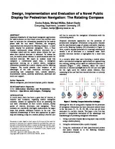
Table 1. Our performance goals for the prototype DOT imager.
PARAMETER DYNAMIC RANGE NONLINEARITY SETTLING TIME CROSSTALK DIGITAL RESOLUTION SOURCE CHANNELS SOURCE OPTICAL POWER DETECTORS MODULATION TECHNIQUE POSTDETECTION BANDWIDTH STRAY LIGHT REJECTION PACKAGING ISSUES POWER REQUIREMENTS PATIENT SAFETY
GOAL 10,000:1 (80dB)