123. Design and Implementation of Peripheral Nerve Stimulator with.
Electromyography using PIC Microcontroller. Shweta A. Borkar1, Ramesh.T.
Patil2 , Dr. A.V. ...
International Journal of Emerging Technology and Advanced Engineering Website: www.ijetae.com (ISSN 2250-2459, ISO 9001:2008 Certified Journal, Volume 2, Issue 12, December 2012)
Design and Implementation of Peripheral Nerve Stimulator with Electromyography using PIC Microcontroller Shweta A. Borkar1, Ramesh.T.Patil2 , Dr. A.V.Nadkarni3 1
2
Master of engineering, Asso.Prof, Department of Electronics, Rajarambapu Institute of technology, Sakharale, M.S., India 3 Professor, Department of Anesthesiology, Krishna Institute of Medical Sciences, Karad, M.S., India It provides different types of stimulation which are needed for neuromuscular monitoring with parameters such as pulse duration, pulse frequency and adjustable current which are programmed using PIC microcontroller.
Abstract— A simple peripheral nerve stimulator is used for assessment of neuromuscular block by drug (muscle relaxant) during anesthesia by visual impression of finger movement which is only a subjective assessment. We proposed the peripheral nerve stimulator with electromyography which is used for monitoring of muscle relaxation during the conduct of anesthesia for surgical procedure which is a integral part of patient safety. The innovation consists in using EMG sensor for sensing electrical signal from a muscle that is action potential which will show the magnitude of muscle contraction leading to the finger movement and is more precise, calibrated and hence perfect, objective in nature qualitatively and quantitatively. We used microcontroller as PWM generator to apply various stimulation modes which stimulates the motor nerve.
II. NERVE CONDUCTION Nervous system is basically divided into Peripheral nervous system and Central Nervous system. The Central Nervous system consist of brain and spinal cord while the Peripheral nervous System consist of all the nerves outside brain and spinal cord. The nervous system consist of a vast number of cells called neurons[1]. It is a cell present mainly within the CNS that is brain & spinal cord and is biologically the main functional unit of nervous system. Each neuron consist of a cell body and its processes, one axon and many dendrites. Each axon is an tubular extension of a neuron. The bundle of axons in large number when packed together form a single peripheral nerve. Each nerve consist of sensory and motor axons & hence it is called mixed peripheral nerve. Each axon is a tubular structure of semi permeable nature containing protein and fluid and is electrically charged. In a living state, the axonal membrane is charged positively on the surface and negatively charged from inside the membrane. With the stimulus generated by the axon or external stimulus these charges are reversed thus creating a temporary reversal of potential which in medical terminology is called an impulse(stimulus).The axon of motor nerve coming from spinal cord ends at or near the muscle. It transmits impulse to muscle fiber . The junction between the terminal fibers of motor axon and the muscle fiber is called neuromuscular junction. This process of transmitting impulse from motor axon to muscle fiber occurs with the help of release of a chemical substance called acetylcholine from nerve ending, leading to contraction of muscle[2]. When the muscle relaxant is administered to patient, the molecules of this drug competitively occupy the receptors on the surface of muscle fibre where normally only acetylcholine acts, thereby preventing the later causing muscle contraction resulting into muscle relaxation.
Keywords— Electromyography (EMG), Monitoring Neuromuscular function, Nerve Stimulation, Peripheral Nerve Stimulator (PNS), Precision.
I. INTRODUCTION The progresses achieved by electronics in medical field during these last decades are undoubtedly completely remarkable. Use of simple Peripheral Nerve Stimulator is a common practice in Anesthesia where drugs are used to paralyze patients under anesthesia so that they will not move during a surgery and hence the operation result is successful. The present practice today is assessment of degree of neuromuscular block produced by these drugs is by simple visual impression of finger movement due to muscle contraction secondary to motor nerve stimulation by Peripheral Nerve Stimulator and hence merely subjective in nature. Muscle relaxants are employed in anesthesia to provide muscle relaxation and/or abolish patient movement. Monitoring the magnitude of neuromuscular block is accomplished by delivering an electrical stimulus externally near a peripheral motor nerve and evaluating the evoked response of muscle contraction innervated by that nerve. In this paper, we proposed PNS with electromyography to stimulate motor nerve which leads to contraction of a muscle of hand or leg and thus the resultant magnitude of muscle contraction (action potential) is sensed by EMG sensor more precisely and displayed on LCD screen. 123
International Journal of Emerging Technology and Advanced Engineering Website: www.ijetae.com (ISSN 2250-2459, ISO 9001:2008 Certified Journal, Volume 2, Issue 12, December 2012) The magnitude of muscle relaxation or contraction therefore is directly proportional to the number of neuro muscular blocking drug molecules against the naturally secreted neurotransmitter Acetylcholine, respectively. Stimulation of a motor nerve artificially by PNS, leads to transient increase in Acetylcholine at neuro muscular junction leading to muscle contraction which is assessed by anesthetist to judge the magnitude of N-M junction guiding him to manage the patient as needed but at the same time safely. When in doubt with a single twitch stimulus, he has the option of using higher modes of stimulation of different types to diagnose the quality and depth of N-M block. Normally, stimulation of motor nerve will thus cause muscle contraction due to transient depolarization of its surface(just like axonal surface) resulted from production of a wave of action potential on surface of muscle. Picking up the potential at two different points along the fiber length of muscle is the electromyogram[3]. The change in the magnitude of potential will indicate how many of the muscle fibers are still contracting effectively and how many remain silent(relaxed) thus indicating the magnitude of muscle relaxation.
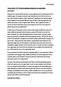
LCD display
Keyboard
Microcontroller
Surface electrodes to Human body
sensor with preamplifier
Fig.1. Block diagram of Peripheral Nerve Stimulator with EMG using PIC Microcontroller
A. PNS Keyboard It is used to set current and provide various stimulations modes such as Twitch, Train of four, Tetanus and Double burst etc. which are required to stimulate the motor nerve[5]. Facility is also provided for incrementing and decrementing the current to be set.It has a facility to maintain a repetitive stimulation for various modes over a duration of time.
III. PERIPHERAL NERVE STIMULATOR Peripheral Nerve Stimulator (PNS) is one of the medical instruments used in the field of anesthesia that uses current pulses to externally stimulate the motor nerve thus generating contraction of muscle, for knowing the time at which muscle relaxant has to be given[4]. It is used to monitor the degree of neuromuscular block produced by muscle relaxant just by observing visually the finger movements. Stimulation of nerve is achieved by placing surface electrodes near motor nerve and passing a current through them. Electromyography is used to detect and record the evoked response of muscle contraction using the EMG sensor.
B. Surface Electrodes These are silver chloride electrodes generally used in ECG which are easily available in market. The impulse delivered by the proposed PNS is applied to skin via these electrodes to stimulate the motor nerve. C. EMG sensor with preamplifier The design of preamplifier is most critical aspect of this device. It is desirable to obtain an EMG signal that contains maximum amount of information from EMG electrodes and minimum amount of contamination from electrical noise.
IV. DESIGN The architecture of stimulator is as described as follows.
124
International Journal of Emerging Technology and Advanced Engineering Website: www.ijetae.com (ISSN 2250-2459, ISO 9001:2008 Certified Journal, Volume 2, Issue 12, December 2012) For this purpose AD620 instrumentation amplifier which acts as an differential amplifier is used. In order to eliminate the potentially much greater noise signal from power line sources, a differential detecting configuration is employed. The signal is detected at two sites by two EMG electrodes which are placed on the muscle belly. The preamplifier circuit subtracts the two signals and then amplifies the difference. As a result any signal that is “common” to both detection sites will be removed and signals that are different at the two sites will have a “differential” that will be amplified which are usually in the range of mV[6].
a)
D. PIC Microcontroller PIC30F3010/3011 Enhanced Flash 16 bit Digital Signal Controller is used since it has features like 1kbytes of on chip RAM, 10bit ADC. The following points describe the function of PIC microcontroller in the proposed work. 1) It generates PWM with the help of PWM driver circuit based on selection of different modes of stimulation. 2) It acquires the EMG signal from EMG sensor that is action potential in the form of voltage and displays it as response in graphical format. 3) It even displays the battery status in graphical form. 4) It checks for various failures associated with this device. 5) It displays the required magnitude of muscle contractions of different stimulation in the form of percentage.
b)
E. LCD Display 128x64 LCD display is divided into 2 parts. Upper part displays battery status, current to be used, type of stimulation selected and percentage of muscle contraction in digital format. Lower part displays graphical representation of strength of electrical stimulation for various modes and muscle contraction as per selected stimulation showing magnitude and duration of electrical activity (action potential ).
c) Fig.2. a) Proposed Instrument b) Train of four stimulation mode c) Tetanus stimulation
VI. CONCLUSION V. RESULT
In PIC microcontroller based Peripheral Nerve Stimulator with electromyography, PIC microcontroller enables to make easy, fast and flexible design and implementation. It generates PWM depending on selection of stimulation modes which stimulates motor nerve. The action potential obtained from muscle contraction is sensed by EMG sensor, amplified and displayed on LCD in graphical format.
Here we carried out test with proposed instrument on patient during surgery under anaesthesia shown in Fig.2.We relaxed the patient by giving muscle relaxant drug so that various stimulation modes can be used to stimulate motor nerve . Surface electrodes were applied to skin of hand and EMG electrodes were applied to muscle belly. 125
International Journal of Emerging Technology and Advanced Engineering Website: www.ijetae.com (ISSN 2250-2459, ISO 9001:2008 Certified Journal, Volume 2, Issue 12, December 2012) [3]
The magnitude of contraction is displayed in terms of percentage thereby making the device highly objective in terms of getting precise results during anesthesia and surgery.
[4] [5] [6]
REFERENCES [1] [2]
Anne Waugh and Allison Grant,Anatomy and Physiology in Health and Illness. Angela Forster and Nigel Palastanga,2002.Clayton’s Electrotheraphy Theory and Practice .
126
UK Misra and J Kalita 1999.Clinical Neurophysiology B.I.Churchill Livingstone Pvt.Ltd. J.A,Dorsch,S.E.Dorsch 1998 4th Edition Understanding Anesthesia Equipment Indian Journal of Anaesthesia Volume:46,August 2002 Carlo J. De Luca Surface Electromyography:Detection and Recording