Home
Search
Collections
Journals
About
Contact us
My IOPscience
Design of a decision support system, trained on GPU, for assisting melanoma diagnosis in dermatoscopy images
This content has been downloaded from IOPscience. Please scroll down to see the full text. 2015 J. Phys.: Conf. Ser. 633 012079 (http://iopscience.iop.org/1742-6596/633/1/012079) View the table of contents for this issue, or go to the journal homepage for more
Download details: IP Address: 85.1.243.28 This content was downloaded on 03/03/2016 at 01:59
Please note that terms and conditions apply.
4th International Conference on Mathematical Modeling in Physical Sciences (IC-MSquare2015) IOP Publishing Journal of Physics: Conference Series 633 (2015) 012079 doi:10.1088/1742-6596/633/1/012079
Design of a decision support system, trained on GPU, for assisting melanoma diagnosis in dermatoscopy images Dimitris Glotsos1, Spiros Kostopoulos1, Stella Lalissidou1, Konstantinos Sidiropoulos2, Pantelis Asvestas1, Christos Konstandinou3, George 1 4 Xenogiannopoulos , Eirini Konstantina Nikolatou , Konstantinos Perakis5, Thanassis Bouras5 and Dionisis Cavouras1 1
Department of Biomedical Engineering, Technological Educational Institute of Athens, Greece European Bioinformatics Institute (EMBL-EBI), European Molecular Biology Laboratory, Welcome Trust Genome Campus, Hinxton, Cambridge, UK 3 Department of Medical Physics, University of Patras, 26504, Rio, Patras, Greece 4 Department of Economic Sciences, University of Patras, Patras, Greece 5 UBITECH Research Department, UBITECH Ltd., Athens, Greece 2
E-mail:
[email protected] Abstract. The purpose of this study was to design a decision support system for assisting the diagnosis of melanoma in dermatoscopy images. Clinical material comprised images of 44 dysplastic (clark’s nevi) and 44 malignant melanoma lesions, obtained from the dermatology database Dermnet. Initially, images were processed for hair removal and background correction using the Dull Razor algorithm. Processed images were segmented to isolate moles from surrounding background, using a combination of level sets and an automated thresholding approach. Morphological (area, size, shape) and textural features (first and second order) were calculated from each one of the segmented moles. Extracted features were fed to a pattern recognition system assembled with the Probabilistic Neural Network Classifier, which was trained to distinguish between benign and malignant cases, using the exhaustive search and the leave one out method. The system was designed on the GPU card (GeForce 580GTX) using CUDA programming framework and C++ programming language. Results showed that the designed system discriminated benign from malignant moles with 88.6 % accuracy employing morphological and textural features. The proposed system could be used for analysing moles depicted on smart phone images after appropriate training with smartphone images cases. This could assist towards early detection of melanoma cases, if suspicious moles were to be captured on smartphone by patients and be transferred to the physician together with an assessment of the mole’s nature.
1. Introduction Melanoma comprises one of the most lethal and difficult to treat forms of cancer with more than 100 000 cases worldwide each year [1]. The annual incidence rate of the disease has an increasing tendency, which is attributed to modern lifestyle and long exposure to ultraviolet radiation that has been identified as among the most influential factors for triggering the disease. Early stage detection of melanoma has been shown as the most crucial step for treating the disease, since at early phases melanoma seems to be more vulnerable to available treatments [2, 3]. Diagnosis of melanoma is done using special observation instruments called dermoscopes. Dermoscopes use polarized light in order to enhance the visibility of pigmented lesions [4]. The Content from this work may be used under the terms of the Creative Commons Attribution 3.0 licence. Any further distribution of this work must maintain attribution to the author(s) and the title of the work, journal citation and DOI. Published under licence by IOP Publishing Ltd 1
4th International Conference on Mathematical Modeling in Physical Sciences (IC-MSquare2015) IOP Publishing Journal of Physics: Conference Series 633 (2015) 012079 doi:10.1088/1742-6596/633/1/012079
accuracy in melanoma detection using dermoscopes has been shown significantly higher (>90%) as compared to conventional visual clinical examination (approximately 65%) [5], rendering dermoscopy the standard every day clinical tool of dermatologists. The evaluation of suspected skin lesions follows various guidelines, among which the ABCD rule is probably the most popular [6]. According to the ABCD guidelines the diagnostic conclusion is derived based on estimations regarding the suspected lesion’s asymmetry (A), border irregularity (B), color (C) and diameter (D). Although dermoscopy has improved melanoma detection rates, it requires training and experience for seizing the benefits that this technology may offer to the dermatologist [6]. Moreover, even with experienced dermatologists, difficulties may arise due to differential diagnosis issues, since melanoma may be mixed with other skin lesion types, such as seborrheic keratosis [7, 8]. The above factors may comprise potential sources of diagnostic misleads that might guide inappropriate treatments with controversial efficacy [9-11]. The purpose of this study was to design a decision support system to be used as a second opinion tool available to the dermatologist in order to safeguard from potential sources diagnostic misleads in melanoma detection. 2. Material and Methods Clinical material comprised images of 44 dysplastic (clark’s nevi) and 44 malignant melanoma lesions, obtained from the dermatology database Dermnet [12]. Initially, images were processed for hair removal and background correction using the Dull Razor algorithm [13]. This method consists of three phases: hair are identified using an operation of closure over each of the color segments, hair pixels are then replaced by interpolation with neighbourhooding tissue pixels and finally, a median filter with a 7×7 kernel is applied in order to eliminate the impulse noise. Processed images were segmented to isolate moles from surrounding background, using a combination of level sets/gradient flow vector snakes and an automated thresholding approach [14]. Morphological and textural features were calculated from each segmented mole. Morphological features comprised measurements of the area, perimeter, roundness and eccentricity [15] describing the size, the shape and asymmetry of segmented moles. Textural features consisted of measurements regarding the mole’s gray-level histogram by means of the mean value, standard deviation, skeweness and kurtosis. Moreover, the spatial distribution of the mole’s illumination was investigated with the aid of the co-occurrence matrix and run-length matrixes [16, 17]. The co-occurrence matrix encodes the frequency of appearance of the same pairs of pixel intensities, whereas the run-length matrix describes the frequency of appearance of a set of consecutive pixel- intensities having the same grey value. In total, twenty (20) textural features were computed (4 histogram-based, 11 co-occurrence matrix-based and 5 run-length matrix-based). Thus, each case was represented by 24 features, 4 morphological and 20 textural. Extracted features were fed into a pattern recognition system, assembled with the Probabilistic Neural Network (PNN) classifier [18], which was trained to distinguish between benign and malignant cases, using the exhaustive search and the leave one out method. The exhaustive search generates all possible combinations of available features. Each feature combination is then evaluated by the classifier using the leave-one-out method that constructs the model using all cases but one. The left-one-out case is considered as an unknown case. The classifier is then asked to classify the left-one-out case to either the benign or malignant class. If the classifier decides that the left-one-out case belongs, for example, to the benign class and the left-one-out case comes from the benign class, then the classifier has a successful/correct classification result, else an unsuccessful/error classification result. By measuring the correct and error classifications then it is possible to determine the prediction accuracy of the model for each feature combination. Then, the feature combination, which provides the highest classification result, is selected as the most informative one [19]. The system was designed on the GPU card (GeForce 580GTX) using CUDA programming framework and C++ programming language.
2
4th International Conference on Mathematical Modeling in Physical Sciences (IC-MSquare2015) IOP Publishing Journal of Physics: Conference Series 633 (2015) 012079 doi:10.1088/1742-6596/633/1/012079
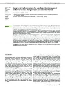
3. Results and Discussion Figure 1 illustrates an example of the application of the proposed image processing strategy for segmentation of melanoma mole. Figure 1(a) is the original color dermoscopy image. Figure 1(b) is image following the application of the DullRazor hari removal algorithm,. It is clear that the effect of darker hairs covering the mole has been greatly reduced. Figure 1(c) is the gray scale transformed image with corrected background. Figure 1(d) is the thresholded image that separated brighter from darker pixels based on automated threshold calculation based on the Otsu’s method [20]. Figure 1(e) illustrates the elimination of stray pixels outside and inside the mole’s region. Figure 1(f) depicts the result following the application of morphological opening and closing filters for smoothing of boundaries. Figure 1(g) shows the border of the mole, which was obtained following the application of an edge-detection Roberts filter. The result of Figure 1(g) was considered as the initialization for the level set method that converged to the final boundaries of the mole using the gradient flow vector approach (Figure 1(h)).
(a)
(b)
(c)
(d)
(e)
(f)
(g)
(h)
Figure 1. (a) Original Image, (b) Application of the DullRazor algorithm for hair removal, (c) Background corrected grey scale image, (d) Thesholded Image, (e) Elimination of stray objects outside and inside the mole’s region, (f) Morphological closing and opening, (g) retaining of the mole’s boundary using Roberts edge detection filter, (h) Final delineation of mole’s boundary following the application of the gradient flow vector algorithm Results showed that the designed system discriminated benign from malignant moles with accuracy higher than 88.6% employing three (3) morphological features (perimeter, roundness and eccentricity) and six (6) textural features extracted from the co-occurrence matrix according to the leave-one-out method. This particular feature combination was also tested on an external cross validation scheme to investigate the generalization of the model to new cases [21]. According to this set-up, the performance of the system was estimated 84%±5%. The proposed system could be used for analysing moles depicted on smart phone images after appropriate training with smartphone images cases. This
3
4th International Conference on Mathematical Modeling in Physical Sciences (IC-MSquare2015) IOP Publishing Journal of Physics: Conference Series 633 (2015) 012079 doi:10.1088/1742-6596/633/1/012079
could assist towards early detection of melanoma cases, if suspicious moles were to be captured on smartphone by patients and be transferred to the physician together with an assessment of the mole’s nature. Acknowledgments The research activities that led to this work, were co-financed by National Funds and by the European Regional Development Fund (ERDF) under the Hellenic National Strategic Reference Framework (NSRF) 2007-2013, concerning the project “MARK1” with ref. number (ISR_3233) within the Bilateral Cooperation between Greece & Israel action. 4. References [1] Holterhues C, Hollestein LM, Nijsten T, Koomen ER, Nusselder W, de Vries E. 2013 The British journal of dermatology 169 389-97. [2] Ruiz D, Berenguer V, Soriano A, Sánchez B. 2011 Expert Sys Appl 38 15217-23. [3] Belli F, Santinami M, Baldini MT, Testori A, Maio A, Cascinelli N. 1989 International journal of radiation applications and instrumentation Part B, Nuclear medicine and biology 16 621-4. [4] Tenenhaus A, Nkengne A, Horn JF, Serruys C, Giron A, Fertil B. 2010 Skin research and technology : official journal of International Society for Bioengineering and the Skin 16 85-97. [5] Schein O, Westreich M, Shalom A. 2009 Harefuah 148 820-3, 55. [6] Abbasi NR, Shaw HM, Rigel DS, Friedman RJ, McCarthy WH, Osman I, Kopf AW, Polsky D. 2004 Jama 292 2771-6. [7] Sahin MT, Ozturkcan S, Ermertcan AT, Gunes AT. 2004 The Journal of dermatology 31 884-9. [8] Deshabhoina SV, Umbaugh SE, Stoecker WV, Moss RH, Srinivasan SK. 2003 Skin research and technology : official journal of International Society for Bioengineering and the Skin 9 348-56. [9] Veierod MB, Parr CL, Lund E, Hjartaker A. 2009 Melanoma research 19 61. [10] Pfahlberg AB, Gefeller O. 2008 Melanoma research 18 300-1. [11] Lorentzen HF, Weismann K, Larsen FG. 2001 Melanoma research 11 495-501. [12] Dermnet-Skin-Disease-Atlas, www.dermnet.com. [13] Lee T, Ng V, Gallagher R, Coldman A, McLean D. 1997 Computers in biology and medicine 27 533-43. [14] Erkol B, Moss RH, Stanley RJ, Stoecker WV, Hvatum E. 2005 Skin research and technology : official journal of International Society for Bioengineering and the Skin 11 17-26. [15] Wolberg WH, Street WN, Mangasarian OL. 1999 Clinical cancer research : an official journal of the American Association for Cancer Research 5 3542-8. [16] Haralick RM, Shanmugam K, Dinstein I. 1973 IEEE Trans Systems Man and Cybernetics SMC-3 610-21. [17] Galloway MM. 1975 Computer Graphics and Image Processing 4 172-9. [18] Specht DF. 1990 IEEE transactions on neural networks / a publication of the IEEE Neural Networks Council 1 111-21. [19] Jain AK, Duin RPW, Mao JC. 2000 IEEE Transactions on Pattern Analysis and Machine Intelligence 22 4-37. [20] Nyma A, Kang M, Kwon YK, Kim CH, Kim JM. 2012 Journal of biomedicine & biotechnology 2012 830252. [21] Ambroise C, McLachlan GJ. 2002 Proceedings of the National Academy of Sciences of the United States of America 99 6562-6.
4