In CDD, one applies a continuous, resonant field to iso- late the driven spin sensor from its ..... Reson.77, 389 (1988). [22] E. C. Reynhardt et al. J. Chem. Phys.
Detecting and polarizing nuclear spins with double resonance on a single electron spin P. London,1 J. Scheuer,2 J.-M. Cai,3 I. Schwarz,3 A. Retzker,4 M.B. Plenio,3 M. Katagiri,5, 6 T. Teraji,6 S. Koizumi,6 J. Isoya,5 R. Fischer,1 L. P. McGuinness,2 B. Naydenov,2 and F. Jelezko2 1
Department of Physics, Technion, Israel Institute of Technology, Haifa, 32000, Israel 2 Institut f¨ ur Quantenoptik, Universitat Ulm, 89073 Ulm, Germany 3 Institut f¨ ur Theoretische Physik, Albert-Einstein Allee 11, Universitat Ulm, 89069 Ulm, Germany 4 Racah Institute of Physics, The Hebrew University of Jerusalem, Jerusalem, 91904, Israel 5 Graduate School of Library, Information and Media Studies, University of Tsukuba, 1-2 Kasuga, Tsukuba, Ibaraki 305-8550, Japan 6 National Institute for Materials Science, Tsukuba, Ibaraki 305-0044, Japan We report the detection and polarization of nuclear spins in diamond at room temperature by using a single nitrogen-vacancy (NV) center. We use Hartmann-Hahn double resonance to coherently enhance the signal from a single nuclear spin while decoupling from the noisy spin-bath, which otherwise limits the detection sensitivity. As a proof-of-principle we: (I) observe coherent oscillations between the NV center and a weakly coupled nuclear spin, (II) demonstrate nuclear bath cooling which prolongs the coherence time of the NV sensor by more than a factor of five. Our results provide a route to nanometer scale magnetic resonance imaging, and novel quantum information processing protocols. PACS numbers: 67.30.hj, 76.70.Fz, 03.67.Lx, 76.30.Mi, 76.90.+d
Measurements of nuclear spin moments are essential to numerous fields including medicine [1], chemistry [2], metrology [3], and quantum information processing (QIP) [4]. Within these, detection and manipulation of single or few nuclear spins may revolutionize microscopy of biological systems with the possibility to reveal the structure of single molecules. Moreover, the potential of single nuclear spins as long-lived quantum memory units is of intense current interest [5]. However, measurements on single or small ensembles (∼ 102 − 104 ) of nuclear spins are extremely challenging due to the small nuclear magnetic moment, leading to typically low polarizations, especially at room temperature. Essentially, one must employ a probe close enough to establish the required sensitivity, since the coupling of the probe and the target spin decreases with the distance between them. A further complication arises due to the difficulty in isolating the signal of a single spin from the background signal produced by the spin-bath. So far single or few nuclear spin measurements have only been achieved with magnetic resonance force microscopy [6], quantum dots [7], and recently with the nitrogenvacancy (NV) center in diamond [8, 9]. The NV center is an attractive system for this task: its optical polarization and spin-dependent photoluminescence along with long ground-state coherence time, make it a perfect probe for sensing nuclear spins coupled to it via dipole-dipole interaction [10]. As the large background noise originating from the spin-bath makes dynamical decoupling techniques a necessity [11], the optimal way to uncover the target signal is not yet fully clear. Recently, three studies have demonstrated the use of pulsed dynamical decoupling to isolate the signal of a single nuclear spin from the nuclear bath [12, 13, 14]. Moreover, measurements of small ensembles have used the mere observation of the effect of statistical fluctuations from nuclear spins state [15, 16]. These can be greatly enhanced by hyperpolarization of the nuclei,
if such is at disposal. Here we experimentally show that one can use continuous dynamical decoupling (CDD) [17, 18, 19] to overcome both challenges, namely to separate a single nuclear spin signal from the bath noise, and to actively enhance the nuclear polarization of the surrounding bath. In CDD, one applies a continuous, resonant field to isolate the driven spin sensor from its environment. The sensor spin is then insensitive to the surrounding spins, however specific frequency components can be selected through a phenomenon known in nuclear magnetic resonance as the Hartmann-Hahn double resonance (HHDR) [19, 20]. We use this technique to experimentally implement an imaging scheme recently proposed in reference [19], and to study existing limitations. We further demonstrate that CDD can be used (through the spinlocking sequence [2]), to enable direct polarization of the target nuclear systems [21, 22]. HHDR occurs when two spins with distinct energy separation are simultaneously driven so that their oscillation (Rabi) frequencies become resonant, or alternatively, when one species is driven with a Rabi frequency that is equal to the natural energy scale of the other spin [20]. Polarization exchange between the two spin systems can then occur via cross-relaxation, which is usually suppressed by their energy mismatch. In our experiments, corresponding to the latter case, we drive a single NV electronic spin with a Rabi frequency that matches the Zeeman energy of a nearby nuclear spin. This enhances the coherent exchange interaction between the two spins, which would otherwise be prohibited due to the three orders of magnitude energy difference (Fig. 1b,c). By adjusting the intensity of the driving field, the NV spin sensor can be used as a tunable, narrow-band spectrometer [19], with spectral resolution limited only by the decoupling efficiency and interrogation time. Hartmann-Hahn dynamics with a single NV-center.
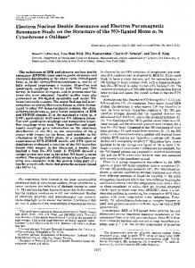
FIG. 1: (color online) Experimental setup and Hartmann-Hahn double resonance. (a) Single NV centers a few microns deep in a (111)-surface oriented ultra-pure diamond were isolated and measured using a home-built confocal microscope. A magnetic field from a permanent magnet located ∼100 µm from the diamond face was aligned to the NV-center axis (z-axis). Microwave fields were delivered to the sample with a co-planar wave guide, fabricated on a cover slip by conventional photolithography. (b) Ground-state energy level structure of the NV center as a function of an axial magnetic field. At a magnetic field of 0.54 T, the NV center ground states ms =0,-1 have an energy separation of 12 GHz, and the nuclear system is split by 6 MHz (Red box). (c) Energy-level diagram of the NV center electronic ground-state and a single 13 C nuclear spin. The coupling term, Ahyp , induces flip-flops between the electron spin and a coupled nuclear spin (green arrow), which are suppressed by the mismatch of the electronic and nuclear energies (blue and red arrows, respectively). (d) Same as in (c), but in the presence of a resonant MW field. The electronic spin is described by the dressed basis |±i (defined in the main text). At a correct MW amplitude, given by Eq. (1), the energy of the electronic dressed-NV center is equal to the energy of the nuclear spin (red arrow) and coherent oscillations between the |+, ↓i level and the |−, ↑i level (green curve) are enhanced .
The interaction of the NV electronic spin, S, with an additional 13 C nuclear spin, I, can be described by the dipole-dipole term HN V −13 C = Sz · Ahyp · I, where Ahyp is the hyperfine vector (defined in [19, 23]), and nonsecular terms are neglected due to the energy mismatch of the two spins [24](Fig. 1b,c). We consider a continuous microwave (MW) field applied to the NV center, whose frequency is resonant with the ms = 0, −1 transition, and with Rabi frequency Ω. The NV center can be described in the MW-dressed basis by |±i = √12 (|0i±|−1i), and its energy scale now becomes Ω (Fig. 1d). When the intensity of the driving field is adjusted, the Hartmann-Hahn energy matching condition given by Ω = γN |Bef f | = γN |B − (1/2) Ahyp | ,
(1)
can be engineered. When Eq. (1) is satisfied, the energy scale of the dressed NV center, Ω, matches the local energy scale of the nuclear spin, γN |Bef f |, where γN is the gyromagnetic ratio, and Bef f is the effective local magnetic field at the nuclear spin position (B is the external magnetic field). If the difference between their energy scales becomes smaller than their mutual coupling strength, the electron-nuclear interaction dominates the dynamics and coherent oscillations between the |±i dressed-states and the (|↑z0 i , |↓z0 i nuclear states (where z 0 is defined along the direction of Bef f (Fig. 1d)) may occur. The probability of finding the dressed NV center, initially set to the state |+i, in the opposite state |−i, after time τ , is p (τ, Ω) =
J2
2 × J 2 + (Ω − γN |Beff |) µq ¶ 2τ , sin2 J 2 + (Ω − γN |Beff |) 2
(2)
where J, given by J=
1 γN |Ahyp | sin θ, 4
(3)
is proportional to the coupling strength, and depends on θ, the angle between Bef f and Ahyp (See [23]). The transition probability (Eq. (2)) shows temporal oscillatory behavior, and a spectral dependence (Lorentzian shape of width J). The former manifests the coherent nature of these oscillations; Starting in the |+, ↓i state, the system evolves according to |Ψi = |+, ↓i cos (Jt) + |−, ↑i sin (Jt). Thus, at time t = π/2J the two spins become maximally entangled, and after a t = π/J a full population transfer occurs; i.e. the states of the two spins are swapped. The latter, i.e. the spectral dependence in Eq. (2), reflects that coherent oscillations between the NV center and weakly coupled nuclear spins are extremely sensitive to detuning from the Hartmann-Hahn condition. Single nuclear spin spectroscopy and imaging. In our experiments, HHDR is performed with single NV centers in a natural abundance (13 C 1.11%) diamond. (Fig. 1a; Details on the diamond sample, and the experimental procedure can be found in the supplementary material, [23]). In order to increase the decoupling efficiency, we apply a high Rabi frequency of ∼ 6 MHz which is matched by the Larmor frequency of the 13 C nuclear spins in a magnetic field of 0.54 T (Fig. 1b). The transition probability in Eq.(2) can be measured in a straightforward way by applying a spin-locking sequence (Fig. 2a) [20, 21]. In the spin-locking sequence, the electronic spin is polarized by illumination with green light for 4 µs, rotated around the x-axis with a π/2 pulse, and then a continuous driving field along the y-axis is applied by introducing a 90◦
Ω (MHz)
NV−center population
6
0.3 0.2 0.1
5.8 5.6 0
5
10
15
20
25
Locking time (µs) 350
300
0
15
0.1
30 250 0
100
350
0 30
5.8 5.6 0
(c)
25 0
200 150
200
Ω [MHz]
6
0.2 0.3 Frequency [MHz]
0.4
0.5
FIG. 2: (Color online) Weakly coupled nuclear spin spectroscopy. (a) The conventional spin-locking sequence (denoted as non-alternating, polarizing sequence) and the alternating spin-locking sequence. 532nm-light pulses are marked in green, MW pulses in blue and pink, for X and Y pulses, respectively. (b) The population of the ms =0 state as a function of the MW driving field Ω and the spin-locking time τ . Colormap is normalized by the contrast observed in a Rabi experiment (performed with a strong driving, Ω > 13MHz). (c) Fourier analysis of the the spin-locking signal for various MW driving fields Ω. Coupling to a single nuclear spin is apparent as a dark spot around 200 kHz, while coupling to the bath produces the lower frequency signal. The blue lines correspond to the predicted values of the resonance driving amplitude γ13 C |Beff | (Eq.(1)) and the flip-flop rate J (Eq.(3)), respectively, for a constant coupling strength and various angles (values are in kHz). The red line is the value of the “bare”Hartmann-Hahn term Ω = γ13 C |B|.
phase shift between the two MW pulses [23]. Readout of the spin state is performed with an additional π/2 rotation around the x-axis and observation on the spindependent fluorescence of the NV center. As will be described later, this protocol produces polarization of the nuclear bath, inhibiting further interaction between the NV center and nearby nuclear spins. Therefore, for spectroscopy measurements, i.e characterization of the coupling strength and orientation, an alternating version of the spin-locking sequence was used, which produces the same experimental signal but without polarization of the nuclear bath. It comprises two similar sequences “+”and “-”, which induce nuclear polarization in alternating di-
rections, thus the net nuclear polarization is zero (Fig. 2a). Fig. 2b shows the measurement of the transition probability for a single NV center interacting with the surrounding spin-bath. Two features which correspond to interaction with nuclear spins can be seen at Ω ' 5.75 MHz and at Ω ' 5.9 MHz. The first agrees well to the expected Larmor frequency for 13 C spins in the applied field (|B| = 5375 G), and shows loss of coherence of the |+i state due to the interaction with many nuclear spins. The second feature is the realization of HHDR with a single nuclear spin, whose coupling strength with the NV center (∼200 kHz) is 2.5 times smaller than the measured inhomogeneous (T2∗ ) linewidth, which characterizes the phase-detection sensitivity without decoupling. Since for our experimental parameters HHDR is efficient for only one hyperfine projection of the nitrogen nuclear spin of the NV-center (a spin half, 15 N nucleus in this case), the high oscillation contrast of 40% indicates an almost complete polarization exchange with the single 13 C spin. The two-dimensional nature of Eq. (2), i.e. the spectral and temporal dependencies, also allows for nuclear spin imaging. Both the optimal Rabi frequency Ωopt which satisfies Eq. (1), and the oscillation rate at double-resonance (Eq. (3)) contain information about the interaction strength, |Ahyp | and its orientation, θ. Inverting Eq. (1) and Eq. (3) for this electron-nuclear pair (Ωopt = 2π × (5.88 ± 0.03) MHz, (J = 2π × (188 ± 30) kHz), we deduce that the coupling for this pair is (1/4)γN |Ahyp | = 2π × 220±40 kHz (which corresponds to a nuclear spin located ∼ 0.5 nm from the NV center, assuming the contribution from the contact term in the interaction is negligible), and the orientation is θ = 56 ± 10◦ . The measured coupling, ∼ 200 kHz, does not mark the ultimate sensitivity of our scheme. Coherent oscillations of the NV-nuclear pair last for more than 25 µs, implying that a 40 kHz coupling could have been detected if such a nuclear spin was present in the vicinity of this NV center. In principle, the interrogation time and hence the sensitivity of the HHDR scheme, is limited by T1ρ - the longitudinal relaxation time of the NV center in the rotating frame [2]. T1ρ times exceeding one millisecond have been measured for NV centers at room temperature [26], which translates to sub-kHz resolution. However both practical and fundamental aspects limit the sensitivity of the scheme. First, fluctuations in the applied MW and static magnetic fields cause broadening and reduce the achieved interrogation time. This may be overcome with improved concatenated continuous driving schemes which mitigate the impact of MW instabilities [27]. Second, the decoupling efficiency of CDD depends on the spectral overlap of the noise spectral density with the decoupling filter function [11]. The overlap may be reduced by modifying either the filter function or the bath spectrum to achieve optimal decoupling performance. For example, to target detection of protons [15, 16], the NV-center can be tuned to the proton spectral region, which is detuned from 13 C nuclear spins in moderate magnetic fields. However, in
1400
127
1200
2
159
1.5
0
2 4 Frequency (MHz)
212
1
318
0.5
637
FWHM [kHz]
T*2 [µs]
2.5
Alternating sequence
(b) 106 FFT Amplitude [Arb]
FWHM [kHz]
3
(a)
IPL
Non−Alternating, polarzing sequence
(c)
1000 800 600 400
0
2 4 6 8 10 Free precession time (µs)
12
0
0
0.5
1
Polarization bias
200 0
200 400 600 Number of polarization sweeps
800
FIG. 3: (color online) Dynamical polarization of the nuclear spin-bath. (a) Free induction decay measured on a single NV center while applying the alternating sequence (blue curve - T2? ∼0.6 µs) and while applying the polarization sequence (red curve T2? ∼3 µs). (b) Dependence of the dephasing time T2? on the polarization bias. The inset shows the Fourier amplitudes of the corresponding FID signals, showing a monotonic narrowing as the bias is increased. The lower blue curve and the upper red curves correspond to polarization balance of 1 and 0, respectively, as plotted in Fig. 3a. (c) Build up of the nuclear spin-bath polarization is reflected in a narrowing of the full-width at half-maximum (FWHM) linewidth in the Ramsey measurements as the number of polarization sweeps is increased. The experimental data (circles) qualitatively agree with simulation of direct polarization mechanism under the spin-temperature approximation (red solid line). To illustrate the effect of magnet drifts on the linewidth, we add an additional offset of 100 kHz to the calculated linewidth (red dashed line). In (b),(c) the T2? values are extracted by fitting the function Y= Λ1 exp(−((f − µ1)/σ1 )2 ) + Λ2 exp(−((f − µ2 )/σ2 )2 ) to the Fourier transform spectra (Fig. 3c inset), and by T∗2 = 1/πσ. The error bars are 95% confidence level from the fitting function.
our experiment we aimed to separate the signal of individual 13 C nuclear spins from a bath comprised from the same nuclear species. Then the spectral density of the bath is peaked near the interrogated frequencies (shifted by only the coupling interaction between the sensor and target spin) leading to a reduced T1ρ coherence time [28]. For a detailed discussion on the sensitivity of the scheme, see [29, 30], and [23]. Nuclear spin-bath polarization. In addition to the detection of single or few nuclear spins, one can utilize the direct flip-flops between the NV center and nuclear spins to polarize the surrounding bath (Figure 3). Under HHDR, the |+, ↓i ←→ |−, ↑i transition allows transfer of polarization from the NV electronic spin to resonant nuclear spins. Therefore, when optical polarization of the NV spin is established at the beginning of each sweep (Fig. 2a, non-alternating, polarizing sequence), an efficient cooling mechanism of nuclear spin-bath is provided. We note that other transitions between the dressedelectronic spin and the nuclear spin can lead to a reversal of polarization (For example, the |+, ↑i ←→ |−, ↓i transition which is shown in Fig. 1d, diagonal cyan arrow). However these transitions are suppressed by an energy mismatch described by ∼ (J/Ω)2 , and in our high-field experiments are of the order 1 × 10⁻³ . As a circumstantial observation, the spin-locking signal shows no oscillations when employing the non-alternating, polarizing sequence [23], entailing that the system was driven into a unharmed state in which all the nuclear spins are polarized to their up state. However, the bath polarization itself can be observed directly from the free-inductiondecay (FID) signal of the NV center, measured using a Ramsey sequence. We note that, in contrast to previous studies of HHDR [21, 22], our use, of an optically pumped NV center as the polarization source allows high efficiency room-temperature nuclear polarization to be
performed. Our experiments also demonstrate this polarization mechanism at the single-spin level for the first time, allowing the associated polarization dynamics to be investigated. The results show that when the bath is polarized (by performing HHDR with Ω ' 5.8MHz and spin-locking time of 7.3µs [23]) the NV phase memory time, T2? , increases five-fold in comparison with a non-polarized bath (Fig. 3a). Further improvements in T2? are limited by magnet drifts of our setup. Since the polarization process now carries a statistical nature, due to the probability of flipping the state of the nearby spins, we can investigate its characteristics. First, we demonstrate a precise control on the degree of polarization and by biasing the rates of polarization towards the up and down state using N+ sweeps of the “+”sequence and N− sweeps of the “-”sequence, with N+ 6=N− . (We define the polarization bias as (N+ -N− )/(N+ +N− )). The smooth transition of T2? times in the range 0.6–3 µs when adjusting the bias from zero to unity, indicates that precise control over spin-bath polarization is achievable (Fig. 3b). Finally, we measure the dynamics of the bath polarization by varying the number of polarization sweeps and measure the FID signal (Fig. 3c). [31] The experimental results are in qualitative agreement with a numerical simulation of a master equation for a single NV center surrounded by 500 13 C spins [32]. The simulation indicates that this scheme polarizes close lying nuclear spins very efficiently (almost one spin per sweep). These spins have the greatest influence on the FID linewidth, but their effect can vary for different NV centers due to variations in the configuration of nearby nuclear spin positions. Polarization of spins which are farther away is much slower and unresolved in our measurement, but it would be of great interest to discriminate this process from spin-diffusion induced polarization [33]. Initializing and probing the
nuclear-bath polarization as demonstrated here, provides an experimental route for characterizing and measuring fundamental processes such as inter-nuclear interactions. Conclusions. We have shown that using continuous dynamical decoupling, a single NV-center can sense minute magnetic fields originating from a single nuclear spin, in spite of the large background noise produced by its environment. We have also demonstrated that a careful tuning of the protocol may bring forth room-temperature hyperpolarization among nuclei in the surrounding bath. The interaction between the NV electronic spin and the nuclear spins preserves its coherent nature, i.e. it can support quantum information protocols using dressed qubits [34]. In biological measurements which are characterized with an extremely disruptive environment, CDD can become an optimal tool: First, it allows improving the decoupling through high Rabi frequencies and high magnetic field with efficient energy-consumption compare to pulsed techniques. In our experiments Rabi frequencies of few MHz were introduced, but Rabi frequencies of hundreds of MHz have been demonstrated using ondiamond MW-waveguides. Second, target spins signal can be amplified using room-temperature nuclear polarization √ which improves the signal-to-noise ratio according to N where N is the number of nuclear spins. Moreover, for diamond-based QIP, the initialization of the nuclear bath to a given state is essential, for example in quantum simulators [35]. We also note that HartmannHahn double resonance can be applied for the detection of electron spins as was demonstrated recently [36]. The authors thank Rainer Pfeiffer and Kay Jahnke for assistance with the experiments. The authors are grateful to Philip Hemmer, J¨org Wrachtrup, Vyacheslav Dobrovitskii and Philipp Neumann for fruitful discussions. The research was supported by DFG (FOR1482, SPP1601 and SFB TR21), EU (DIAMANT), DARPA (QUASAR) and the Alexander von Humboldt Foundation. J.-M.C acknowledges the support of Marie-Curie fellowship and FP7.
[1] P. Mansfield, (Nobel Lecture) Angewandte Chemie International Edition 43, 5456 (2004). [2] Slichter, C. P. Principles of Magnetic Resonance(SpringerVerlag,1990). [3] J. H. Simpson, J. T. Fraser, and I. A. Greenwood.IEEE Trans. Aerosp. Support 1, 1107-1010 (1963). [4] P. Neumann et al. Science 320, 1326 (2008).
[5] [6] [7] [8]
Maurer et al. Science 336, 1283-1286 (2012). J. A. Sidles et al. Rev. Mod. Phys. 67, 249-265(1995). A. Greilich et al. Science 317, 1896-1899 (2007). F. Jelezko, T. Gaebel, I. Popa, A. Gruber, and J.Wrachtrup, Phys. Rev. Lett. 92, 076401 (2004). [9] L. Childress et al. Science 314, 281-284 (2006). [10] M. V. Gurudev Dutt et al., Science 316, 1312-1314 (2007). [11] J.M. Taylor et al., Nature Phys. 4, 810-816 (2008). [12] S. Kolkowitz, Q.P. Unterreithmeier, S.D. Bennett,and M.D. Lukin, Phys. Rev. Lett. 109, 137601 (2012). [13] T. H. Taminiau et al. Phys. Rev. Lett. 109, 137602 (2012). [14] N. Zhao et al. Nature Nanotechnology 7, 657 (2012). [15] H. J. Mamin, M. Kim, M. H. Sherwood, C. T. Rettner, K. Ohno, D. D. Awschalom, and D. Rugar, Science 339, 557 (2013) [16] T. Staudacher, F. Shi, S. Pezzagna, J. Meijer, J. Du, C. A. Meriles, F. Reinhard, and J. Wrachtrup, Science 339, 561 (2013) [17] P. Facchi, D. A. Lidar, and S. Pascazio, Phys. Rev. A 69,032314 (2004). [18] F. F. Fanchini, J. E. M. Hornos, and R.d.J. Napolitano, Phys. Rev. A 75, 022329 (2007). [19] J.M. Cai et al. New J. Phys. 15, 013020 (2013). [20] S.R. Hartmann, and E.L. Hahn, Physical Review 128, 5 (1962). [21] A. Henstra, P. Dirksen, J. Schmidt, and W. Wenckebach, J. Magn. Reson.77, 389 (1988). [22] E. C. Reynhardt et al. J. Chem. Phys. 109, 4100 (1998). [23] Supplementary information. [24] The energy mismatch can result from the NV-center’s zero-field splitting at zero magnetic field, or from the presence of an external magnetic field B (provide that the NV’s is not at the level anti-crossing [25]) [25] G. D. Fuchs, and G. Burkard, and P. V. Klimov and D. D. Awschalom, Nature Phys. 7, 789 (2011). [26] B. Naydenov et al. Phys. Rev. B. 83, 081201(R)(2011). [27] J.M. Cai et al. New J. Phys. 14, 113023 (2012). [28] E. van Oort and M. Glasbeek, Phys. Rev. B. 40, 10, 6509 (1989). [29] M. Loretz, T. Rosskopf, and C. L. Degen, Phys. Rev. Lett. 110, 017602 (2013) [30] M. Hirose, C. D. Aiello, and P. Cappellaro, Phys. Rev. B. 86, 062320(2012). [31] To initialize (depolarize) the nuclear bath before each measurement, we have used many sweeps of the alternating sequence. [32] H. Christ, J.I. Cirac and G. Giedke, Phys. Rev. B. 75, 155324 (2007). [33] R. Fischer et al, http://arxiv.org/abs/1211.5801 (2012). [34] Bermudez, A. and Jelezko, F. and Plenio, M. B. and Retzker, A., Phys. Rev. Lett.107 150503 (2011) [35] J.M. Cai, A. Retzker, F. Jelezko and M.B. Plenio, Nature Physics 9, 168173 (2013). [36] C. Belthangady et al, http://arxiv.org/abs/1211.2749(2012).