in the tick gut and include detection of pathogens and identiï¬cation of the host origin of the blood meal. For host identiï¬cation, a universal primer pair was used ...
VECTOR-BORNE DISEASES, SURVEILLANCE, PREVENTION
Detection and Identification of Pathogens and Host DNA in Unfed Host-Seeking Ixodes ricinus L. (Acari: Ixodidae) BRUNO PICHON, DAMIAN EGAN, MARK ROGERS,1
AND
JEREMY GRAY
Department of Environmental Resource Management, University College of Dublin, BelÞeld, Dublin 4, Ireland
J. Med. Entomol. 40(5): 723Ð731 (2003)
ABSTRACT In this study, we have developed molecular methods for the identiÞcation of reservoir hosts of sylvatic tick-borne zoonoses. The methods are based on the analysis of the blood meal remnant in the tick gut and include detection of pathogens and identiÞcation of the host origin of the blood meal. For host identiÞcation, a universal primer pair was used to amplify part of the vertebrate 18S rRNA gene followed by reverse line blot hybridization using subgroup-speciÞc probes. Analyses of DNA from whole blood of vertebrates identiÞed the correct subgroup of a broad range of vertebrate species (e.g., Ruminantia, Leporidea, Canidae, Murinae, Arvicolinae, Insectivora, Galliformes, Passeriformes) using probes based on the 18S rDNA sequences. Host DNA in the remnants of larval blood meals was detected in the gut of Ixodes ricinus nymphs maintained under natural conditions up to 9 mo after molting. For pathogen identiÞcation, a multiplex polymerase chain reaction was used that targeted parts of the 18S rRNA gene of piroplasm protozoa, the 16S rRNA gene of bacteria, and the intergenic spacer of the Borrelia burgdorferi genospecies complex. The utility of both methods was demonstrated under laboratory conditions by detecting Babesia microti (Franca) and gerbil DNA in 3-mo-old I. ricinus nymphs that had fed on B. microti-infected gerbils as larvae, and under Þeld conditions by analyzing unfed ticks that were collected in a forest. The Þeld study showed that the majority of ticks had fed on ruminants or birds and few on rodents, which is in accord with our knowledge of the fauna in this forest. Few pathogens were detected but the discovery of Borrelia valaisiana and B. burgdorferi s.s. in ticks that had fed on deer and Borrelia afzelii in a tick that had fed on a bird raises questions about the mode of transmission of these spirochetes and possibly about their host speciÞcity. KEY WORDS tick-borne zoonoses, Ixodes ricinus, blood meal analysis, reservoir host identiÞcation
IN THE NORTHERN HEMISPHERE, ticks are considered to be the most important hematophagous arthropods able to transmit disease to humans and animals. Ixodes ricinus, the most common European tick, transmits numerous pathogens including viruses (the cause of tick-borne encephalitis and louping-ill), bacteria (Lyme borreliosis and human granulocytic ehrlichiosis), and protozoa (babesiosis). IdentiÞcation of the reservoir host is essential for understanding the circulation of pathogens in a wild focus and for identiÞcation of possible measures for the control of transmission of these tick-borne diseases. Traditional investigations require the use of inconvenient methods, such as trapping large numbers of wild animals or the extrapolation of experimental data obtained in the laboratory to the natural Þeld situation. In consequence, the true picture tends to emerge slowly and may not be valid for some habitats. Analysis of the vectorÕs blood meal in tandem with the 1 Department of Zoology and Conway Institute of Biomedical and Biomolecular Research, University College of Dublin, BelÞeld, Dublin 4, Ireland
detection of a pathogen in the same tick could identify the pathogen reservoir host much more directly and efÞciently than conventional methods, especially in the absence of signiÞcant transovarial transmission. This approach has been used for hematophagous insect vectors in which the blood meals are relatively fresh and it has been possible to detect host speciesspeciÞc plasma proteins with immunological methods such as enzyme immunoassays and precipitin tests (Magnarelli 1977, Tarry and Boreham 1977, Clausen et al. 1998) In the case of ticks, analysis of relatively fresh blood is not possible because captured host-seeking stages will have fed many months earlier as the previous instar. Kirstein and Gray (1996), using primers that ampliÞed part of the cytochrome B (cytB) gene, demonstrated the feasibility of using polymerase chain reaction (PCR) to identify host DNA in ticks containing remnants of the previous instar blood meal. In the only published Þeld study involving the use of tick blood meal analysis (Gray et al. 1999), PCR product was obtained for most of the ticks analyzed, but determination of the host origin of those infected with Borrelia burgdorferi s.l. failed. Subsequent analysis
0022-2585/03/0723Ð0731$04.00/0 䉷 2003 Entomological Society of America
724
JOURNAL OF MEDICAL ENTOMOLOGY
(not published) showed that because of the high degree of variation in cytB, primers that are sufÞciently universal to cover the full range of possible tick hosts could not be produced. To improve the coverage of putative reservoir hosts for a range of tick-borne pathogens, we have incorporated an additional step for the identiÞcation of major host groups by ampliÞcation of part of the 18S rRNA gene (18S rDNA) of vertebrates. The 18S rDNA coding for the small subunit of ribosomes is a very conserved gene within the vertebrate phylum (Wada and Satoh 1994), and allows the design of universal primers for the ampliÞcation of DNA of most tick-host species. In parallel to the host identiÞcation, a multiplex PCR was developed targeting pathogen 16S or 18S rDNA for detection and identiÞcation of a wide range of tick-borne pathogens. In this report, we demonstrate the relevance of these molecular methods to detect and identify pathogens and hosts associated with experimentally and naturally infected I. ricinus. Materials and Methods Tick Infestations and Maintenance. Larval I. ricinus from a pathogen-free laboratory colony were applied by brush to gerbils [Meriones unguiculatus (Milne Edwards)] (⬇300 per animal) and mice (⬇200 per animal) that had been lightly anesthetized by intraperitoneal injection of sodium pentobarbitone, resulting in sedation for ⬇1 h. The animals were maintained in cages suspended over trays of water from which detached engorged ticks were harvested. A rabbit was infested with the progeny of a single female I. ricinus (⬇1500 larvae) by placing the larval tube inside a 5-cm diameter plastic screw-top capsule attached to the clipped fur of the rabbit with Copydex adhesive (Pritt (Henkel), Winsford, Cheshire, UK). The gerbils used in this study were obtained from Harlan UK, Bicester, Oxon, UK. The mice and rabbits were from the breeding colonies of the University animal house. Their care and use complied with European Community Directive 86/609/EC. Replete larvae were recovered from the trays twice daily, placed in cotton-plugged tubes, and stored at 20⬚C until molting (approximately 6 wk after detachment from the host). Newly emerged nymphs were kept in tubes placed in a Þeld at the soil surface under bushes to prevent high sun exposure. Ticks were subject to natural soil temperatures (5Ð15⬚C) and relative humidities in excess of 85% from the beginning of March 2001 to December 2001. Each month ticks were withdrawn from the batch and stored for short periods at ⫺20⬚C until DNA extraction. Field Tick Collections. To determine the speciÞcity of the host identiÞcation test, nymphal I. ricinus ticks engorging on hosts were collected (in Ireland, Italy, and Spain) into 70% ethanol and stored at 4⬚C until analysis. Unfed host-seeking I. ricinus nymphs, required for assessment of the Þeld utility of the test, were collected in April 2002 by ßagging the vegetation at Portumna Forest Park, County Galway, Republic of Ire-
Vol. 40, no. 5
land. The sample site consists of a mixed deciduous forest of ⬇600 hectares in which the vegetation and a thick, humid litter layer permits survival of a large I. ricinus population. The vertebrate fauna consists mainly of a large herd of fallow deer (Dama dama), which varies in size between 200 and 400 animals depending on culling policy. Small numbers of wood mice (Apodemus sylvaticus), bank voles (Clethrionomys glareolus), pygmy shrew (Sorex minutus), hedgehogs (Erinaceus europaeus), red squirrels (Sciurus vulgaris), foxes (Vulpes vulpes), stoats (Mustela erminea), and badgers (Meles meles) are also present. Pheasants (Phasianus colchicus) and many passerine bird species are present, the blackbird, Turdus merula, being the most abundant ground-feeding species. Preparation of DNA from Ticks. DNA of unfed ticks was puriÞed using a modiÞed method described previously by Guy and Stanek (1991). Ticks were individually placed in 1.5-ml tubes containing 5 l of 0.7 M ammonium hydroxide. Tubes were frozen in liquid nitrogen and ticks were ground with a Micropestle (Eppendorf, Cambridge, United Kingdom). The homogenates were mixed with 95 l of 0.7 M ammonium hydroxide and heated to 100⬚C for 15 min. The solutions of DNA were evaporated at 100⬚C for 15 min to reduce the volume to 50 l and then centrifuged for 10 min at 10,000 g. DNA was stored at ⫺20⬚C until PCR analysis. Vertebrate DNA from engorged ticks was extracted using the QIAamp DNA blood kit (QIAGEN) according to the manufacturerÕs protocol. DNA was eluted in 200 l of ddH2O. Ten microliters of the DNA solution were used for PCR ampliÞcation. Preparation of DNA from Vertebrate Samples. For mammal species, 200 l samples of whole blood were treated using the QIAamp DNA blood kit (QIAGEN) according to the manufacturerÕs protocol. DNA was eluted in 200 l of ddH2O. For tissue samples, 20 Ð50 mg of muscle were extracted using the QIAamp DNA tissue kit according to the manufacturerÕs protocol. DNA was eluted in 200 l of ddH2O. For bird species, DNA from 20 l of whole blood diluted in 200 l of 0.22 M-Þltered phosphate-buffered saline (PBS) was extracted by the phenol/chloroform method and ethanol precipitation. Pellets of DNA were resuspended in 50 l of ddH2O. Extraction of Pathogen DNA. Strains of spirochetes [Borrelia afzelii (Canica et al.) NE1272, B. burgdorferi sensu stricto (s.s.) NE1250, Borrelia garinii (Baranton et al.) NE1216, and Borrelia valaisiana (Wang et al.) NE1301] were cultivated in BSKH medium (Sigma) completed with 6% vol:vol rabbit serum to a concentration of ⬇1 ⫻ 106 bacteria per ml. A 1-ml sample of culture was washed twice in 0.22 M-Þltered PBS (centrifugation for 10 min at 10,000g). Cells were homogenized in 100 l of ddH2O and boiled for 15 min at 100⬚C. After lysis, cell debris was pelleted by centrifugation at 15,000g for 5 min. Babesia spp. DNA was obtained by infecting gerbils with a European strain of Babesia microti (HK strain) (Gray et al. 2002) or with Babesia divergens Mac Fadyean and Stockman (TX-8 strain). Parasitemias were assessed by microscopic
September 2003
PICHON ET AL.: DETECTION OF PATHOGEN AND HOST DNA IN TICKS
725
Table 1. Oligonucleotide sequences of primers used in PCR for amplification of the 18S rDNA of vertebrates and for amplification of the 18S rDNA, the 16S rDNA and the intergenic spacer 23S–5S rDNA of pathogens Primer ID 0033 0049 0066 0067 0202 0204 0206 0209 23SN2 5SCB a
Sequences 5⬘-⬎3⬘
Targeted genes
Origins
Positions
ttctagagctaatacatgccga biotin-ycgaggttatctagagtcacc acctggttgatcctgcca taccatcgaaagttgataggg acggtagggtattggcct biotin-ctaagaatttcacctctgacagt tcctacgggaggcagca biotin-ctcgttgcgggacttaacc accatagactcttattactttgacca biotin-gagagtaggttattgccaggg
Vertebrata 18S rDNA Vertebrata 18S rDNA Vertebrata 18S rDNA Vertebrata 18S rDNA Piroplasmida 18S rDNA Piroplasmida 18S rDNA Eubacteria 16S rDNA Eubacteria 16S rDNA Borrelia burgdorferi s.l. 23S-5S spacer Borrelia burgdorferi s.l. 23S-5S spacer
X00686 X00686 X00686 X00686 AB050732 AB050732 X85199 X85199
160Ð181 309Ð289 2Ð19 377Ð357 284Ð301 854Ð832 308Ð324 1070Ð1052
a a
Designed by Rijpkema et al. 1995.
examination of Giemsa-stained blood smears. Blood samples (200 l) with parasitemias of 7.0% and 24% for B. microti and B. divergens, respectively, were collected in tubes containing heparin. To obtain Babesia bovis (Babes) DNA (unknown South African strain from an infected bovine), a stained thin blood smear of parasites was scraped with a sterile scalpel blade and the dried infected blood was homogenized in 200 l of ddH2O and extracted using the QIAamp DNA blood kit. PuriÞed DNA was eluted in 200 l of ddH2O. Anaplasma phagocytophilum (Foggie) DNA of strains TBF lephamore, TBF 538 Perth; TBF R153; Sheep 471 Dpi four OS; AB and Cairn, were kindly provided by Dr N. Ogden (University of Liverpool, Liverpool, UK). DNA Amplification. Vertebrate DNA was ampliÞed by PCR using the universal primers 0033 and 0049 (Table 1), which were designed to generate 150-bp (mammals) or 120-bp (birds) 18S rDNA PCR fragments. The Þnal reaction volume used for ampliÞcation was 50 l (dNTP 200 M, primers 0.2 M, PCR buffer, and Taq polymerase 1 U [FastStart Taq polymerase; Roche Molecular Biochemicals, Roche Diagnostic, Lewes, United Kingdom]). Before adding the DNA sample, the reaction mixtures were incubated for 60 min at 37⬚C in the presence of the restriction enzyme MspI (5 U) to eliminate traces of human DNA. MspI was degraded by heating at 70⬚C for 15 min followed by chilling on ice, and then 10 l of DNA sample was added to the reaction mixture. AmpliÞcation was started by an initial incubation at 94⬚C for 4 min to activate TaqDNA polymerase, followed by 50 cycles of denaturation (30 s at 94⬚C), annealing (30 s at 59⬚C) and extension (30 s at 72⬚C), and ended by 5 min at 72⬚C. PCR products were stored at 4⬚C until reverse line blot (RLB) analysis. DNA of pathogens was ampliÞed by multiplex PCR. Universal primers 0202 and 0204 (Table 1) were selected for ampliÞcation of a fragment (⬇550 bp) containing the V4 region of the 18S rDNA of piroplasmid protozoa. A fragment (⬇770 bp) of bacterial 16S rDNA was ampliÞed using universal primers 0206 and 0209 (Table 1), and the 23S-5S intergenic spacer of the B. burgdorferi complex was ampliÞed using primers 23SN2 and 5SCB (Table 1) previously described by Rijpkema et al. (1995). A Þnal reaction volume of 50 l (1 U of Taq polymerase [GIBCO BRL, INVITRO-
GEN-Life Technologies, Paisley, United Kingdom], PCR buffer, 2 mM MgCl2, 0.2 M of each primer, and 200 M of each dNTP) was used for the multiplex PCR, according to the following procedure: 94⬚C for 2 min, then 14 cycles of 94⬚C for 30 s, 62⬚C for 30 s with a decrease of 0.5⬚C at each cycle, and 72⬚C for 45 s, followed by 45 cycles of 94⬚C for 30 s, 55⬚C for 30 s, 72⬚C for 45 s, and, Þnally, 72⬚C for 5 min. The sensitivity of the detection was assessed by amplifying serially diluted DNA solutions of quantiÞed 18S or 16S rDNA PCR product of respectively Babesia microti HK or B. burgdorferi s.s. NE1250 and A. phagocytophilum (TBF lephamore). PCR products were quantiÞed by spectrophotometry and the DNA was serially diluted to a Þnal concentration of one copy of gene/l. Competition between targeted DNA and unspeciÞc DNA was tested by mixing unspeciÞc DNA (from other pathogen species) or tick extract, with serially diluted DNA solution of targeted DNA. PCR negative controls (10 l of Molecular Grade H2O, Sigma-Aldrich, Dublin, Ireland) were added in all series of PCR Detection of Biotin-Labeled DNA by RLB. 5⬘-End amino-link probes (Tables 2 and 3) were blotted in lines using a Miniblotter 45 (Immunetic, Cambridge, MA) on an activated Biodyne C membrane (Pall, Ann Arbor, MI), as described by Rijpkema et al. (1995). The membranes were stored at 4⬚C before use. Before hybridization, the blots were placed in the miniblotter with the slots perpendicular to the probe lines, and the slots were Þlled with 150 l of hybridization buffer (2⫻ SSPE, 0.1% wt:vol sodium dodecyl sulfate) and incubated for 45 min at 42⬚C for pathogen DNA or 62⬚C for vertebrate DNA. The buffer was aspirated, the slots Þlled with 10 l of denatured biotinylated PCR products diluted in 140 l of hybridization buffer, and the miniblotter reincubated at 42⬚C or 62⬚C for 90 min. The membrane was then washed twice for 5 min using the hybridization buffer at room temperature and twice for 5 min in the same buffer preheated to 50⬚C (pathogen DNA) or 62⬚C (vertebrate DNA). Biotin-labeled DNA was detected using streptavidinalkaline phosphatase conjugate and CDP-start (Roche) chemiluminescent substrate according to the manufacturerÕs procedure. Sequencing of Vertebrate 18S rDNA. Universal primers 0066 and 0067 (Table 1) were designed to
726 Table 2.
JOURNAL OF MEDICAL ENTOMOLOGY
Vol. 40, no. 5
Oligonucleotide sequences of probes used in RLB for detection and identification of amplified vertebrate DNA
Probes sequence
Probe ID
TM
Level of identiÞcation
gggctcgcccggcggct gggcccgcccggcggct
PRNA029 PRNA012
72.7 72.7
Galliformes Passeriformes
ccgacctccggggacgc
PRNA010
72.7
agcctccccccggcccc agcctccctccggcccc ctccctccggctccggc agcctccccccggcccc
PRNA019 PRNA020 PRNA030 PRNA019
74.7 75 72.6 74.7
Aves (Galliformes and Passeriformes) Leporidae/Carnivora Canidae Insectivora Leporidae/Canivora
cgggggggtgggcgccg No probe designed acctctcggctccggccgc gagctcccccgcggccc ccctcccggctccggccg gctccggccgtgggggcg cggtcagcttccccccgg agcctcctcccggcccc
PRNA018 Ð PRNA015 PRNA017 PRNA011 PRNA023 PRNA026 PRNA013
77.4 Ð 74.9 75 72.7 77.2 72.7 77.2
Leporidae Primates Apodemus sp Clethrionomys sp Murinae/Gerbilinae Meriones unguiculatus Sciurus sp Ruminantia
Accession numbers of sequences used for alignment and design D38359a (Coturnix coturnix L); D38360a (Gallus gallus L) AY150550b(Erithacus rubecula L); D38344a(Passer montanus); L D38343a(Parus major L); AY150544b(Turdus merula L); D38342a(Troglodytes troglodytes L) as above AY150542b(Felis catus L); AY150554b (Meles meles L) AY150549b(Vulpes vulpes L) AY150551b and AJ311675a(Erinaceus europaeus L) AY150553b, X00640a and X06778a(Oryctolagus cuniculus L); (Lepus europaeus Pallas AY150540b) X03205a(Homo sapiens L); AY150541b(Apodemus sylvaticus L) AY150543b(Clethrionomys glareolus Schreber) X00686a (Mus musculus); X01117a and K01593a(Rattus norvegicus); AY150552b(Meriones unguiculatus) AY150546b(Sciurus vulgaris L) AB016657a and AF176811a(Bos taurus L); AY150545b(Capreolus capreolus L); AY150547b(Cervus elaphus L); AY150548b(Cervus nippon Temmnick)
a
Sequence obtained from Genbank database at the NCBI site http://www.ncbi.nlm.nih.gov. Sequence obtained in this study. Accession numbers in bold correspond to the sequences used for designing probes. b
generate PCR fragments of 400 bp (mammal species) and 350 bp (bird species). A Þnal reaction volume of 50 l (dNTP 200 M, primers 0.2 M, PCR buffer and Taq polymerase 1 U [FastStart Taq polymerase; Roche Molecular Biochemicals]) was used for the ampliÞcations. Treatment with restriction enzyme MspI and heat inactivation at 70⬚C for 15 min before the addition of the DNA sample ensured the removal of any contaminating human DNA. AmpliÞcation was started by an initial incubation at 94⬚C for 4 min followed by 35 cycles of 30 s at 94⬚C, 30 s at 57⬚C, and 45 s at 72⬚C, and ended by 5 min at 72⬚C. PCR products were puriÞed from 1% wt:vol agarose gel by the UltraFree DNA Kit (Millipore, Watford, United Kingdom) and doublestrand sequenced by Genome Express (Meyland, France) using primers 0066 and 0067. Sequence Analysis. DNA sequences were aligned using ClustalW multiple-alignments software provided in the BioEdit package, version 5.0.7 (Hall 1999).
Table 3. Probe ID Ptg001 Ptg002 Ptg019 Ptg003 Ptg009 Ptg010 Ptg011 Ptg012 Ptg013 Ptg007
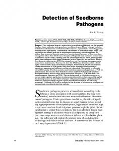
Results Design of the Vertebrate Subgroup-Specific Probes. DNA from 15 vertebrate species (for which sequences of 18S rDNA were not available in the EMBL/GenBank database) were ampliÞed for sequencing. For each species, a unique PCR fragment ranging from 350 to 400 bp was directly sequenced. The sequences obtained together with those available in the database were aligned to design subgroup speciÞc probes (Table 2). DNA from blood samples or tissue samples was ampliÞed using the primers 0033 and 0049, and the PCR products tested against the array of subgroupspeciÞc probes by RLB. Figure 1 shows an RLB assay of 16 species against an array of nine probes). Eventually, 25 vertebrate species were identiÞed at the correct subgroup level (Table 4). The speciÞcity of the test was further conÞrmed by correct determina-
Oligonucleotide sequences of probes used in RLB for detection and identification of amplified pathogen DNA Sequence of 5⬘ primaryamined oligonucleotides
Level of identiÞcation
TM (⬚C)
cttccgagcgtttttttatt gttaatattgactaatgtcga aggtttcgcctgtataattga cgaacttctgggtcaagac ctttgaccatatttttatcttcca aacaccaatatttaaaaaacataa aacatgaacatctaaaaacataaa aacatttaaaaaataaattcaagg cattaaaaaaatataaaaaataaatttaagg tggggattttttatctctgtg
Babesia microti (18S rDNA) Babesia divergens (18S rDNA) Babesia bovis (18S rDNA) Borrelia burgdorferi s.l. (16S rDNA) Borrelia burgdorferi s.l. (23SÐ5S spacer) Borrelia burgdorferi s.s. (23SÐ5S spacer) Borrelia garinii (23SÐ5S spacer) Borrelia afzelii (23SÐ5S spacer) Borrelia valaisiana (23SÐ5S spacer) A. phagocytophilum (16S rDNA)
57.7 57.3 61.2 64.1 61.1 56.0 57.7 56.0 58.7 61.0
TM, melting temperatureÑ calculated with the formula of Bolton and Mc Carthy (Sambrook et al., 1989). Designed by Gubbel et al. 1999. Designed by Rijpkema et al. 1995.
a
b
Reference sequences
Positions
AB050732
618Ð637
a a
X85199 b
701Ð719
b b b b
AF084907
780Ð800
September 2003
PICHON ET AL.: DETECTION OF PATHOGEN AND HOST DNA IN TICKS
Fig. 1. AmpliÞcation and RLB hybridization against vertebrate subgroup-speciÞc probes (Table 2) targeting the 18S rDNA of 16 vertebrate species: lanes 1 and 2: negative controls; lanes 3Ð18: DNA puriÞed from total blood: lane 3: Phasianus colchicus, lane 4: Alectoris rufa, lane 5: Perdix perdix, lane 6: Erithacus rubecula, lane 7: Turdus merula, lane 8: Lepus europaeus, lane 9: Meles meles, lane 10: Felis catus, lane 11: Vulpes vulpes, lane 12: Canis familiaris, lane 13: Capreolus capreolus, lane 14: Cervus elaphus, lane 15: Clethrionomys glareolus, lane 16:.Apodemus sylvaticus, lane 17: Rattus norvegicus, lane 18: Mus musculus.
tion of blood meal identity in engorging ticks collected from seven host species (Table 5). Persistence and Detection of Vertebrate DNA in Tick Blood Meal Remnants. Vertebrate DNA present in the blood meal remnants of nymphs developed from larvae fed on mice and rabbits was ampliÞed using primers 0033 and 0049 at different times after the molt. PCR product was detected and correctly identiÞed by RLB hybridization in all nymphs tested in each of the Þrst 7 mo after molting and 4 of 10 were still positive after 9 mo. No host DNA was detected at 10 mo (Table 6). Amplification and Identification of Pathogen DNA. The multiplex PCR was developed using DNA extracted from known strains of pathogens. The speciÞcity of detection was assessed by amplifying DNA of Table 4.
727
different species of bacteria and protozoa including B. burgdorferi s.l., A. phagocytophilum, B. microti, B. divergens, and B. bovis. When using the set of primers (0202/0204/0206/0209/23SN2/5SCB) for the PCR reaction, a PCR product was observed for all DNA samples by electrophoresis on agarose gel. RLB analysis of biotin-labeled DNA using the probe design shown in Table 3 demonstrated that hybridizations with the probe array against protozoal 18S rDNA, bacterial 16S rDNA, and the 23S-5S rDNA intergenic spacer of the B. burgdorferi group were able to distinguish and successfully identify the PCR products (Fig. 2). Moreover, no cross-hybridization of PCR products between unrelated species of pathogens was observed. The sensitivity of the multiplex PCR was tested with a range of DNA concentrations of A. phagocytophilum (TBF lephamore), B. burgdorferi s.s. (NE1250), and B. microti (HK). The sensitivity of detection by RLB hybridization was determined to be approximately 10 copies of DNA for the three species. The presence of nonspeciÞc DNA (Fig. 3) or tick extract from pathogen-free laboratory-reared ticks (not shown) does not seem to affect the sensitivity of the reaction. Detection of Pathogens and Identification of the Origin of the Infecting Blood Meal in Experimentally Infected Ticks. Larvae were allowed to feed until repletion on gerbils with patent infections of B. microti. Three months after the molt, nymphs that developed from these engorged larvae were tested for the presence of B. microti and for host identiÞcation. Pathogens were detected by multiplex PCR of extracts of tick DNA; PCR products were analyzed by RLB hybridization and the presence of B. microti DNA was detected in each sample (Fig. 4A). In a second step, primers 0033 and 0049 were used in PCR of the extracts, and the PCR products were analyzed by RLB
Identification of vertebrate species by RLB hybridization of amplified 18S rDNA
Species
Common name
Probes hybridized
Level of identiÞcation
Alectoris rufa L. Phasianus colchicus L. Perdix perdix L. Turdus merula Turdus pilaris L. Erithacus rubecula Apodemus sylvaticus Clethrionomys glareolus Mus musculus Rattus norvegicus Meriones unguiculatus Sciurus carolinensis Sciurus vulgaris Oryctolagus cuniculus Lepus europaeus Felis catus Meles meles Canis familiaris Vulpes vulpes Erinaceus europaeus Bos taurus Ovis aries L. Cervus elaphus Cervus nippon Capreolus capreolus
red-legged partridge pheasant grey partridge blackbird thrush robin wood mouse bank vole mouse rat gerbil grey squirrel red squirrel rabbit European hare cat badger dog red fox hedgehog cow sheep red deer sika deer roe deer
PRNA010 ⫹ PRNA029 PRNA010 ⫹ PRNA029 PRNA010 ⫹ PRNA029 PRNA010 ⫹ PRNA012 PRNA010 ⫹ PRNA012 PRNA010 ⫹ PRNA012 PRNA015 PRNA017 PRNA011 PRNA011 PRNA011 ⫹ PRNA023 PRNA026 PRNA026 PRNA018 PRNA018 PRNA019 PRNA019 PRNA020 PRNA020 PRNA030 PRNA013 PRNA013 PRNA013 PRNA013 PRNA013
Aves/Galliformes Aves/Galliformes Aves/Galliformes Aves/Passeriformes Aves/Passeriformes Aves/Passeriformes Apodemus spp (Muridae) Clethrionomys spp (Arvicolinae) Muridae Muridae Muridae/M. unguiculatus Sciurus spp Sciurus spp Leporidea Leporidea Lagomorpha/Carnivora Lagomorpha/Carnivora Canidae Canidae Erinaceus spp Ruminantia Ruminantia Ruminantia Ruminantia Ruminantia
728
JOURNAL OF MEDICAL ENTOMOLOGY
Table 5.
Vol. 40, no. 5
Identification of the origin of the blood meal in engorged ticks collected from vertebrate hosts
Blood meal origin
Common name
No. of ticks
Probes hybridized
IdentiÞcation
Apodemus sylvaticus Sciurus vulgaris Erinaceus europaeus Lepus europaeus Felis catus Capreolus capreolus Phasianus colchicus
Wood mouse Red squirrel Hedgehog Brown hare Domestic cat Roe deer Pheasant
5 3 2 3 1 4 2
PRNA015 PRNA026 PRNA030 PRNA18 ⫹ PRNA019 PRNA019 PRNA013 PRNA010 ⫹ PRNA029
Apodemus sp Sciurus sp Insectivora Leporidae Carnivora Ruminantia Galliformes
against the set of vertebrate subgroup-probes. The presence of gerbil DNA was successfully detected in the tick blood meal remnants (Fig. 4B). Host and Pathogen Identification in Field-Collected Ticks. Host identiÞcation analysis was attempted on 49 nymphs collected from vegetation and PCR product was obtained for 26 of them. Ruminant, bird, and rodent DNA were identiÞed in 14, 10, and 2 nymphs, respectively. Host DNA could not be detected in the remaining 23 ticks as determined by electrophoresis on agarose gel. For pathogen detection, multiplex PCR followed by RLB hybridization against an array of 10 probes (Table 3) of the PCR products revealed the presence of Þve species of pathogens (A. phagocytophilum, B. afzelii, B. burgdorferi s.s., B. garinii, and B. valaisiana) in Þve ticks (Table 6). One nymph carried a simultaneous infection with B. afzelii and B. burgdorferi s.s. No B. divergens or B. microti were detected. Ruminant DNA was detected in nymphs infected by A. phagocytophilum, B. valaisiana and B. burgdorferi s.s. Passerine bird DNA was detected in a nymph infected by B. garinii, in a nymph infected by B. valaisiana, and in the nymph co-infected by B. afzelii and B. burgdorferi s.s.
brate group and its sequence presents some highly homologous regions suitable for designing universal primers. The sequence analysis of this part of the 18S rDNA revealed that one variable region is speciÞc to subgroups of vertebrates. By using a set of subgroupspeciÞc probes, DNA extracted from blood and also from engorging ticks collected from animals was correctly identiÞed to the subgroup level for all species of vertebrates tested. These results demonstrate that the use of 18S rDNA as a molecular target for the study of many tick-borne zoonoses may be very useful for the identiÞcation of the host subgroup acting as the pathogen reservoir. However, within subgroups, genera or species cannot be discriminated using the 18S marker because of the low level of variability in sequences. If identiÞcation of host species is required, additional analysis must be performed, consisting of ampliÞcation of part of the cytochrome B gene, followed by the use of speciÞc probes in a RLB assay (Kirstein and Gray 1999). Work in this laboratory using cytochrome B species-speciÞc primers has shown that it is possible to include Þve pairs of primers simultaneously for the identiÞcation of hosts at species level (data not shown).
Discussion Molecular identiÞcation of the vertebrate host by analyzing the gut contents of unfed host-seeking ticks was achieved in this study by targeting 18S rDNA. This particular gene is very conserved within the verteTable 6.
Persistence of vertebrate DNA in the gut of ticks No. positive ticks/ No. analyzed ticka
Age of the tick
Week after the blood meal
Months after the molt
1 1 3 4 5 6 7 8 9 10
Larvae fed on mice
Larvae fed on rabbit
5/5 5/5 5/5 5/5 5/5 5/5 5/5 5/5 ND ND
5/5 5/5 5/5 5/5 5/5 5/5 5/5 8/10 4/10 0/10
a The 18S rDNA of vertebrates were ampliÞed by PCR using primers 0033 and 0049 and identiÞed by RLB hybridization. ND, not done.
Fig. 2. Detection and identiÞcation of pathogen species by multiplex-PCR and RLB hybridization against oligonucleotide probes (Table 3). Lane 1: Babesia microti, lane 2: Babesia divergens, lane 3: Babesia bovis, lane 4: Borrelia burgdorferi s.s. (NE1250), lane 5: Borrelia garinii (NE1216), lane 6: Borrelia afzelii (NE1272), lane 7: Borrelia valaisiana (NE1301), lanes 8Ð13: Anaplasma phagocytophilum (lane 8: TBF lephamore, lane 9: TBF 538 Perth, lane 10: TBF R153, lane 11: Sheep 471 Dpi four OS, lane 12: AB, lane 13: Cairn), and lane 14: negative control
September 2003
PICHON ET AL.: DETECTION OF PATHOGEN AND HOST DNA IN TICKS
Fig. 3. Competition between Babesia microti and Borrelia burgdorferi s.s. DNA for ampliÞcation by multiplex PCR and reverse line blot hybridization. Lane 1: negative control, lanes 2Ð7: PCR mixture contains 500 copies of the 16s rDNA of Borrelia burgdorferi s.s. (NE1250) and the following amounts of copies of the 18s rDNA of Babesia microti (HK): lane 2, 10 copies; lane 3, 25 copies; lane 4, 50 copies; lane 5, 100 copies; lane 6, 250 copies; and lane 7, 500 copies.
The method developed here is sufÞciently sensitive for the detection of minute amounts of DNA (of the larval host) in unfed nymphs. When fed larvae were maintained in the Þeld under natural temperature conditions, DNA was detected in all analyzed ticks 7 mo after their molt to nymphs. After 9 mo, DNA detection was reduced to 4 (40%) of 10 nymphs and had declined to 0% at 10 mo, indicating that by this time the host DNA in the tick gut had become too degraded to be ampliÞed by PCR. The duration of development of I. ricinus from the larval blood meal to host-seeking activity can vary from a few weeks to more than 1 yr (Gray 1991). This duration depends, in
Fig. 4. Experimental identiÞcation of the reservoir host of Babesia microti by analysis of Ixodes ricinus nymphs developed from larvae fed on infected gerbils. (A) Detection of Babesia microti in nymphs by multiplex PCR and reverse line blot hybridization. Lane 1Ð4: nymphs of I. ricinus, lane 5: negative control, lane 6: Babesia microti (HK). (B) IdentiÞcation by PCR and reverse line blot hybridization of the nutritive host for the nymphs analyzed in A. Lane 1Ð4: nymphs of I. ricinus, lane 5: negative control, and lane 6: Meriones unguiculatus.
729
particular, on the climatic conditions and the time of year when the larval blood meal was taken. Blood meal analysis of spring/summer-active I. ricinus nymphs, which are the most important cohort for the transmission of disease, will thus present greater problems than autumn-active nymphs because of the increased length of time before host-seeking activity after the larval molt. The method of host identiÞcation described here sacriÞces some sensitivity in the interest of improved speciÞcity (demonstrated by analysis of ticks collected from animals). Any decreased sensitivity compared with the earlier method (Kirstein and Gray 1996) could be a result of the abandonment of the nested PCR because of contamination dangers, and to the selection of a nuclear gene target, 18S rDNA, rather than the mitochondrial cytochrome B gene, which cannot be used for the detection of host groups. Nevertheless, sufÞcient sensitivity remained to detect vertebrate DNA in the blood meal remnants of a proportion (53%) of host-seeking ticks collected in April 2002 at a site (Portumna Forest Park) where mammal hosts, especially deer, are dominant. Results from ongoing analyses indicate that DNA from birds can be detected in ticks collected much later in the year (late JuneÑ by which time much DNA degradation would be expected to have occurred), presumably because bird erythrocytes contain nuclei, so that birds provide more host nuclear DNA in a tick blood meal than do mammals. To detect mammal DNA in a greater proportion of ticks, it may be necessary to collect them from the vegetation soon after their emergence from winter torpor in early March. To determine the host origin of pathogens, a method for pathogen detection must be integrated with the method for host identiÞcation. Many PCR methods for the detection and identiÞcation of pathogens in ticks have already been described (reviewed by Sparagano and Jongejan 1999). It has been shown elsewhere (Schouls et al. 1999, Gubbels et al. 1999, Georges et al. 2001) that related pathogen species may be discriminated using PCRs that target highly conserved genes such as 16S rDNA for bacteria and 18S rDNA for protozoa. Universal primers are used for ampliÞcation of the DNA and speciÞc probes for identiÞcation of the PCR products. In the current study, we developed a multiplex-PCR method targeting 16S rDNA of bacteria and 18S rDNA of piroplasms. Eight pathogen species (six bacteria and three babesias) were tested and successfully ampliÞed by the set of universal primers used in the multiplex PCR. By designing speciÞc probes within the ampliÞed region, all pathogen species were identiÞed in the RLB assay. This approach permits the identiÞcation of a broad range of pathogen species (including both prokaryotic and eukaryotic species) in a single assay, but problems arise in the discrimination of some species belonging to the same genomic group because of high homology, for example, the genomic species of the B. burgdorferi complex. To overcome this particular problem, we included the primers 23SN2/5SCB in the multiplex PCR, to amplify a more variable region that distin-
730
JOURNAL OF MEDICAL ENTOMOLOGY
Vol. 40, no. 5
Table 7. Pathogen detection and origin of the blood meal in questing nymphs sampled in April 2002 at Portumna Forest Park (Co. Galway, Republic of Ireland) Pathogen detection
Origin of the blood meal
BA ⫹ BBss
BBss
BG
BV
AP
Noninfected
Not done
Passerifomes Galliformes Apodemus sp Sciurus sp Ruminants Undetermined
1 Ð Ð Ð Ð Ð
Ð Ð Ð Ð 1 Ð
1 Ð Ð Ð Ð Ð
1 Ð Ð Ð 1 Ð
Ð Ð Ð Ð 1 Ð
1 3 1 1 3 10
3 Ð Ð Ð 8 13
BA, B. afzelii; BBSS, B. burgdorferi sensu stricto; BG, B. garinii; BV, B. valaisiana, AP, A. phagocytophilum.
guishes the genospecies of B. burgdorferi s.l. (Rijpkema et al. 1995). The sensitivity of the multiplex PCR was tested by assessing the threshold of detection of the RLB and showed that a small amount of DNA, corresponding to 10 copies of genes (bacteria or protozoa) per PCR reaction, could be detected. The presence of nonspeciÞc DNA or tick extract does not seem to affect the sensitivity of the method. In recent years there have been many reports of the presence of more than one pathogen species in a single tick. Ixodes ricinus co-infections can involve two species of bacteria belonging to the same genus (LeubaGarcia et al. 1994, Pichon et al. 1995) or two separate genera (Magnarelli et al. 1991, Schouls et al. 1999, Cinco et al. 1997) or can be a mix of species of bacteria and protozoa (Varde et al. 1998, Piesman et al. 1986). The particular usefulness of the multiplex PCR lies in the detection of possible co-infections in a single assay. For large-scale studies, this technique should decrease manipulation of the sample and therefore reduce the risk of contamination. Combining pathogen and host identiÞcation for the same sample could also help solve the problem of the origin of co-infections detected in nymphs. The integrated host and pathogen identiÞcation methods were validated under controlled laboratory conditions using B. microti in gerbils (M. unguiculatus) and their utility under Þeld conditions was investigated in a study in which nymphal ticks collected from vegetation were analyzed. Five pathogen species were detected and some of them were distinctly associated with particular vertebrate groups; A. phagocytophilum with ruminants (deer in this case), and B. burgdorferi s.s., B. garinii, and B. valaisiana with birds. These associations have already been made with conventional methods such as xenodiagnosis, PCR, and culture (Stuen et al. 2001, Humair 2002). Three other weak associations were less expected: B. afzelii with birds, B. burgdorfer s.s. with ruminants, and B. valaisiania with ruminants. These latter results do not necessarily imply that the hosts involved are reservoir competent for the particular pathogens and the associations may be a result of less signiÞcant phenomena in B. burgdorferi s.l. transmission such as transovarial transmission (Zhioua et al. 1994), co-feeding transmission (Gern and Rais 1996), or interrupted feeding followed by attachment to another host (Shih and Spielman 1993).
This Þeld study demonstrated the feasibility of determining the host origin of the larval blood meal in nymphs collected from vegetation, and thus the host origin of certain pathogens. More data are required to assess the signiÞcance of some of the unexpected host/ pathogen associations we observed. Acknowledgments We are grateful to Drs. L. Gern, P.-F. Humair (University of Neuchaˆtel, Neuchaˆtel, Switzerland), N. Ogden (University of Liverpool, Liverpool, United Kingdom), A. Mannelli (University of Turin, Turin, Italy), and B. Olse´ n (University of Ume´ a, Ume´ a, Sweden) for gifts of biological samples for this study, and to Ste´ phanie Ballaux for technical assistance. This study was funded by the Wellcome Trust.
References Cited Cinco, M., D. Padovan, R. Murgia, M. Maroli, L. Frusteri, M. Heldtander, K. E. Johansson, and E. O. Engvall. 1997. Coexistence of Ehrlichia phagocytophila and Borrelia burgdorferi sensu lato in Ixodes ricinus ticks from Italy as determined by 16S rRNA gene sequencing. J. Clin. Microbiol. 35: 3365Ð3366. Clausen, P. H., I. Adeyemi, B. Bauer, M. Breloeer, F. Salchow, and C. Staak. 1998. Host preferences of tsetse (Diptera: Glossinidae) based on bloodmeal identiÞcations. Med. Vet. Entomol. 12: 169 Ð180. Georges, K., G. R. Loria, S. Riili, A. Greco, S. Caracappa, F. Jongejan, and O. Sparagano. 2001. Detection of haemoparasites in cattle by reverse line blot hybridisation with a note on the distribution of ticks in Sicily. Vet. Parasitol. 99: 273Ð 86. Gern, L., and Rais, O. 1996. EfÞcient transmission of Borrelia burgdorferi between cofeeding Ixodes ricinus ticks (Acari: Ixodidae). J. Med. Entomol. 33: 189 Ð192. Gray, J. S. 1991. The development and seasonal activity of the tick, Ixodes ricinus: a vector of Lyme borreliosis. Rev. Med. Vet. Entomol. 79: 323Ð333. Gray, J. S., F. Kirstein, J. N. Robertson, J. Stein, and O. Kahl. 1999. Borrelia burgdorferi sensu lato in Ixodes ricinus ticks and rodents in a recreational park in south-western Ireland. Exp. Appl. Acarol. 23: 717Ð729. Gray, J. S., L-V. von Stedingk, M. Gu¨ rtelschmid, and M. Granstro¨ m. 2002. Transmission studies on Babesia microti in Ixodes ricinus ticks and gerbils. J. Clin. Microbiol. 40: 1258 Ð1263. Gubbels, J. M., A. P. De Vos, M. van der Weide, J. Viseras, L. M. Schouls, E. De Vries, and F. Jongejan. 1999. Simultaneous detection of bovine Theileria and Babesia
September 2003
PICHON ET AL.: DETECTION OF PATHOGEN AND HOST DNA IN TICKS
species by reverse line blot hybridization. J. Clin. Microbiol. 37: 1782Ð1789. Guy, E. C., and G. Stanek. 1991. Detection of Borrelia burgdorferi in patients with Lyme disease by the polymerase chain reaction. J. Clin. Pathol. 44: 610 Ð 611. Hall, T. A. 1999. BioEdit: a user-friendly biological sequence alignment [ed.], and analysis program for Windows 95/98/NT. Nucl. Acids. Symp. Ser. 41: 95Ð98. Humair, P. F. 2002. Birds and borrelia. Int J Med Microbiol. 291 Suppl. 33: 70 Ð74. Kirstein, F., and J. S. Gray. 1996. A molecular marker for the identiÞcation of the zoonotic reservoirs of Lyme borreliosis by analysis of the blood meal in its European vector Ixodes ricinus. Appl. Environ. Microbiol. 62: 4060 Ð 4065. Kirstein, F., and J. S. Gray. 1999. Blood meal identiÞcation in ticks: a promising tool in ecological research on tickborne diseases. Zent. bl. Bakteriol. 289: 760 Ð764. Leuba-Garcia, S., M. D. Kramer, R. Wallich, and L. Gern. 1994. Characterization of Borrelia burgdorferi isolated from different organs of Ixodes ricinus ticks collected in nature. Zentralbl. Bakteriol. 280: 468 Ð 475. Magnarelli, L. A., T. G. Andreadis, K. C. Stafford 3rd, and C. J. Holland. 1991. Rickettsiae and Borrelia burgdorferi in ixodid ticks. J. Clin. Microbiol. 29: 2798 Ð2804. Magnarelli, L. A. 1977. Host feeding patterns of Connecticut mosquitoes (Diptera: Culicidae). Am. J. Trop. Med. Hyg. 26: 547Ð552. Pichon, B., E. Godfroid, B. Hoyois, A. Bollen, F. Rodhain, and C. Perez-Eid. 1995. Simultaneous infection of Ixodes ricinus nymphs by two Borrelia burgdorferi sensu lato species: possible implications for clinical manifestations. Emerg. Infect. Dis. 1: 89 Ð90. Piesman, J., T. N. Mather, S. R. Telford 3rd, and A. Spielman. 1986. Concurrent Borrelia burgdorferi and Babesia microti infection in nymphal Ixodes dammini. J. Clin. Microbiol. 24: 446 Ð 447. Rijpkema, S.G.T., M.J.C. Molkenboer, L. M. Schouls, F. Jongejan,. and J.F.P. Schellekens 1995. Simultaneous detection and genotyping of three genomic groups of Bor-
731
relia burgdorferi sensu lato in Dutch Ixodes ricinus ticks by characterization of the ampliÞed intergenic spacer region between 5S and 23S RNA genes. J. Clin. Microbiol. 33: 3091Ð3095. Sambrook, J., E. F. Fritsch, and T. Maniatis. 1989. Molecular cloning. A laboratory manual, 24th ed. Cold Spring Harbor Laboratory Press, New York. Schouls, L. M., I. Van De Pol, S. G. Rijpkema, and C. S. Schot. 1999. Detection and identiÞcation of Ehrlichia, Borrelia burgdorferi sensu lato, and Bartonella species in Dutch Ixodes ricinus ticks. J. Clin. Microbiol. 37: 2215Ð2222. Shih, C. M., and A. Spielman. 1993. Accelerated transmission of Lyme disease spirochetes by partially fed vector ticks. J Clin Microbiol. 31: 2878 Ð2881. Sparagano, O., and F. Jongejan. 1999. Molecular characterization of ticks and tick-borne pathogens. Parassitologia Suppl. 41: 101Ð105. Stuen, S., E. O. Engvall, I. van de Poll, and L. M. Schouls. 2001. Granulocytic ehrlichiosis in a roe deer calf in Norway. J Wildl. Dis. 37: 614 Ð 616. Tarry, D. W., and P. F. Boreham. 1977. Studies on the feeding patterns of the sheep headßy Hydrotaea irritans (Diptera: Muscidae) in Great Britain. Vet. Rec. 101: 456 Ð 458. Varde, S., J. Beckley, and I. Schwartz. 1998. Prevalence of tick-borne pathogens in Ixodes scapularis in a rural New Jersey County. Emerg. Infect. Dis. 4: 97Ð99. Wada, H., and N. Satoh. 1994. Details of the evolutionary history from invertebrates to vertebrates, as deduced from the sequences of 18S rDNA. Proc. Natl. Acad. Sci. U.S.A. 91: 1801Ð1804. Zhioua, E., A. Aeschlimann, and L. Gern. 1994. Infection of Þeld-collected Ixodes ricinus (Acari: Ixodidae) larvae with Borrelia burgdorferi in Switzerland. J. Med. Entomol. 31: 763Ð 6. Received for publication 23 October 2002; accepted 27 May 2003.