remaining one was treated with mebendazole (40 mg/kg per day for courses of 2 to 3 months each). One patient (no. 1), who presented with hepatic and.
JOURNAL OF CLINICAL MICROBIOLOGY, Nov. 1983, p. 1021-1026
Vol. 18, No. 5
0095-1137/83/111021-06$02.00/0 Copyright © 1983, American Society for Microbiology
Detection and Partial Characterization of Circulating Immune Complexes in Hydatid Disease R. D'AMELIO,'t
PONTESILLI,2 L. PALMISANO,2 M. PEZZELLA,3 V. VULLO,3 S. DELIA,3 F. DE ROSE,4 F. SORICE,3 AND F. AIUTI2* Department of Hygiene and Immunology, Italian Air Force Aerospace Medical Centre'; and Department of Clinical Immunology, Institute of Internal Medicine II1,2 Institute of Infectious Diseases I,3 and Department of Infectious Diseases 11,4 University of Rome, Rome, Italy 0.
Received 6 October 1983/Accepted 8 August 1983
Thirty sera from eight patients with disseminated or localized hydatid disease have been examined for the presence of circulating immune complexes (CICs) by the conglutinin-binding assay and for immunoglobulin levels. The highest levels of CICs were of the immunoglobulin A (IgA) class, with lower values of IgG-CIC and IgM-CIC; these results did not correlate, except for IgG, with the free immunoglobulin levels. Efforts to identify parasitic antigen(s) involved in the CIC formation with different methods have been unsuccessful. In the follow-up of each patient, CIC appeared to be better correlated to clinical conditions than to hemagglutination titers. We have concluded that the presence of CIC in hydatid disease is probably an expression of B-cell polyclonal activation and that these complexes are valuable in the clinical monitoring of the disease.
Circulating immune complexes (CICs) may occur in several pathological conditions (17). In some diseases, CICs could be responsible for the clinical symptomatology, whereas in others, they probably only represent an epiphenomenon not directly related to specific symptomatology. In this latter situation, however, the identification of CICs seems to be important because they could exert a modulating effect on immunological reactions (15). CICs have been identified in many pathological conditions; in parasitic diseases, particularly, they seem to play an important role in the pathogenesis of clinical manifestations. Some authors, in fact, have demonstrated that a B-cell polyclonal activation is induced by parasitic
antigens (7). In hydatid disease, B-cell polyclonal activation is not yet documented, but a strong specific antibody response is normally associated with the disease, and a hypothesis of CIC formation thus can be made. We studied 30 sera from eight patients with localized or disseminated hydatid disease by the conglutinin-binding assay to detect and characterize CICs. MATERIALS AND METHODS Immunological reagents. Rabbit immunoglobulin G (IgG) was prepared by DEAE-cellulose chromatograt Address reprint requests to: R. D'Amelio, Department of Hygiene and Immunology, Italian Air Force Aerospace Medical Centre, Viale Piero Gobetti 2/A-00185 Rome, Italy.
phy (Whatman DE-52) from normal rabbit serum. Anti-human hydatid cyst fluid (HHCF) was raised in rabbits. Purified anti-HHCF antibodies were obtained by affinity chromatography. A 50-mg amount of sheep hydatid cyst fluid was coupled with 3 g of CNBrSepharose 4B (Pharmacia Fine Chemicals) by the method of Cuatrecasas (5). The antibody-containing fraction was eluted with 0.1 M glycine hydrochloride buffer (pH 2.6). Sheep hydatid cyst fluid antigen has been used to obtain only antibodies specifically directed against parasite antigen. F(ab')2 fragments from specific IgG and normal rabbit IgG were obtained by pepsin (Worthington Diagnostics) digestion at a concentration of 1 mg/100 mg of IgG in acetate buffer (0.1 M, pH 4.5) by the method of Nisonoff et al. (12). Radiolabeled reagents. Rabbit anti-HHCF antibodies, normal rabbit IgG, rabbit anti-HHCF F(ab')2, normal rabbit F(ab')2, Staphylococcus aureus protein A (Pharmacia Fine Chemicals) rabbit anti-human IgG, and rabbit anti-human IgM were radiolabeled with 1251 (Radiochemical Centre) by the chloramine T method (11). The specific activity of the labeled material was about 1 mCi/mg. Bovine conglutinin. A conglutinin-enriched fraction was obtained by the absorption of heated bovine serum on yeast and subsequent elution by the method of Lachmann and Hobart (8). Conglutinin was further purified by pepsin digestion of contaminants by the method of Maire et al. (9). Conglutinin purification was monitored by conglutination of an EAC43 intermediate (8) and immunochemical analysis with rabbit anti-conglutinin and anti-whole bovine serum. Furthermore, the lot used was previously tested to rule out binding of free human immunoglobulin. Conglutinin-binding assay. The conglutinin-binding assay was performed as described by Casali et al. (3), with an amplification by anti-immunoglobulin antibod-
1021
1022
D'AMELIO ET AL.
J. CLIN. MICROBIOL.
ies (M. Barnet, A. Carpentier, and P. Lambert, Abstr. Intl. Congr. Immunol. 4th, Paris, 1980). Briefly in step one, 100 ,ul of a 1:20 dilution in Veronal-buffered saline (VBS)-Tween 20 (0.035%) of the samples was incubated for 2 h at room temperature in polypropylene tubes precoated with purified bovine conglutinin. Coating of the tubes was accomplished by incubating conglutinin (5 ,ul/ml) in carbonate buffer (0.05 M, pH 9.6) for 3 h at 37°C. The tubes were washed three times with VBSTween 20 just before use. Immune complexes which carried fixed C3bi reacted with the solid-phase conglutinin. After incubation, the tubes were washed three times with VBS-Tween 20 to remove all unbound serum proteins. In step two, 100-pI volumes of rabbit anti-human IgG, rabbit anti-human IgA, and rabbit anti-human IgM (Behring Corp.) diluted 1:50 in VBSTween 20 were incubated in different tubes for 30 min at room temperature. After three more washes with VBS-Tween 20, the amount of rabbit antibodies fixed to the complexes was revealed by incubating 100 p.l (0.5 p.g/ml in VBS-Tween 20) of 125I-protein A for 3 h at room temperature, and finally, after three more washes, by measuring the residual radioactivity in the tubes in a gamma counter. Results were expressed as nanograms of 125I-protein A bound. Procedure for antigen identification in CICs. For antigen identification, we used a modification of the conglutinin-binding assay (6). Briefly, after step one above, the conglutinin-bound complexes were incubated for 3 h at 37°C with 100 p.1 of a dilution ranging from 1 to 10 pg/ml in VBS-Tween 20 of radiolabeled IgG or F(ab')2 specifically directed against HHCF. After three more washes with VBS-Tween 20, the
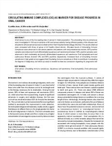
Igm-dCI
In addition, we also used another method. Briefly, 100 p.l each of unlabeled F(ab')2 anti-HHCF and unlabeled F(ab')2 from normal rabbit serum, both at a concentration of 20 p.g/ml in carbonate buffer (0.5 M, pH 9.6), were incubated separately in polypropylene tubes for 3 h at 37°C and overnight at 4°C. After three washes with VBS-Tween 20, the F(ab')2-coated tubes were filled with 100 pL. of a 1:20 dilution in VBSTween 20 and 100 pL. of 125I-protein A (0.5 p.g/ml in VBS-Tween 20). After 3 h of incubation at room temperature, the tubes were washed three times with VBS-Tween 20 and counted in a gamma counter. The purity of the F(ab')2 preparation was indirectly confirmed by the low background obtained in this test. Quantitation of immunoglobulins. Immunoglobulin levels were measured by single radial immunodiffusion (10), using commercial plates (Kallestad). Antigen preparation. The anti-HHCF used as antigen was collected from hepatic and pulmonary cysts of sheep infected with Echinococcus granulosus; this material was subsequently filtered, centrifuged, and concentrated by perventilation to a concentration of 2 mg/ml of protein. Hemagglutination test. The hemagglutination test was performed by the method of Boyden (2). Titers of 1:400 or more were considered significant (4-15). Patients. Thirty sera from eight patients with hepatic
IgG-CIC
p(0.01
24
tubes were counted in a gamma counter. In these
experiments, the specificity of the binding was checked by using radiolabeled IgG or F(ab')2 from rabbit serum in parallel with the antigen-specific IgG or F(ab')2, at the same concentration and specific activity.
p P > 0.001).
DISCUSSION In our patients we found different classes of immunoglobulins included in CICs, with a prevalence of IgA > IgG > IgM; this polyclonal response probably reflects the physiological switch from IgM to IgG and IgA, as may be expected in chronic diseases. Regarding the antigenic component of CICs, we were unable to identify parasitic antigens; this could probably be explained on the basis of one or more of the following hypotheses: (i) the
gPA No.
6
CR Hepatic
HT
mebendazole
)O
\
"25600
203
/
3200
,\ \I
10I00 .i:.::ii.
10
A
0
0
4
8 HT
HT
ngPA
No. 7
g Pulmonary
ngPA1 No. 8
mebendazole
9
Disseminated
25600
'25600 20
20. mebendazole
,3200
1
1 5.
1 5-
mebendazole
3200
"l~ ~ ~ ~ .
5-
5
15
0 IgA-C-C IgM-CIC....---__ IgG-CIC_--
1
4
7
Months of disease
HT
FIG. 3. Follow-up of IgM-CIC, IgG-CIC, and IgA-CIC and of hemagglutination titer (HT) in patients through 8 with hydatid disease. ngPA, Nanograms of protein A.
5
VOL. 18, 1983
absence of specific parasitic antigen(s) in the CICs, at least as recognized by our antiserum (The complex antigenic mosaicism of the Echinococcus may partially account for these difficulties.); (ii) the steric inhibition of the antigen within the complex due to the possible large size of the CICs; (iii) the molar ratio of the CIC components (in fact, the method we employed shows a good performance only in conditions of equivalence or slight antigen or antibody excess); (iv) the possibility that CIC formation is a consequence of a B-cell polyclonal activation, with consequent antibody response against hetero- and autoantigens, but not against Echinococcus antigens. Even if we failed to demonstrate the presence of a specific hydatid antigen(s) in CICs, the follow-up of our patients strengthens our hypothesis about the value of CICs in monitoring the disease. In fact, CICs seemed to present a more strict parallelism with clinical course when compared with the hemagglutination titer. In patient no. 5, for example, we observed a stable decrease of CIC levels after surgery during a 48month follow-up, whereas the hemagglutination titer showed an irregular behavior; in patient no. 4, who was affected by a disseminated hydatidosis and was successfully treated with mebendazole, CIC levels gradually decreased, while hemagglutination titers exhibited an unexplained oscillation; in patient no. 1, the increase of CIC values between the first and second serum samples was probably related to an episode of cyst rupture into a bronchus, associated with massive liberation of antigenic material from the cyst and subsequent passage of the material into the bloodstream; and finally, in patient no. 8, a girl with disseminated hydatidosis successfully treated with mebendazole, an increase of CIC levels in month 7 of follow-up was observed associated with a rise of serum glutamic oxalacetic transaminase and glutamic pyruvic transaminase, probably due to toxic hepatitis, a condition in which other authors have demonstrated the appearance of circulating specific antibodies to altered hepatocytes (16). These observations suggest that, in our patients, CICs belong to different populations, some involving hydatid antigens and specific responses, and others probably consisting of auto- or heteroantigens as documented by the high titer of anti-rabbit F(ab')2 activity [studies are in progress to further characterize this antiF(ab')2 activity]. The discordance between our data and previous data relative to the low positivity of CIC levels in hydatid disease (13) or to the significant increase of CIC levels after treatment with mebendazole (1) could probably be explained on the basis of the different methods employed to
CICs IN HYDATID DISEASE
1025
detect CICs. Finally, the positive significant correlation observed between the levels offree IgG and IgGCIC is probably not due to a possible interference of free immunoglobulin levels in the conglutinin-binding assay (U. DiMario and K. Guy, personal communication), but likely to the clonal expansion of IgG, with possible involvement of IgG auto- and heteroantibodies in the CICs. In conclusion, the study of our patients with hydatid disease showed that CICs can be useful for monitoring clinical conditions since they reflect both specific and nonspecific symptomatologies more reliably than does hemagglutination titer. Furthermore, even if in this condition the typical features of immune complex diseases are lacking, the presence of CICs is probably relevant from a pathogenic point of view because they could exert a modulating effect through the interaction with receptors for IgGFc fragments and C3 on the surface of circulating cells. LITERATURE CITED 1. Bekhti, A., J.-P. Schaaps, M. Capron, J.-P. Dessaint, F. Santoro, and A. Capron. 1977. Treatment of hepatic hydatid disease with mebendazole: preliminary results in four cases. Br. Med. J. 2:1047-1051. 2. Boyden, S. V. 1951. The adsorption of protein on erythrocytes, treated with tannic acid, and subsequent hemagglutination with antiprotein sera. J. Exp. Med. 93:107-120. 3. Casali, P., A. Bossus, N. A. Carpentier, and P. H. Lambert. 1977. Solid phase enzyme immunoassay or radio immunoassay for the detection of immune complexes based on their recognition by conglutinin: conglutinin binding test. A comparative study with '25I-labelled Clq binding and Raji cell RIA tests. Clin. Exp. Immunol.
29:342-354. 4. Castagnari, L., and A. Tolu. 1964. L'emagglutinazione indiretta nella diagnosi biologica dell'idatidosi. Policlinico
Sez. Med. 71:395-399. 5. Cuatrecasas, P. 1971. Affinity chromatography. Annu. Rev. Biochem. 40:259-278. 6. D'Amelio, R., G. Brighouse, M. Barnet, and P. H. Lambert. 1981. Antigen specific detection of soluble immune complexes in conglutinin binding assay. Clin. Exp. Immunol. 45:283-289. 7. Greenwood, B. M. 1974. Possible role of B cell mitogen in hypergammaglobulinaemia in malaria and trypanosomiasis. Lancet i:435-438. 8. Lachmann, P. J., and K. J. Hobart. 1978. Complement technology. In D. M. Weir (ed.), Handbook of experimental immunology-1978, vol. 1. Blackwell Scientific Publications, Ltd., Oxford. 9. Maire, M. A., M. Barnet, and P. H. Lambert. 1981. Purification of bovine conglutinin using pepsin digestion. Mol. Immunol. 18:85-90. 10. Mancini, G., A. 0. Carbonara, and J. F. Heremans. 1965. Immunochemical quantitation of antigens by single radial immunodiffusion. Immunochemistry 2:185-189. 11. McConahey, P. H., and F. J. Dixon. 1966. A method for trace iodination of proteins for immunological studies. Int. Arch. Allergy Appl. Immunol. 29:185-189. 12. Nisonoff, A., F. C. Wissler, L. N. Lipman, and D. L. Woernley. 1960. Separation of univalent fragments from the bivalent rabbit antibody molecule by reduction of disulfide bonds. Arch. Biochem. Biophys. 89:230-244. 13. Richard-Lenoble, D., M. D. Smith, M. Loisy, and P. J. Verroust. 1978. Human hydatidosis: evaluation of three
1026
D'AMELIO ET AL.
serodiagnostic methods, the principal subclass of specific immunoglobulin and the detection of immune complexes. Ann. Trop. Med. Parasitol. 72:553-560. 14. Sorice, F., L. Castagnari, and A. Tolu. 1966. La diagnosi biologica dell'idatidosi umana. G. Mal. Infett. Parassit. 18:192-197. 15. Theofilopoulos, A. N., and F. J. Dixon. 1980. Immune complexes in human disease. A review. Am. J. Pathol.
J. CLIN. MICROBIOL. 100:529-594. 16. Vergani, D., G. Mieli-Vergani, A. Alberti, J. Neuberger, A. L. Eddleston, M. Davis, and R. Williams. 1980. Antibodies to the surface of halothane-altered hepatocytes in patients with servere halothane-associated hepatitis. N. Engl. J. Med. 303:66-71. 17. WHO Scientific Group. 1977. The role of immune complexes in disease. W.H.O. Tech. Rep. Ser. 606.