Quantification vs. Holder Tilt Angle. Figure 1. Energy Resolution of G1 and G2 Electronics. Energy resolution (FWHM) of Mn Kα X-ray peak) calculated from a.
Detection and Quantification Capabilities of X-ray Energy Dispersive Spectrometry Data acquired Using ThermoFisher G2 Electronics processed in Velox C.A. Wade1, 2, T. Withaar2, M. Ovsyanko2, B. Freitag2 and M.G. Burke1 1. Materials Performance Centre, The University of Manchester, Manchester, UK 2. Thermo Fisher Scientific, Materials and Structural Analysis, Eindhoven, Netherlands INSTRUMENTATION:
RESULTS
The Talos F200A analytical scanning transmission electron microscope (STEM) with a high brightness XFEG electron source and a SuperXTM X-ray energy dispersive spectrometry (XEDS) detection system at the University of Manchester has recently been equipped with the new Thermo Fisher G2 pulse processing electronics. Accompanying the hardware upgrade the Velox software for STEM image and XEDS data acquisition has been updated to Velox 2.1. With this poster we illustrate and test the new features of the combination of new hardware and software like :
Basic performance test with NiOX Film
• Comparison of the G2 electronics with the previously installed G1 electronics and Bruker Esprit 1.9 SW.. • New Velox quantification engine for post processing of spectra and spectrum images taking holder absorption into account (please see poster IM4.P011) • The ability to record time resolved spectrum images to observe changes in composition that occurred during multi frame chemical mapping. • 4 independent channel readout of the four sensors of the SuperX detector in mapping applications
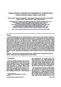
Figure 1. Energy Resolution of G1 and G2 Electronics
Energy resolution (FWHM) of Mn Kα X-ray peak) calculated from a NiOx thin film as a function of alpha tilt for each G2 electronics pulse processor setting and the G1 electronics ‘exhaustive’ setting
Figure 2. STEM Imaging and XEDS Mapping of Alloy 718
Figure 4. NiOx Quantification vs. Holder Tilt Angle
XEDS Quantification results for spectra obtained on a NiOx film across various alpha tilt values. Cliff-Lorimer analysis was performed on the spectra with and without the absorption effects of the holder taken into account. The result of accounting for the absorption of O X-rays by the holder can be seen to increase the calculated O composition.
Table 1. Alloy 718 Matrix Composition
MATERIALS: Element
Alloy 718 is a Fe- and Cr-rich Ni-superalloy which may be precipitation hardened by the formation of semicoherent the γ’ (ordered - L12) and γ” (ordered - DO22) phases in the cubic matrix (γ) when an appropriate heat treatment is applied. This alloy has long been of industrial significance due to its high strength at elevated temperature with excellent corrosion resistance. Costreducing low-loss fabrication methods for the creation of Alloy 718 parts, such as the additive manufacturing technique of selective laser melting (SLM), require the full characterization of microstructure features before these fabrication techniques can replace traditional fabrication and post process heat treatment methods. The ability to eliminate the expensive heat treatment step is dependent on a suitable distribution of γ” and γ’ precipitates in the matrix. In Alloy 718 the nm-scale and precipitates, nominally Ni3Nb and Ni3Al, respectively, have complex compositions first revealed via atom probe field-ion microscopy analysis [2]. The presence of measurable Ti and Al in the precipitates, as well as the presence of Nb and Ti in the precipitates can now be directly measured in the analytical electron microscope (AEM) with SuperX XEDS configuration.
Figure 1 provides a comparison of energy resolution across alpha tilt values for the three available pulse processor settings of the G2 electronics with the best energy resolution acquisition setting of the G1 electronics. An improvement in energy resolution is achievable using the G2 electronics compared to the energy resolution of the G1 electronics with either the optimal or high resolution process time. The high P/B number of 4000 is untouched by the change of electronics.
G1 (at%)
G2 (at%)
Burke & Miller (at%) [2]
Ni
44.8
46.6
47.4
Cr
27.1
26
22
Fe
19.2
21.6
23.8
Nb
0.9
1.2
2
Ti
0.1
0.2
0.5
Al
1.0
0.8
0.7
Electronics
Quantification of the γ matrix was performed using data collected with G1 and G2 electronics and processed through the Bruker Esprit 1.9, and ThermoFisher Velox 2.1 software packages, respectively. The resulting compositions of selected alloying elements are given in Table 1 with atom probe compositional results from literature provided for comparison. The G2 electronics and quantification through Velox provides an improved compositional analysis.
Figure 3. Chemical Distribution in Alloy 718
Beginning with Velox version 2.1 an independent channel readout from each detector may be recorded simultaneously. This data is then available for processing though the selection of which detector signals to add together and process. To demonstrate this effect a carbide present in Alloy 718 was mapped, with the resulting C at% maps shown in Figure 5. The maps b and c can be seen to have a lower carbon concentration while a and d have a higher concentration. This is likely due to partial shadowing of detectors 2 and 3 while detectors 1 and 4 are not shadowed. The decrease in the peak to background ratio in the data collected on the partially shadowed detectors leads to poor quantification results. This is easily seen and the data may be adjusted by excluding these detectors from analysis allowing better quantification results to be obtained.
• The detection capabilities of the SuperXTM with the G1 and G2 electronics were determined using spectra from a 52 nm thick NiOx thin film and the XUtils plugin for DigitalMicrograph [1].
• XEDS maps of SnOx / Pd nanoparticles were acquired to illustrate the capability to observe changes in composition that occurred during multi frame chemical mapping.
HAADF image of γ” in γ (a) and a composite map of Al (green) and Nb (red) marking the location of γ’ on γ” (b)
Figure 5. New Simultaneous 4 independent channel readout on SuperX detector using G2 electronics
XEDS maps of the distribution of Al, Nb, Ti, and Ni in AM Alloy 718
Figure 6. Solute Clusters Formation Under Electron Beam captured by time resolved spectroscopy in Velox
CONCLUSIONS • The new SuperX G2 pulse processing electronics provide more flexibility with variable process time to improve either the energy resolution or lower the dead time for high throughput applications. • The new Independent channel read out feature allows data from each X-ray detector to be acquired both separately and concurrently. The benefit of correct quantification by easy detection of shadowing is illustrated in the alloy 718 example.
C a
• Specimen and holder absorption effects can be corrected in the new Velox quantification software and the XEDS quantification rivals the results of atom probe examinations. • Frame resolution in mapping allows specimen changes and beam damage effects to be removed from XEDS data sets, allowing more accurate quantification or more general enable time resolved spectroscopy applications. REFERENCES [1] M. Watanabe, Microsc. Microanal., 16, Suppl. 2, (2010), pp. 260-261 [2] M.G. Burke, and M. Miller, Superalloys 718, (1991), pp. 337-343.
b
C c
a
b
c
d
C d
XEDS quantitative maps showing the distribution (at%) of C in an area of Alloy 718 using data collected simultaneously from each SuperX detector 1-4, a-d, respectively. The calculated composition can be seen to be dramatically reduced for detectors 2 and 3, suggesting that these detectors are heavily shadowed and should most likely be excluded from analysis.
Using the new Quantification software in Alloy 718 Figure 2a shows a high-angle annular dark-field (HAADF) image of γ” in the matrix, γ. The presence of γ’ is seen in the combined chemical map, Figure 2b, where the Al corresponds to γ’ forming on the sides of the γ”. The chemical distribution of major components of γ’ and γ” are displayed in the elemental maps in Figure 3.
METHODS
• Quantification of the matrix and precipitates in additively manufactured (AM) Alloy 718 has been investigated using the two electronics and software configurations. These results are compared to previous STEM EDX experiments [2].
The Velox acquisition and analysis software allows the X-ray absorption path through the holder to be calculated for each detector, effectively tracking the shadowing of the detectors by the stage. With this path length calculated, and a complete knowledge of stage material and dimensions, the effect of both specimen Xray absorption and holder X-ray absorption may be corrected for. Figure 4 shows the effect of correcting the O composition in a NiOx thin film across multiple alpha tilt values using the implementation of this software in Velox.
Time resolved spectroscopy on SnOx / Pd particles Often the areas of interest in an XEDS map may be influenced by their observation with the electron beam. Melting, diffusion, and contamination often occur and must be accounted for in any analysis. Velox offers the ability to obtain frame resolved data. In post processing frames may be removed from the end of the data allowing the evolution of the sample under the beam as a function of exposure time to be observed. Figure 6 shows solute clustering in SnOx / Pd nanoparticles. These begin their analysis with Pd uniformly distributed throughout the SnOx. As electron beam increases the Pd will begin to aggregate, forming small clusters under the electron beam. Figure 6 a-d show Pd at% compositional maps at different total frame numbers, 20, 40, 70, and 140. Data acquired after solute aggregation has begun may be omitted from the final analysis by removing any frames taken after the solute clustering becomes visible. This allows beam damage effects to be removed in post processing, no longer leading to unusable data if the acquisition is left running too long.
XEDS maps of SnOx / Pd nanoparticles acquired with a 160 µs dwell time showing the map at frame number 20 (a), 40 (b), 70 (c), and 140 (d). Pd is shown in red superimposed on a HAADF-STEM image of the nanoparticles. Solute clustering is circled in the center of the nanoparticles suggesting that the once uniform distribution of Pd has changed due to the high energy of the beam and that results obtained after frame 40 do not reflect the original structure of the material.
Thermo Fisher Scientific • 5791 Van Allen Way • Carlsbad, CA 92008 • www.thermofisher.com