GAF, glucocorticoid antagonizing factor; LPS, lipopolysaccharide; SAA, serum amyloid A. THE JOURNAL OF ... Materials and Methods. Mice. Female ... Company, Grand Island, N. Y.) was supplemented with penicillin (100 U/ml), streptomycin.
DETECTION
OF A MEDIATOR
ENDOTOXIN-STIMULATED THE ACUTE
PHASE SERUM
DERIVED
MACROPHAGES
FROM
THAT
AMYLOID A RESPONSE
INDUCES IN MICE
By J. D. SIPE, S. N. VOGEL, J. L. RYAN, K. P. W. J. McADAM, AND D. L. ROSENSTREICH From the Laboratory of Experimental Pathology, National Institute of Arthritis, Metabolism, and Digestive Diseases, and the Laboratory of Microbiology and Immunology, National Institute of Dental Research, National Institutes of Health, Bethesda, Maryland 20205; and the Department of Medicine, Tufts University School of Medicine, Boston, Massachusetts 02111 Amyloid A fibrils accumulate in human tissues during amyloidosis secondary to diseases characterized by recurrent inflammation, and in tissues of experimental animals that have been subjected to repeated or chronic inflammatory stimulation (1-3). Antibodies to the amyloid A fibril protein (AA) 1 cross-react with a protein in serum which is called serum amyloid A (SAA) (4, 5). SAA has an apparent mol wt of 160,000-180,000 (6, 7) and has the same density as high density lipoproteins, 1.15 g/ cc in man (8) and 1.13 g/cc in mouse (J. Sipe, unpublished observations). SAA is converted to a species of 12,000-13,000 mol wt upon gel filtration in dissociating agents (6-8), and there is considerable homology between the amino acid sequences of this denatured SAA and that of the 8,000- to 10,000-mol wt AA polypeptide which is solubilized from amyloid A fibrils (2, 9). SAA concentrations are elevated in chronic inflammatory and neoplastic diseases (10, 11), and it has been documented in man (12) and mouse (13) that SAA is an acute phase protein whose concentration in serum is highly increased after an inflammatory stimulus. Despite study, the mechanism by which SAA is produced remains unknown. SAA has been found in fibroblasts and connective tissues (14), and in vitro SAA synthesis by cultured polymorphonuclear leucocytes has been observed (15). During an acute phase response to inflammation, SAA synthesis (16) occurs in hepatocytes (17), as does synthesis of the quintessential acute phase reactant, C-reactive protein (CRP) (18). In a previous study (19), we demonstrated that lipopolysaccharide (LPS)sensitive lymphocytes and macrophages were necessary for LPS-induced SAA production, indicating a primary role for these cells in the acute phase SAA response. LPS has been shown to induce production of a mediator, glucocorticoid antagonizing factor (GAF), that acts on hepatocytes (20). We have therefore investigated whether a similar type of mediator might be involved in SAA production. C 3 H / H e J mice do not produce a significant SAA response to phenol extracted Escherichia coli K235 LPS, in contrast to C 3 H / H e N mice which exhibit a several hundred-fold increase in SAA-concentration at the peak of the acute phase response to endotoxin (13). We have therefore utilized this strain pair to investigate the i Abbreviationsusedin this paper:AA, antibodies to the amyloid A fibril protein; CRP, C-reactive protein; GAF, glucocorticoidantagonizing factor; LPS, lipopolysaccharide;SAA,serum amyloid A. THE JOURNAL OF EXPERIMENTAL MEDICINE • VOLUME 150, 1979 597
598
DETECTION OF A MEDIATOR THAT INDUCES MOUSE SAA RESPONSE
possibility of a n SAA i n d u c e r a n d have found that macrophages make a n SAA i n d u c e r in response to LPS. These findings d e m o n s t r a t e the m e c h a n i s m by which LPS produces increased SAA concentrations a n d lay a logical framework for explaining how chronic i n f l a m m a t i o n m a y result in amyloidosis.
Materials and Methods Mice. Female C3H/HeN mice, aged 6-10 wk, were obtained from the Division of Research Resources, National Institutes of Health, Bethesda, Md. Female and male C3H/HeJ mice, aged 4-8 wk, were obtained from The Jackson Laboratory, Bar Harbor, Maine. No significant sex differences have been observed in the acute phase response of C3H or other mice to endotoxin although males tend to fight and elevated SAA concentrations have been observed in these injured mice (K. McAdam and J. Sipe, unpublished results). Serum containing SAA inducer was obtained by bleeding C3H/HeN mice from the orbital plexus between 2 and 2.5 h after the i.p. administration of K235 LPS (1 /ag in 0.2 ml of pyrogen-free saline). Control serum was obtained from untreated or saline-injected animals as specified. Adoptive transfer of bone marrow cells was carried out with C3H/HeJ or C3H/HeN bone marrow cells prepared from 12-wk-old mice by the method of Michalek and co-workers (Suzanne Michalek, personal communication). The method consists of grinding femurs and tibias in RPMI-1640 medium, filtering the suspended cells through gauze to remove small pieces of bone, and suspending the cells at a concentration of 10S/ml for injection. Chimeric mice were tested 5 wk after adoptive transfer for their ability to produce SAA inducer. Reagents. Endotoxin was prepared from E. coli K235 by the phenol-water extraction procedure of Mclntire et al. (21). The dose response curve for SAA induction in C3H/HeN and C3H/HeJ mice was similar to that reported earlier (13). With the present preparation of LPS, 90% macrophages by differential staining of cytospin preparations (Shandown Southern Products Limited, Runcorn, Cheshire, England). Adherent cells were obtained by incubation of 2 X 10⁶ peritoneal exudate cells in serum-free RPMI in TC-24 culture dishes (Costar, Data Packaging, Cambridge, Mass.) at 37°C for 3 h after which the nonadherent cells were removed by washing. Adherent cell monolayers contained >98% macrophages. In some experiments, the nonadherent cells were not removed. Spleen cell preparations were prepared by teasing the spleens and resuspending the washed cells to 2 X 10⁶ spleen cells/ml in serum-free RPMI. Cells were incubated with or without 10/.tg/ml LPS. SAA Measurements. SAA concentration was estimated in serum obtained by bleeding animals from the orbital plexus at various times after injection of serum from LPS-treated C3H/HeN or C3H/HeJ mice, or medium from LPS-treated cell cultures. A 10-/LI aliquot of serum to be tested for SAA was added to 500/A of 10% formic acid and incubated for 24 h at 56°C. Either the entire dilution, or aliquots of it, was lyophilized and resuspended in casein barbital buffer, and the SAA concentration was estimated according to the capacity of the sample to compete with I2SI-AA binding by immobilized anti-AA antibodies (22). Each dilution of sample was assayed in duplicate on at least two separate occasions. At least 10 samples were employed to normalize the data for immunoassays carried out on separate occasions. The SAA concentration for individual animals was averaged for the multiple determinations, and the arithmetic mean and SEM determined. The SEM is indicated after each mean concentration of SAA. Occasionally, as is indicated in the legends, a single value was discarded if its value was greater than the mean plus 1.5 times the SD of the mean.
599
SIPE, VOGEL, RYAN, Mc A D A M, AND R O S E N S T R E I C t t a
b
c
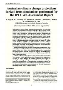
Serum from LPS-Treated C3H/HeN Mice
LPS
Normal C3H/HeN Serum
1.
~ 8o ~
if)
6o
20 ~
q
01~,
k_
2
4
2
4
2
4
HOURS AFTER INJECTION
FIc. 1. Appearance of SAA in C 3 1 | / t t e J mice. (a) 0.2 ml of serum from C 3 t t / H e N mice 2.5 h after i.p. injection of 1 /~g of LPS was administered i.p. to C 3 H / H e J mice. The SAA concentration of the injected serum was 10/~g/ml. (b) LPS, I/~g in 0.2 ml pyrogen free saline was administered to C 3 H / H e J mice. (c) 0.2 ml of serum from untreated C 3 H / H e N mice was administered to C 3 1 t / H e J mice. The SAA concentration of the administered serum was