original articles
Annals of Oncology Annals of Oncology 25: 2042–2047, 2014 doi:10.1093/annonc/mdu271 Published online 23 July 2014
Detection of circulating tumor cells for prediction of recurrence after adjuvant chemoradiation in locally advanced squamous cell carcinoma of the head and neck I. Tinhofer1*, R. Konschak1,2, C. Stromberger1, J.-D. Raguse3, J. H. Dreyer4, K. Jöhrens5, U. Keilholz6 & V. Budach1 1 Translational Radiation Oncology Research Laboratory, Department of Radiation Oncology and Radiotherapy, Charité, Berlin; 2Partner Site Berlin, German Cancer Consortium (DKTK), Deutsches Krebsforschungzentrum (DKFZ), Berlin; 3Clinic for Oral and Maxillofacial Surgery, Charité, Berlin; 4Institute of Pathology, Unfallkrankenhaus Berlin, Berlin; 5Institute of Pathology–Charité, Berlin; 6Department of Hematology and Oncology, Charité, Berlin, Germany
Received 11 April 2014; revised 13 June 2014; accepted 4 July 2014
Background: The prognostic role of persistence of circulating tumor cells (CTC) after upfront tumor surgery for outcome of adjuvant (chemo)radiation in locally advanced squamous cell carcinoma of the head and neck (LASCCHN) was evaluated. Patients and methods: In this prospective study, peripheral blood samples from 144 patients with LASCCHN presenting after tumor resection for adjuvant treatment were analyzed for CTC. Their detection was correlated with tumor site, clinical risk factors, disease-free (DFS) and overall survival (OS). Results: CTC were detected in 42 of 144 patients (29%). CTC detection was higher in cases with nodal involvement and in carcinomas located at the tonsil or base of tongue but was not influenced by age, smoking history, T stage, extracapsular lymph node extension, surgical margins or the human papillomavirus status. Overall, the presence of CTC was not predictive for OS or DFS. However, while in oropharyngeal carcinomas (OPC, n = 63), the detection of CTC was associated per trend with improved DFS [CTC+ versus CTC− (% of patients without evidence of disease at 2 years): 100% versus 79%; log rank: P = 0.059]; the reverse was observed for carcinomas from other sites (non-OPC, n = 81; CTC+ versus CTC−: 29% versus 75%; P = 0.001). In multivariate analysis, CTC remained an independent prognostic marker for DFS [hazard ratio (HR) 4.3, 95% confidence interval (CI) 1.7–10.9, P = 0.002] and OS (HR 2.7, 95% CI 1.2–6.3, P = 0.016) in non-OPC. Conclusions: Assessment of CTC in non-OPC should prove useful for identification of patients who benefit from treatment intensification. The basis for the good prognostic value of CTC in OPC has to be elucidated in future studies. Key words: circulating tumor cells, HPV-associated carcinoma, biomarker, adjuvant treatment, chemoradiation
introduction Even in the more advanced setting, surgery remains a desired treatment of squamous cell carcinomas of the head and neck region (SCCHN) if at least a marginal resection is feasible. The pathologic stage of the primary tumor, extracapsular lymph node extension (ECE) and macro- and microscopically involved surgical margins have been established as major determinants of the relapse risk after tumor resection. Current guidelines
*Correspondence to: Dr Ingeborg Tinhofer, Charité University Hospital Berlin, Department of Radiooncology and Radiotherapy, Translational Radiation Oncology Research Laboratory, Charitéplatz 1, 10117 Berlin, Germany. Tel: +49-30-450-527074; Fax: +49-30-450-527974; E-mail:
[email protected]
implement these factors to identify patients who should benefit from adjuvant treatment. Despite the risk-adjusted multimodal treatment of resectable locally advanced squamous cell carcinoma of the head and neck (LASCCHN), mean 3-year locoregional recurrence rates still exceed 30%. Poor tumor control is mainly observed in non-human papillomavirus (HPV)-associated carcinomas for which 3-year recurrence rates up to 60% have been reported whereas relapse rates of 70% of carcinoma cells showed strong, diffuse cytoplasmic and/or nuclear staining [10].
statistical methods Categorial variables were analyzed with the Fisher’s exact test. The influence of CTC detection on overall survival (OS), in which failure was defined as death due to any cause, and disease-free survival (DFS), in which failure was defined as local, regional or distant progression was determined. Event times were measured from the date of diagnosis (OS) or the date of start of
Volume 25 | No. 10 | October 2014
adjuvant treatment (DFS) to the date of event occurrence, censored by the competing event occurrence or the last follow-up. OS and DFS curves were estimated by the Kaplan–Meier method and compared between groups with the log-rank test. Interactions between risk parameters and survival end points were evaluated using Cox models. Statistical analyses were carried out using the SPSS Statistics software (version 18.0.0, IBM, Armonk, NY). The level of significance was set at P < 0.05.
results frequency of CTC in operable LASCCH after intended curative tumor surgery Blood samples from 144 SCCHN patients were analyzed for CTC. Mean time between tumor surgery and sample collection was 5.1 weeks (range: 0.7–13.7 weeks). Overall, CTC were detected in 42 of 144 patients (29%). No significant association was observed for sex, age and smoking history (supplementary Table S2, available at Annals of Oncology online). Higher frequency of CTC+ cases was observed for OPC located at the tonsil or base of tongue which—in line with results from several studies—comprised the sites with the highest incidence of HPV-driven carcinomas in our cohort. Indeed, p16 IHC staining as surrogate marker for HPV (available for 90 patients) revealed 56% versus 15% of p16+ cases in OPC versus non-OPC (P = 0.00006) and 66% versus 15% of p16+ cases in carcinomas at the tonsil/base of tongue versus any other site (P = 0.000004). However, direct correlation of CTC detection with p16 IHC did not reveal any significant association (supplementary Table S2, available at Annals of Oncology online). We observed a trend for increased frequency of CTC+ cases in patients with nodal involvement compared with patients without lymph node metastasis but no correlation with T stage (supplementary Table S2, available at Annals of Oncology online). CTC were not detected more frequently after R1 resection or in ECE+ cases.
correlation of CTC with DFS and OS after adjuvant (chemo)radiation Median follow-up for OS was 34 months, during which 34 patients (23.6%) died. OS was reduced in patients who were either current smokers, had high pT stage, presented with a carcinoma located at a site other than the oropharynx, an HPVnegative carcinoma or were classified according to pathological risk factors as ‘high-risk’ and received adjuvant chemoradiation (supplementary Table S3, available at Annals of Oncology online). Overall, persistence of CTC after tumor resection did not influence OS after adjuvant (chemo)radiation [hazard ratio (HR) 1.0, 95% confidence interval (CI) 0.5–2.2, P = 0.91]. Median follow-up for DFS was 25 months. Tumor recurrence and/or distant progression were observed in 29 patients (20.1%): 17 patients (11.8%) had a local or regional relapse, 10 patients (6.9%) developed distant metastases without any evidence of locoregional recurrence and 5 patients (3.5%) showed both locoregional and distant progression. In univariate analysis, smoking status, tumor site (oropharynx versus other) and HPV status were identified as prognostic factors for DFS (supplementary Table S3, available at Annals of Oncology online). Again, CTC detection before start of adjuvant treatment did not
doi:10.1093/annonc/mdu271 |
original articles
Annals of Oncology
identify patients with worse outcome (HR 1.4, 95% CI 0.6–3.1, P = 0.35).
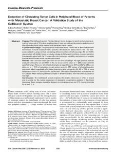
differential prognostic role of CTC in OPC versus non-OPC Since we observed a difference in the frequency of CTC in OPC versus non-OPC, we assessed whether their prognostic value differed according to the tumor site as well. Patients were stratified into four groups based on tumor site (OPC versus non-OPC) and CTC detection (CTC+ versus CTC−). Analysis of DFS (Figure 1A, separate analysis of locoregional and distant DFS shown in supplementary Figure S1, available at Annals of Oncology online) and OS (Figure 1B) revealed an opposite relationship between CTC and outcome according to tumor site: while in non-OPC detection of CTC was significantly associated with shorter DFS [CTC+ versus CTC− (% of patients without evidence of disease at 2 years): 29% versus 75%, log rank: P = 0.001],
A
OPC / CTC+ (n = 23) 1.0 OPC /CTC– (n = 40)
Disease-free survival
0.8
0.6 Non-OPC / CTC– (n = 62) 0.4 Non-OPC / CTC+ (n = 19) 0.2
0.0
Non-OPC: log-rank P = 0.001 OPC: log-rank P = 0.059 0
12
24 36 Time [months]
B 1.0
48
60
OPC / CTC+ OPC / CTC-
Overall survival
0.8
0.6 Non-OPC / CTC0.4 Non-OPC / CTC+ 0.2
0.0
Non-OPC: log-rank P = 0.016 OPC: log-rank P = 0.042 0
12
24 36 Time [months]
48
60
Figure 1. Kaplan–Meier estimates of disease-free survival (DFS) and overall survival (OS) among SCCHN patients, according to CTC status and tumor site. Patients were stratified into four groups based on tumor site (OPC versus non-OPC) and CTC detection (CTC+ versus CTC−). Kaplan–Meier curves for DFS (A) and OS (B) are shown and log-rank P values are given.
| Tinhofer et al.
the reverse was observed per trend for OPC (CTC+ versus CTC−: 100% versus 79%, log rank: P = 0.059). Accordingly, persistence of CTC after tumor surgery was significantly correlated with reduced OS in the group of non-OPC [CTC+ versus CTC− (% of patients alive at 2 years): 36% versus 67%, log rank: P = 0.016] whereas the reverse association was observed for OPC (CTC+ versus CTC−: 100% versus 87%, log rank: P = 0.042).
interaction of surgical margins, ECE or CTC status with tumor progression Given the opposite association of CTC with outcome in OPC versus non-OPC, we next evaluated whether R status and ECE interacted in a distinct manner according to tumor site as well. Patients were stratified into four groups based on tumor site (OPC versus non-OPC) and R status (R1 versus R0, Figure 2A) or ECE (ECE+ versus ECE−, Figure 2B), respectively. R1 resection was associated with prolonged DFS in OPC and no single event was observed in this patient subgroup. In contrast, R1 was negatively associated with DFS in non-OPC, with 2-year DFS rates of 33% and 74% for patients with R1 and R0 resection, respectively (Figure 2A). The differences in DFS were smaller when ECE was used for group stratification. However, the highrisk feature ECE seemed again positively associated with DFS in OPC whereas no influence was observed for non-OPC (Figure 2B). Detection of CTC after tumor resection was independent of R status or ECE and could thus represent an independent marker for minimal residual disease (MRD). We therefore investigated whether the combination of all three markers might be superior for risk assessment than each marker alone. Patients were stratified into three groups defined by (i) the absence of any risk factor, (ii) the presence of CTC or pathological risk factor(s) and (iii) the presence of both CTC and pathological risk factor(s). Again, a clear difference was observed for OPC versus nonOPC: MRD negativity defined by absence of any risk factor was significantly associated with shorter DFS in OPC (Figure 2C, log rank: P = 0.005) whereas no significant difference was observed between the two other OPC patient subgroups. In contrast, MRD positivity defined by either risk factor alone did not significantly influence outcome in non-OPC. However, the presence of residual local (R1) and/or regional disease (ECE+) together with systemic spread (CTC+) was significantly associated with poor outcome, with no evidence of disease in only 13% of patients compared with 72% of patients presenting without any or only one of these risk parameters (Figure 2D, log rank: P = 0.013). In multivariate analysis, CTC detection in non-OPC remained an independent prognostic factor of OS and DFS (Table 1). The Cox model for calculation of hazard ratios (HRs) could not be applied to the group of OPC since no event was observed in the CTC+ group.
discussion Here, we confirm the recently reported poor prognostic value of CTC in non-OPC [9]. In this former study, the frequency of CTC detection in tumors of the oral cavity was only 12.5% which is considerably lower than the rate observed in our study. This could be simply due to differences in inclusion criteria:
Volume 25 | No. 10 | October 2014
original articles
Annals of Oncology
B 1.0
OPC / R1 (n = 14) OPC / R0 (n = 49)
0.8 0.6
Non-OPC / R0 (n = 63)
0.4 Non-OPC / R1 (n = 18) 0.2 0.0
Non-OPC: log-rank P = 0.024 OPC: log-rank P = 0.22 0
12
24
36
C 1.0
48
Disease-free survival
Disease-free survival
A 1.0
OPC / ECE+ (n = 28) OPC / ECE– (n = 35) Non-OPC / ECE– (n = 49)
0.8 0.6
Non-OPC / ECE+ (n = 31) 0.4 0.2 Non-OPC: log-rank P = 0.60; OPC: log-rank P = 0.11
0.0 60
0
12
24
36
48
60
D 1.0
#3 (n = 13)
Disease-free survival
Disease-free survival
#2 (n = 32) 0.8 0.6 #1 (n = 18) 0.4 0.2 0.0
OPC: log-rank P = 0.0005 0
12
24
36
#2 ( n = 38)
0.6 0.4 0.2 0.0
48
#1 ( n = 35)
0.8
60
#3 ( n = 8) Non-OPC: log-rank P = 0.013 0
12
24
36
48
60
Figure 2. Classical pathological risk factors (surgical margins, ECE) and CTC status have distinct prognostic impact in OPC and non-OPC. (A and B) Patients were stratified into four groups based on tumor site (OPC versus non-OPC): (A) R status (R1 versus R0) or (B) ECE (ECE+ versus ECE−), respectively. (C and D) Patients with OPC (C) or non-OPC (D) were stratified into three groups defined by the absence of any risk factor (group #1), the presence of CTC or the indicated pathological risk factor(s) (group #2) or the presence of both CTC and pathological risk factor(s) (group #3). Kaplan–Meier curves for DFS of patient subgroups are presented. P values from comparison of groups using the log-rank test are given.
Table 1. Hazard ratios for OS and DFS in non-OPC, according to Patient Group Covariate Overall survival CTC status (positive versus negative) Tumor stage (pT3–pT4 versus pT1–pT2/pTx) Nodal stage (pN2c–pN3 versus pN0–pN2a) R status (R1 versus R0) ECE status (positive versus negative) Treatment (RCT versus RT) Smoking status (current versus never/ex-smoker) Disease-free survival CTC status (positive versus negative) Tumor stage (pT3–pT4 versus pT1–pT2/pTx) Nodal stage (pN2c–pN3 versus pN0–pN2a) R status (R1 versus R0) ECE status (positive versus negative) Treatment (RCT versus RT) Smoking status (current versus never/ex-smoker)
Univariate model, hazard ratio (95% CI)
P value
Multivariate model hazard ratio (95% CI)
P value
2.6 (1.2–5.6) 1.1 (0.5–2.5) 1.8 (0.8–4.0) 1.9 (0.9–4.3) 1.1 (0.5–2.4) 2.6 (1.2–5.9) 1.1 (0.4–2.8)
0.02 0.73 0.12 0.11 0.79 0.018 0.83
2.7 (1.2–6.3)
0.016
1.6 (0.7–3.6) 0.9 (0.4–2.3)
0.25 0.84
2.4 (0.9–5.8)
0.061
3.9 (1.7–9.2) 0.9 (0.4–2.3) 1.8 (0.8–4.2) 2.8 (1.2–6.5) 1.3 (0.6–3.1) 2.1 (0.9–4.9) 1.2 (0.4–3.3)
0.002 0.91 0.18 0.02 0.51 0.09 0.71
4.3 (1.7–10.9)
0.002
1.4 (0.6–3.4) 2.2 (0.8–5.9)
0.44 0.11
0.9 (0.4–2.7)
0.98
indeed, while we excluded patients with UICC stage I–II disease, 32 of 90 patients (36%) had stage I–II disease in the study of Grobe et al. [9]. Alternatively, CTC detection by PCR amplification of tumor-associated antigens might be more sensitive than single-cell-based detection of tumor cells expressing epithelial
Volume 25 | No. 10 | October 2014
markers [2]. Our previous comparative analysis of PCR versus flow cytometry for detection of CTC in SCCHN [7] speaks against large differences between the two techniques. However, this comparison was carried out in the definitive setting for which limitations in sensitivity might be less relevant than in
doi:10.1093/annonc/mdu271 |
original articles the adjuvant setting, taking into account the reducing effect of tumor surgery on CTC frequency [11]. It is still a matter of debate whether CTC are representing vital tumor cells or whether they just reflect shedding of apoptotic tumor cells. Indeed, signs of apoptosis were detected in a proportion of CTC in prostate [12] and breast cancer [13]. Although the method used in our study did not allow discrimination between vital and apoptotic tumor cells, the fact that these cells could be detected up to 7 weeks after tumor surgery strongly argues against them being passively shed dying cells. Rapid clearance of apoptotic cells by phagocytes [14] would preclude their detection after such a long period of time. Besides confirming previous data in non-OPC, we show here for the first time that CTC represent an independent risk factor for tumor progression beside pathological risk factors. Despite risk-adjusted adjuvant treatment, R1 resection remained a risk factor for tumor progression in non-OPC. Importantly, the prognostic value of surgical margins was significantly influenced by the CTC status and dismal outcome was observed only in non-OPC patients who presented after R1 resection with CTC. In the majority of cases, we observed locoregional but not distant tumor progression. If a causal relationship between CTC and locoregional relapse exists, re-seeding of CTC to the primary tumor site has to be considered. In support of this assumption, re-infiltration of CTC at the primary site was previously demonstrated in xenograft models of breast and colon cancer [15]. In these models, self-seeding was stimulated by tumor-derived cytokines acting as CTC attractants into the original tumor bed [15]. It is tempting to speculate that residual tumor cells at surgical margins could facilitate re-seeding of CTC which would explain the significant interaction of CTC with the prognostic value of the R status. The prognostic impact of CTC was independent of the type of adjuvant treatment. Beside local radiosensitizing effects, concurrent chemotherapy should act systemically and target tumor cells in peripheral blood as well. However, recent studies have shown that adjuvant chemotherapy cannot eliminate CTC [16], probably because of their nonproliferating dormant state [17]. In contrast, chemotherapy-resistant CTC were efficiently eliminated by Her2 blockade, thereby significantly reducing the risk of relapse in early breast cancer [18]. In line with a failure of chemotherapy in inhibiting CTC, we previously observed persistence of CTC expressing activated EGFR after concurrent chemoradiation whereas their frequency was significantly reduced when radiotherapy was combined with cetuximab [8]. A further important finding of our study was that MRD defined by detection of CTC, positive margins and/or ECE was not associated with poor prognosis in OPC but seemed to identify patients with reduced risk of progression. Certainly, larger patient cohorts have to be analyzed to validate this unexpected finding. It has been argued that the favorable prognosis of HPVassociated OPC may at least partly be due to T-cell-mediated immunity against viral antigens [19]. Although information on the HPV status was not available for all cases in our study, the incidence of HPV-driven carcinomas in OPC cases with known HPV status was high, indicating that >55% of OPC should express viral antigens. It is also known that T-cell memory which is induced by chronic antigen exposure in the course of persistent viral infection or cancer is more susceptible to
| Tinhofer et al.
Annals of Oncology
functional exhaustion [20] and long-lasting protective immunity does not develop under these conditions. Mounting evidence indicates that radiotherapy can interfere with the immune system by induction of so-called danger signals, thereby reverting some of these immunosuppressive barriers [21]. Since OPC seem to represent more immunogenic carcinomas than nonOPC surgical resection of the immunosuppressive bulk tumor followed by irradiation of residual tumor cells at surgical margins and/or re-seeded CTC could potentially provide immunogenic stimulation and support the establishment of a protective long-lasting immunity. In conclusion, we established postoperative detection of CTC by nested RT-PCR of EGFR transcripts as independent prognostic ‘liquid’ biomarker in non-OPC. Introduction of EGFRtargeting drugs in adjuvant radiotherapy regimens based on CTC detection might prove useful for treatment optimization in this scenario. The implication of the good prognostic value of CTC in OPC—assuming successful validation of our findings in future larger trials—for novel therapeutic strategies in HPVdriven OPC remains to be determined.
acknowledgements We are grateful to the patients and their families for their participation in this study and to Bettina Kupny, our study nurse and the clinical team of the Clinic of Radiation Oncology and Radiotherapy for their continuous support in the collection of blood samples.
funding This work has been supported by grants from the Berliner Krebsgesellschaft, Berlin, Germany (to IT) and the German Cancer consortium (to IT and VB). The support of the Wilhelm Sander Foundation (2012.029.1, to JHD) is also gratefully acknowledged.
disclosure The authors have declared no conflicts of interest.
references 1. Licitra L, Perrone F, Bossi P et al. High-risk human papillomavirus affects prognosis in patients with surgically treated oropharyngeal squamous cell carcinoma. J Clin Oncol 2006; 24: 5630–5636. 2. Cristofanilli M, Budd GT, Ellis MJ et al. Circulating tumor cells, disease progression, and survival in metastatic breast cancer. N Engl J Med 2004; 351: 781–791. 3. Cohen SJ, Punt CJ, Iannotti N et al. Relationship of circulating tumor cells to tumor response, progression-free survival, and overall survival in patients with metastatic colorectal cancer. J Clin Oncol 2008; 26: 3213–3221. 4. Scher HI, Jia X, de Bono JS et al. Circulating tumour cells as prognostic markers in progressive, castration-resistant prostate cancer: a reanalysis of IMMC38 trial data. Lancet Oncol 2009; 10: 233–239. 5. Brakenhoff RH, Stroomer JG, ten Brink C et al. Sensitive detection of squamous cells in bone marrow and blood of head and neck cancer patients by E48 reverse transcriptase-polymerase chain reaction. Clin Cancer Res 1999; 5: 725–732. 6. Partridge M, Brakenhoff R, Phillips E et al. Detection of rare disseminated tumor cells identifies head and neck cancer patients at risk of treatment failure. Clin Cancer Res 2003; 9: 5287–5294.
Volume 25 | No. 10 | October 2014
original articles
Annals of Oncology 7. Hristozova T, Konschak R, Stromberger C et al. The presence of circulating tumor cells (CTCs) correlates with lymph node metastasis in nonresectable squamous cell carcinoma of the head and neck region (SCCHN). Ann Oncol 2011; 22: 1878–1885. 8. Tinhofer I, Hristozova T, Stromberger C et al. Monitoring of circulating tumor cells and their expression of EGFR/phospho-EGFR during combined radiotherapy regimens in locally advanced squamous cell carcinoma of the head and neck. Int J Radiat Oncol Biol Phys 2012; 83: e685–e690. 9. Grobe A, Blessmann M, Hanken H et al. Prognostic relevance of circulating tumor cells in blood and disseminated tumor cells in bone marrow of patients with squamous cell carcinoma of the oral cavity. Clin Cancer Res 2014; 20: 425–433. 10. Dreyer JH, Hauck F, Oliveira-Silva M et al. Detection of HPV infection in head and neck squamous cell carcinoma: a practical proposal. Virchows Arch 2013; 462: 381–389. 11. Li J, Shi SB, Shi WL et al. LUNX mRNA-positive cells at different time points predict prognosis in patients with surgically resected nonsmall cell lung cancer. Transl Res 2014; 163: 27–35. 12. Larson CJ, Moreno JG, Pienta KJ et al. Apoptosis of circulating tumor cells in prostate cancer patients. Cytometry A 2004; 62: 46–53. 13. Kallergi G, Konstantinidis G, Markomanolaki H et al. Apoptotic circulating tumor cells in early and metastatic breast cancer patients. Mol Cancer Ther 2013; 12: 1886–1895.
14. Ravichandran KS. ‘Recruitment signals’ from apoptotic cells: invitation to a quiet meal. Cell 2003; 113: 817–820. 15. Kim MY, Oskarsson T, Acharyya S et al. Tumor self-seeding by circulating cancer cells. Cell 2009; 139: 1315–1326. 16. Xenidis N, Ignatiadis M, Apostolaki S et al. Cytokeratin-19 mRNA-positive circulating tumor cells after adjuvant chemotherapy in patients with early breast cancer. J Clin Oncol 2009; 27: 2177–2184. 17. Muller V, Stahmann N, Riethdorf S et al. Circulating tumor cells in breast cancer: correlation to bone marrow micrometastases, heterogeneous response to systemic therapy and low proliferative activity. Clin Cancer Res 2005; 11: 3678–3685. 18. Georgoulias V, Bozionelou V, Agelaki S et al. Trastuzumab decreases the incidence of clinical relapses in patients with early breast cancer presenting chemotherapyresistant CK-19mRNA-positive circulating tumor cells: results of a randomized phase II study. Ann Oncol 2012; 23: 1744–1750. 19. Albers A, Abe K, Hunt J et al. Antitumor activity of human papillomavirus type 16 E7-specific T cells against virally infected squamous cell carcinoma of the head and neck. Cancer Res 2005; 65: 11146–11155. 20. Nolz JC, Harty JT. Protective capacity of memory CD8+ T cells is dictated by antigen exposure history and nature of the infection. Immunity 2011; 34: 781–793. 21. Formenti SC, Demaria S. Systemic effects of local radiotherapy. Lancet Oncol 2009; 10: 718–726.
Annals of Oncology 25: 2047–2052, 2014 doi:10.1093/annonc/mdu368 Published online 4 August 2014
Phase II study of single-agent panitumumab in patients with incurable cutaneous squamous cell carcinoma M. C. Foote1,7*, M. McGrath2, A. Guminski3, B. G. M. Hughes4, J. Meakin5, D. Thomson2, D. Zarate6, F. Simpson7 & S. V. Porceddu1,7 Departments of 1Radiation Oncology; 2Medical Oncology, Princess Alexandra Hospital, Brisbane; 3Department Medical Oncology, Royal North Shore Hospital, Sydney; 4 Department Medical Oncology, Royal Brisbane and Womens Hospital, Brisbane; 5Research Unit, Cancer Services, Princess Alexandra Hospital, Brisbane; 6 Queensland Cancer Control Analysis Team, Queensland Health, Brisbane; 7Diamantina Institute, University of Queensland, Brisbane, Australia
Received 12 November 2013; revised 17 April 2014 and 11 June 2014; accepted 28 July 2014
Background: Although advanced cutaneous squamous cell carcinoma (CSCC) is quite common, there are few prospective trials regarding its optimal management. This study evaluated the efficacy and safety of single-agent panitumumab in the treatment of patients with CSCC not suitable for local therapy. Patients and methods: Sixteen patients received single-agent panitumumab at a dose of 6 mg/kg repeated every 2 weeks for a minimum of three cycles and continued until progression, a maximum of nine cycles or dose-limiting toxicity. The primary end point was the best overall response rate (ORR) as assessed by Response Evaluation Criteria in Solid Tumours (RECIST version 1.1) criteria. Secondary end points included evaluation of safety, toxicity and progressionfree survival (PFS). Results: Between May 2010 and May 2012, 16 patients were recruited. Fourteen patients were male and the median age was 68 years. Fifteen patients had locoregionally advanced or recurrent disease with 14 patients receiving previous radiotherapy and 7 receiving previous cytotoxic chemotherapy. The best ORR [ partial (PR) or complete response (CR)] was 31% (3/16 PR, 2/16 CR) with a further 6 of 16 patients achieving SD. The median PFS and overall survival were 8 and 11 months respectively. Grade 3 or 4 events were observed in five patients (four being skin toxicity) with one
*Correspondence to: Dr Matthew C. Foote, Princess Alexandra Hospital, Radiation Oncology, 199 Ipswich Rd, Woolloongabba, Brisbane, Queensland, 4102 Australia. Tel: +61-7-3176-3067; Fax: +61-7-3176-1983; E-mail:
[email protected]
© The Author 2014. Published by Oxford University Press on behalf of the European Society for Medical Oncology. All rights reserved. For permissions, please email:
[email protected].