ORIGINAL PAPER
Detection of Epstein Barr virus in formalin-fixed paraffin tissues by fluorescent direct in situ PCR N. Marziliano,1 E. Arbustini,1 M. Rossi de Gasperis,2 S. Crovella3 1 Transplant Research Area, Molecular Diagnostic Cardiovascular and Transplant Pathology Laboratory, IRCCS Policlinico S Matteo, Pavia; 2Service of Microbiology and Virology, Paediatric Hospital “Bambino Gesù”, Rome; 3Department of Reproductive and Developmental Sciences, University of Trieste, Italy
©2005, European Journal of Histochemistry Specific viral laboratory diagnosis of primary Epstein-Barr Virus (EBV) infection is usually based on antibody-detection assays. However, molecular detection is also considered the reference standard assay for diagnosis of central nervous system infections and of most cases of nasopharyngeal carcinoma (NPC). One-step or nested polymerase chain reaction (PCR) has rapidly replaced immunological assays based on virus-specific Ig antibodies for the laboratory diagnosis of Herpesvirus infections, even if serological methods are considered an additional tool for defining clinical diagnosis. In this article, we will present a rapid, sensitive and robust molecular tool for the viral detection of EBV (EBNA-1) within tissue specimens by making use of in situ PCR (IS-PCR). Key words: EBV, lymph node, salivary glands, in situ hybridization, in situ PCR, quantitative PCR. Correspondence: Nicola Marziliano, Transplant Research Area, Molecular Diagnostic Cardiovascular and Transplant Pathology Laboratory, IRCCS Policlinico S Matteo, Piazzale Golgi 1, 27100 Pavia, Italy Tel. +39.335.1801042. Fax. +39.1782786483. E-mail:
[email protected] Paper accepted on April 18, 2005 European Journal of Histochemistry 2005; vol. 49 issue 3 (Jul-Sep): 309-312
everal human infections play a role in human diseases. Herpes simplex virus type-1 (HSV1) and type-2 (HSV-2), human cytomegalovirus (HCMV) and Epstein-Barr virus (EBV) are widespread human viruses frequently detected on specific tumours and immunosuppressed individuals. Numerous reports link HCMV and EBV to tumours, lymphoepithelial carcinoma, smooth muscle tumours and oesophageal carcinoma, suggesting thus that these viruses might transactivate cellular oncogenes such as jun, fos and myb (Chang F et al., 2000). Whilst clinical features may indicate a diagnosis of viral infection, a definitive diagnosis relies on the direct demonstration of the presence of the virus by culture or detection by immunohystochemistry. For most viruses, viral culture is extremely time-consuming or impossible to carry out, as for the Human Papilloma Virus (HPV). In this study, a rapid, sensitive and robust protocol for the detection of rare copies of Epstein Barr Virus (EBV), based on direct in situ PCR technology and fluorochromemodified or biotin-labeled nucleotides, was developed to detect EBV on infected tissues (such as lymph nodes and salivary glands). We discuss the potential of our experimental protocol for the rapid detection of EBV infections and which may be suitable for clinical purposes as already elsewhere described (Benkoel L et al., 2004; Preziuso S et al., 2003; Preziuso S et al., 2003). Five µm sections of paraffin-embedded lymph nodes (fifteen) and salivary glands (fifteen), coming from a collection belonging to the Pathology Service of the Maggiore Hospital (Trieste, Italy), were placed onto pre-coated slides (Applied Biosystems, Foster City, CA) and incubated onto a hot plate (60°C) for 24 hours to achieve maximum adhesion. Paraffin-embedded EBV-infected tissues fixed in 10% buffered formalin were dewaxed by xilene treatment at 37°C for two hours. Tissue samples were then rehydrated with ethanol 100%, 95% and PBS (10 minutes each), and then passed through Triton X-100 0.01
S
309
N. Marziliano et al.
% in PBS (1 minute and 30 seconds) and PBS (2 minutes). Tissues were treated with proteinase K (Bothering, Manheim) 2 mg/ml in Tris 20 mM, Tween 20 0.5 % for 30 minutes at 37°C and then transferred into a microwave oven to boil for 30 seconds. In situ hybridization was performed using a biotin-labeled commercial DNA probe towards the EBNA-1 region (Chemicon, Temecula, CA) as described in Temple GK et al., (2004). The hybridized probe was detected by fluoroscein isothiocyanate (FITC) labeled avidin (Vector, Burlingame, CA). Propidium Iodide (PI) was used for counterstaining. The IS PCR was performed as follows: slides were heated at 70°C before starting the reaction; the IS PCR solution (preheated at 70°C) was 10 mM Tris 50 mM KCl, pH 8.3, MgCl2 (4mM), dTTP, dCTP, dGTP 200 mM each, 200 mM dATP and 5 mM FITC-dATP (Boehringer) or Biotin-16dUTP (Boehringer), EBV specific primers for the EBNA-1 region (Forward 5’-CCACCAGCAGCACCA GCA-3’; Reverse 5’-CAGGGCCACCATGGTGGC-3’; amplicon length of 300 bp) 0.5 mM each and 10 units for reaction of IS-Amplitaq (Applied Biosystems). IS PCR was performed in a GeneAmp PCR System 1000 (Applied Biosystems) repeating the following cycle 15 times: 94°C 1 minute, 58°C 30 seconds and 72°C 2 minutes. Fluorescent detection made use of DAPI (Vectashield, Burlingame, CA) counterstaining, while alkaline phosphataseconjugated anti-biotin antibody, 100 ml of antibody solution diluted 1:300 in PBS per tissue spot (Boheringer Mannheim), NBT/BCIP chromogen (Boerhinger) and Mayer’s solution counterstaining allowed colorimetric target visualization. To check the presence of aspecific amplifications, experiments of IS PCR were performed with the exclusion of either the primers or the target (a human lymph nodes from a healthy donor was used). EBV was quantitatively evaluated by real time PCR on DNA extracted from lymph node and salivary glands sections, adjacent to those used for IS PCR, with 5700 ABI Sequence Detection System (Applied Biosystems) and SYBR Green I chemistry. For real time amplifications, the protocol was the same as in IS PCR with the exception of 1X Universal SYBR Green I Master Mix (Applied Biosystems) and 900 nM forward/reverse primers added to the reaction mixture. Two types of PCR controls were used: no template controls (NTC) with no target DNA and no amplification control 310
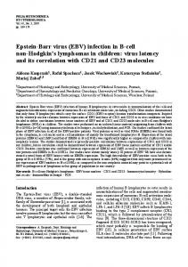
(NAC) with the addition of 1 ml SDS (0.5% w/v) in order to ensure the denaturation of the DNA polymerase. Standard controls were purchased from Clonit Srl (PN CEQ02). The results of in situ amplification of EBV in paraffin-embedded tissues are reported in Figure 1. Colorimetric and direct FITC labeling of the amplification product in lymph nodes (Figure 1a, 1b) and salivary glands (Figure 1c) produced a very clear and bright signal with no backdiffusion of the amplicon. EBV was localized in epithelial cells of acini and ducts of salivary glands, while in the lymph node, the localization at the inner cortex level led us to speculate a vehiculation via T cells (this hypothesis must be proven by co-localizations experiments with specific T cells antibodies and EBV DNA). The negative controls were respectively “no primers” and “no target”: they showed no positive signal of amplification, thus highlighting the absence of background noise due to gap filling and nick repair activity of Taq polymerase (data not shown). Once achieved the Proteinase K digestion conditions, the morphology of the tissues (remained intact) was preserved despite the multiple steps of permeabilization and thermal cycling. Conventional fluorescent in situ hybridization (ISH) on paraffin-embedded lymph node and salivary gland tissues was also performed using a commercial probe specific for EBV. The signal of hybridization was weak, indicating a very low copy number of EBV (Figure 1d). Solution phase PCR on the paraffin-embedded tissues of both the lymph node and the salivary glands successfully amplified EBV DNA and no aspecific amplicon was detected. In order to better understand the sensitivity of our IS PCR protocol, we performed an end-point quantitative PCR on DNA extracted from lymph node and salivary gland sections adjacent to those used for in situ techniques, using the same primers as for the IS PCR and PCR amplification was carried out as described by Mancuso et al., (1997). A very low copy number (approximately 15) of EBV was detected in both the lymph node and the salivary glands by SYBR Green I-ABI 5700 sequence detection system technology and this finding allowed us to check the presence of EBV and thus confirming the reliability of our IS PCR protocol. A quantitation was easily achieved by running a standard curve together with the DNA extracted from the tissues (see Figure 1e). No increase in fluorescence was detected in both the “no template con-
Original Paper
Figure 1.a) Colorimetric labeling of EBV amplicon in Lymph node: the amplification signal is localized in the inner cortex (arrows). The magnification is 1200X.b) FITC-labeling detection of Epstein Barr Virus in Lymph node DAPI counterstained: EBV is localized at the inner cortex level. Arrows indicate the amplification signals. The magnification is 1500X.c) Colorimetric labelling of the amplification product in salivary glands produced a very bright, clear signal with no backdiffusion of the amplicon. The magnification is 1200X.d) Fluorescent in situ hybridisation with FITC labelled probe for EBV detection in Lymph node PI counterstained: the hybridisation signal is weak (arrow) indicating the presence of low EBV copy number. The magnification is 1200X.e) Amplification plot of the DNAs extracted from lymph node and salivary glands tissues and the standard controls. The Cycle threshold (Ct) for the amplicon from EBV-infected tissues was 35 (dark circle), whereas the Ct values for the standards (500, 250, 50, 25 and 2.5 EBV copies as from the CEQ02 reference clone) were respectively 28.5, 29.5, 32, 33 and 36. The EBV copy number predicted by the Gene Amp 5700 SDS Software was approximately 15; this result was calculated by plotting the Ct values of the EBV-infected tissues against the standard curve made with the reference samples. 311
N. Marziliano et al.
trols (NTC)” with no target DNA or the “no amplification control (NAC)” after 50 cycles. Several molecular biological data suggest that certain types of some members of herpesviridae family such as Epstein-Barr virus, human herpes simplex virus type 2 and human cytomegalovirus may play an oncogenic role (Koffa et al.,1995) and also induce neurological syndromes such as acute encephalitis. We herein describe a simple and reliable molecular assay that localizes in situ the EBV presence; we also tested the sensitivity of our method by comparing it with the real time PCR on the same virus target region (EBNA-1). In situ PCR technology was already used for Epstein Barr Virus (EBV) detection in paraffin sections of nasopharyngeal carcinomas (Takeuchi et al., 1997), in epithelial cells and lymphocytes of non-neoplastic tonsils (Kobayashi et al., 1998) and in cells in suspension (Fares et al., 1998). PCR and ISHwas also used by Barkholt and coll. (1998) for the diagnosis of EBV hepatitis in liver biopsies, after liver transplantation. In our study, we obtained very clear results for EBV detection - without background noise or nonspecific amplification - by optimizing the steps of permeabilization and morphology preservation of the paraffin-embedded tissue samples. Our direct IS PCR protocol is very rapid (three hours with FITC and six hours with Biotin 16dUTP), robust (it worked on tissue specimens dating back to more than a year before the experiment), reproducible and able to detect rare copies of EBV. Quantitative SYBR Green I PCR performed on the same tissues sections - adjacent to those used for in situ techniques - allowed us to establish the sensitivity of our method, which is able to detect about 15 copies in our paraffin-embedded EBV infected tissues.
Acknowledgements This work was partially supported by grants from the MIUR (PRIN 2003057187_002) and by grants: "Ricerche Finalizzate e Correnti" IRCCS Policlinico S Matteo, Pavia, Italy. We also thank Mrs Barbara Bernato for her precious work in the English revisions of the manuscript.
312
References Barkholt L, Reinholt FP, Teramoto N, Enbom M, Dahl H, Linde A. Polymerase chain reaction and ISH of Epstein-Barr virus in liver biopsy specimens facilitate the diagnosis of EBV hepatitis after liver transplantation. Transpl Int 1998; 11: 336-44. Benkoel L, Biagini P, Dodero F, De Lamballerie X, De Micco P, Chamlian A. Immunohistochemical detection of C-100 hepatitis C virus antigen in formaldehyde-fixed paraffin-embedded liver tissue. Correlation with serum, tissue and in situ RT-PCR results. Eur J Histochem 2004;48:185-90. Chang F, Syrjanen S, Shen Q, Cintorino M, Snatopietro R,Tosi P et al., Evaluation of HPV, CMV, HSV and EBV in esophageal squamous cell carcinome from high-incidence area of China. Anticacer Res 2000; 20:3935-40. Fares F, Habib M, Verniol C, Drouet E, Niveleau A. IS amplification of the Epstein-Barr virus genome in cell suspensions. J Virol Methods 1998; 71: 211-8. Kobayashi R, Takeuchi H, Sasaki M, Hasegawa M, Hirai K. Detection of Epstein-Barr virus infection in the epithelial cells and lymphocytes of non-neoplastic tonsils by ISH and IS PCR. Arch Virol 1998; 143:803-13. Koffa M, Koumantakis E, Ergazaki M, Tsatsanis C, Sapndidos DA. Association of herpesvirus infection with the development of genital cancer. Int J Cancer 1995; 63:58-62. Mancuso T, Crovella S, Casazza S, Pescarolo MP, Russo P, Clerico L, et al., A fast and simple method to detect human papillomavirus infection in archival paraffin-embedded tissues. Int J Oncol 111997; 527-31. Preziuso S, Taccini E, Rossi G, Renzoni G, Braca G. Experimental Maedi Visna Virus Infection in sheep: a morphological, immunohistochemical and PCR study after three years of infection. Eur J Histochem 2003;47:373-8. Preziuso S, Sanna E, Sanna MP, Loddo C, Cerri D,Taccini E, Mariotti F, Braca G, Rossi G, Renzoni G. Association of Maedi Visna virus with Brucella ovis infection in rams. Eur J Histochem 2003;47:151-8. Takeuchi H, Kobayashi R, Hasegawa M, Hirai K. Detection of latent Epstein-Barr virus (EBV) DNA in paraffin sections of nasopharyngeal carcinomas expressing no EBV-encoded small RNAs using IS PCR. Arch Virol 1997; 142:1743-56. Temple GK, Sales M, Kernohan N, Scott F, Meiklejohn D, Pratt N. Application of combined immunofluorescence and fluorescence in situ hybridisation on paraffin-embedded sections to characterize Tcell lymphoma with EBV-infected B-cell blasts. Genes Chromosomes Cancer 2004; 41:405-9.