Free Radical Biology & Medicine 45 (2008) 321–328
Contents lists available at ScienceDirect
Free Radical Biology & Medicine j o u r n a l h o m e p a g e : w w w. e l s ev i e r. c o m / l o c a t e / f r e e r a d b i o m e d
Original Contribution
Detection of mitochondrial dysfunction by EPR technique in mouse model of dilated cardiomyopathy Martyna Elas a,⁎, Joanna Bielanska a, Katarzyna Pustelny a, Przemyslaw M. Plonka a, Lukasz Drelicharz b, Tomasz Skorka c, Urszula Tyrankiewicz c, Miroslaw Wozniak b, Sylwia Heinze-Paluchowska c, Michal Walski d, Leszek Wojnar e, Dominique Fortin f, Renee Ventura-Clapier f, Stefan Chlopicki b,⁎ a
Laboratory of Radiospectroscopy of Cancer and Radiobiology, Department of Biophysics, Faculty of Biochemistry, Biophysics and Biotechnology, Jagiellonian University, Gronostajowa 7, 30-387 Krakow, Poland b Department of Experimental Pharmacology, Chair of Pharmacology, Jagiellonian University Medical College, Grzegórzecka 16, Krakow, 31-531 Poland c H. Niewodniczanski Institute of Nuclear Physics, Polish Academy of Sciences, Krakow, Poland d M. Mossakowski Medical Research Centre, Polish Academy of Sciences, Warsaw, Poland e Institute of Applied Computer Science, Cracow University of Technology, Krakow, Poland f INSERM, U-769, F-92296 Châtenay-Malabry, France; Univ Paris-Sud, IFR141, F-92296 Châtenay-Malabry, France
A R T I C L E
I N F O
Article history: Received 26 November 2007 Revised 21 March 2008 Accepted 9 April 2008 Available online 20 April 2008 Keywords: Heart failure Mitochondria EPR Semiquinones Iron
A B S T R A C T Tgαq⁎44 mice with targeted overexpression of activated Gαq protein in cardiomyocytes mimic many of the phenotypic characteristics of dilated cardiomyopathy in humans. However, it is not known whether the phenotype of Tgαq⁎44 mice would also involve dysfunction of cardiac mitochondria. The aim of the present work was to examine changes in EPR signals of semiquinones and iron in Fe-S clusters, as compared to classical biochemical indices of mitochondrial function in hearts from Tgαq⁎44 mice in relation to the progression of heart failure. Tgαq⁎44 mice at the age of 14 months displayed pulmonary congestion, increased heart/body ratio and impairment of cardiac function as measured in vivo by MRI. However, in hearts from Tgαq⁎44 mice already at the age of 10 months EPR signals of semiquinones, as well as cyt c oxidase activity were decreased, suggesting alterations in mitochondrial electron flow. Furthermore, in 14-months old Tgαq⁎44 mice loss of iron in Fe-S clusters, impaired citrate synthase activity, and altered mitochondrial ultrastructure were observed, supporting mitochondrial dysfunction in Tgαq⁎44 mice. In conclusion, the assessment of semiquinones content and Fe(III) analysis by EPR represents a rational approach to detect dysfunction of cardiac mitochondria. Decreased contents of semiquinones detected by EPR and a parallel decrease in cyt c oxidase activity occurs before hemodynamic decompensation of heart failure in Tgαq⁎44 mice suggesting that alterations in function of cardiac mitochondria contribute to the development of the overt heart failure in this model. © 2008 Elsevier Inc. All rights reserved.
Introduction It is well known that heart failure, regardless of its cause, is associated with mitochondrial dysfunction of cardiomyocytes. Abnormalities in mitochondrial function are characterized by an impairment of ATP
Abbreviations: ATP, adenosine triphosphate; DPPH –1, 1-diphenyl-2-picryl hydrazyl (the EPR free-radical standard); EDTA, ethylenediaminetetraacetic acid; EDA, end-diastolic area; ESA, end-systolic area; EM, electron microscopy; EPR, electron paramagnetic resonance; FAC, fractional area change, FAC = (EDA-ESA)/EDA; FVB, wild type FVB mice (strain sensitive to Friend leukemia virus B); Gαq*, guanine nucleotide-binding protein, alpha subunit; LV, left ventricle; MRI, Magnetic Resonance Imaging; NADPH, reduced form of nicotinamide adenine dinucleotide; Q·- and QH·-, semiquinones; ROS, reactive oxygen species; Tgαq⁎44, Transgenic mice with targeted overexpression of activated Gαq⁎ protein in cardiomyocytes. ⁎ Corresponding authors. M. Elas is to be contacted at Fax: +48 12 664 6902. S. Chlopicki, Fax: +48 12 421 72 17. E-mail addresses:
[email protected] (M. Elas),
[email protected]. 0891-5849/$ – see front matter © 2008 Elsevier Inc. All rights reserved. doi:10.1016/j.freeradbiomed.2008.04.016
production, switch from fatty acid to glucose metabolism, accumulation of metabolic intermediates (e.g. lactate, long-chain acylCo-A), increased production of ROS, and finally the release of cytochrome c, a powerful signal of apoptosis [1]. However, it is not entirely clear whether mitochondrial dysfunction of cardiomyocytes precedes the development of heart failure or is rather a consequence of the heart failure, or if both are true. In fact, on the one hand it has been demonstrated that patients suffering from inherited mitochondrial syndromes may develop heart failure due to mitochondrial dysfunction [2]. On the other hand, an impairment of mitochondrial function in cardiomyocyte was observed only at the advanced stage of heart failure in animal models as well as in humans [3–5]. Interestingly, structural and functional mitochondrial alterations can provide an adaptive mechanism protecting endangered myocardium against cardiac overload [5,6]. With time, however, this compensatory mechanism may become maladaptive. Then, excessive ROS production in mitochondria and impairment of function of electron transport chain leads to the deficiency
322
M. Elas et al. / Free Radical Biology & Medicine 45 (2008) 321–328
in ATP and further increases generation of ROS, leading to the oxidative damage of lipid membranes, proteins, as well as mtDNA mutations [7] that accelerates the end-stage heart failure. Mitochondrial function is most frequently evaluated in the isolated mitochondrial preparation. Assessment of mitochondria function in whole tissue ex vivo may however offer a better insight into their functional status [8]. Interestingly, electron paramagnetic resonance EPR signals of semiquinones and iron in mitochondrial Fe-S clusters may provide an indirect assessment of mitochondrial function. Indeed, the free-radical EPR signal (g= 2.004) from the aerobic heart originates mainly from a pool of semiquinones (Q·- and QH·-), which represent partially reduced derivatives of coenzyme Q [9,10], shuttling reducing equivalents from dehydrogenases to cytochromes. The alteration in the semiquinone pool is typical for disturbances in mitochondrial chain electron flow and its measurement was used to indicate the dysfunction of the mitochondria [10–14]. In turn, an alteration in the EPR signal of iron in Fe-S clusters was previously shown to be associated with changes in mitochondria induced by ischemia/reperfusion [10,15]. Moreover, EPR may be used to detect oxidant stress-induced changes in cellular equilibrium of iron in cardiomyocytes. Indeed, the release of iron from iron-containing molecules by superoxide anion generation in mitochondria was detected using this technique [16–19]. A non-protein bound iron signal at g = 4.3, arising from high spin, rhombic, mononuclear Fe(III) [14,20] such as “loosely bound” iron (i.e. not contained in any specific, e.g. antiferromagnetic clusters) in transferrin, apoferrritin, and low-molecular weight complexes was considered to be a good indicator of easily accessible iron ions pool [17,21–25]. Therefore, in the present work we attempted to use the EPR technique to describe perturbations in mitochondrial function in the animal model of heart failure represented by Tgαq⁎44 mice [26]. Importantly, Tgαq⁎44 mice mimics the phenotype of human dilated cardiomyopathy both at functional and morphological levels [26] and develop heart failure over several months enabling to examine whether the mitochondrial changes precede the development of the disease. The mitochondrial function was assessed by EPR signals representative of semiquinones and iron in some FeS clusters. In addition EPR signals of “loosely-bound” iron was analyzed. In order to correlate the results of the EPR measurements with classical biochemical indices of mitochondrial function we analyzed the activity of cytochrome c oxidase and citrate synthase. The progression of cardiac dysfunction in Tgαq⁎44 mice was characterized in vivo using MRI. Material and methods Animals Tgαq⁎44 mice were generated as described previously [26]. Homozygous Tgαq⁎44 mice and wild-type mice (wt FVB) were bred
in the Animal Laboratory of Polish Academy of Sciences Medical Research Centre in Warszawa or in Animal Facility of Pharmacy Faculty, Jagiellonian University in Krakow. To control the genotype of Tgαq⁎44 mice used for experiments, genomic DNA was extracted from mouse tail biopsies and genotyped by polymerase chain reaction (PCR) according to the previously published protocol [27]. Mice were housed in pathogen-free condition, fed a standard laboratory diet and given water ad libitum. All animal procedures conformed to the Guide for the Care and Use of Laboratory Animals published by the US National Institutes of Health (NIH Publication No. 85-23, revised 1996), and the experimental procedures used in the present study were approved by the local Jagiellonian University Ethical Committee on Animal Experiments. It has been shown previously that Tgαq⁎44 mice with overexpression of activated Gαq protein in cardiomyocytes develop phenotype of dilated cardiomyopathy at the age of 14-16 months [26]. Tgαq⁎44 mice at the age of 4-6, 8, 10, 12, 14-16 months as well as age-matched wild-type FVB mice were used for experiments. Animals were anaesthetized (ketamine 150 mg/kg, Narkamon, Spofa, Prague, Czech Rep. and xylazine 10 mg/kg Rometar, Spofa, Prague, Czech Rep.) and blood, heart and other organs (kidney, spleen, lungs, liver) were excised, weighed, placed into tubes 4.7 mm in diameter, and immediately frozen in liquid nitrogen. Hearts, lungs and kidneys were frozen as one piece, without any chopping or grounding, whereas livers were cut with scissors into two or three parts in order to fit into the freezing tube to limit the possibility of artifacts due to damage to cellular structures, complexes and proteins. Samples were stored in liquid nitrogen until examined by EPR. Assessment of cardiac function in vivo using MRI Cardiac function in vivo in both Tgαq⁎44 and FVB mice was analyzed using MRI at 4.7 T as described previously [28]. Mice were anesthetized with 2% isoflurane (Baxter, IL, USA) via nose cone. Animals were positioned in the probehead supine and their temperature was stabilized with the use of the warm air at 35 °C. MR imaging was performed using an ECG triggered fast gradient echo (cine-like flow compensated FLASH) sequence. For the assessment of the left ventricle (LV) dynamics [29] at least 20 images per cardiac cycle were acquired in the midventricular short-axis projection. The measurements were performed with the following parameters: echo time 2.5 ms, acquisition matrix 128 × 128, field of view 30 mm2, slice thickness 1.5 mm, number of scans 8. Flip angle was set to achieve the best contrast between myocardium and blood pool (about 30 degrees) and repetition time was adjusted to the R-R (typically, 5-6 ms). Areas of the LV endocardium were automatically or semiautomatically delineated in all the acquired frames using the Aphelion v.3.2 (ADCIS-AAI, France) package for image analysis, and plotted
Fig. 1. Phenotype of heart failure in Tgαq⁎44 mice. (A) Kaplan-Meyer survival curve of Tgαq⁎44 mice. (B) Increase in the ventricular to body weight index in Tgαq⁎44 mice, ⁎ p b 0.05 vs FVB mice, # p b 0.05 vs 12-month-old Tgαq⁎44 mice, § p b 0.05 vs 14-month-old Tgαq⁎44 mice. (C) Development of pulmonary congestion in Tgαq⁎44 mice, ⁎ p b 0.05 vs FVB, # p b 0.05 vs 12-month-old Tgαq⁎44.
M. Elas et al. / Free Radical Biology & Medicine 45 (2008) 321–328
against the acquisition time. Resulting area-time curve was filtered using running average filter with step 2. Subsequently, the endsystolic (ESA) and end-diastolic (EDA) area were measured. Fractional area change (FAC) was calculated as (EDA-ESA)/EDA. The FAC value is considered representative for the fractional contraction [30].
323
(approximately 3 × 3 × 3 mm3) were post-fixed, dehydrated, embedded in Spur resin (Polysciences, Warrington, USA) by routine procedure [31] and ultrastructure was viewed and photographed using Transmission EM Jem 1200 Ex. EPR spectroscopy
Electron microscopy Animals were anaesthetized (ketamine 150 mg/kg and xylazine 10 mg/kg ) and hearts were excised immediately after opening of the thorax, and fixed using a mixture of 2.5 % glutaraldehyde (SigmaAldrich, St. Louis, USA), 2% freshly prepared paraformaldehyde (SigmaAldrich, St. Louis, USA) in 0.1 mol/L cacodylate buffer (Sigma-Aldrich, St. Louis, USA) at a pH 7.4. Hearts were fixed by immersion for 2 hours at room temperature. Subsequently 2-5 pieces of left ventricle were gently dissected and fixed at room temperature for further 24 hours. After rinsing with 0.1 mol/L cacodylate buffer (12 hours) heart tissue blocks
EPR spectroscopy ex vivo at 77 K was performed using the whole intact excised heart muscles, blood, and selected organs (lung, spleen, kidney, liver) obtained from transgenic Tgαq⁎44 mice at various age and age-matched wild type FVB mice. Measurements were performed using either Varian E-3 or Bruker EMX spectrometer at X-band. Parameters were as follows: magnetic field 3400 ± 200 Gs (or 1600 ± 200 Gs), modulation amplitude 5 Gs, microwave power – 10 mW and frequency 9.45 GHz. Receiver gain was 2 × 10⁵, time constant 0.1 s, and scan time 200 s (average of 3 scans) for Varian measurements, and a gain of 4 × 10³, 1024 points, 21 s sweep time, 3 scans were used for
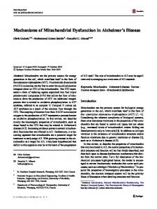
Fig. 2. Ultrastructure of cardiomyocytes from Tgaq⁎44 mouse hearts as compared to age-matched FVB mice. Increased number and size of mitochondria as well as altered mitochondrial structure in terms of less contrasted mitochondrial cristae and more oval shape of mitochondria can be seen in 12 and 14 month-old Tgaq⁎44 mice as compared to FVB and 4 month-old Tgaq⁎44 mice. Representative microphotograph of cardiomyocytes from (A, B) 4 month-old FVB mice and Tgaq⁎44 mice, respectively (C, D) 12 month-old FVB mice and Tgaq⁎44 mice, respectively (E, F) 14 month-old FVB mice and Tgaq⁎44 mice, respectively. Degenerative changes within cardiomyocyte are also visible (F).
324
M. Elas et al. / Free Radical Biology & Medicine 45 (2008) 321–328
Bruker measurements. Non-specific free radical signal seen at g = 2.004 in the heart tissue originates from semiquinones. Mitochondrial Fe-S centers are best measured at 10-30 K [10,32]. At liquid nitrogen temperature the signal at g = 1.94 and 2.025 reflects only a part of mitochondrial Fe-S centers, from complex II. Signal at g = 4.3 originates from high-spin, rhombic, mononuclear Fe(III) [14,20] such as “loosely bound” iron in transferrin, apoferritin, and low-molecular weight complexes. Signals at g = 1.94, 2.004, and 4.3 were quantitated. As some signals had poor signal to noise ratio, and signal double integration would result in a large error, signal amplitude was used to express the signal intensity. Line widths of all signals were measured and they did change neither between groups nor with age (p ≥ 0.1). Cytochrome c oxidase activity and citrate synthase activity Frozen powder was weighed, homogenized in ice-cold buffer (approximately 50 mg wet weight per ml) containing: HEPES 5 mM (pH 8.7), ethyleneglycol-bis (ß-aminoethyl ether) N, N, N′, N′-tetraacetic acid (EGTA) 1 mM, dithiothreitol 1 mM, and Triton X-100 (0.1%) and incubated for 60 min at 4 °C for complete enzyme extraction. Cytochrome c oxidase (COX) was assayed by measuring the disappearance of reduced cytochrome c at 550 nm (pH 7.4, 30°C), and citrate synthase (CS) activity was measured at 412 nm (pH 8, 30 °C) according to Srere [33]. Cytochrome c oxidase activity and citrate synthase activity were measured in Tgαq⁎44 mice at the age of 4, 6, 10, 14-16 months as well as in age-matched wild-type FVB mice. Since the activity of these enzymes was age-independent in FVB mice, the results from wild-type mice were pooled. Statistical analysis Values from samples from the same groups were averaged and SD (n b 5) or SEM (n ≥ 5) was calculated. Independent two-tailed Student's t-test was used to determine the statistical significance, except for cardiac function MRI results, where Kruskal - Wallis test and unpaired t-test with Welch's correction were used. Results Phenotype of heart failure in Tgαq⁎44 mice As shown in the Kaplan-Meyer survival curve in Fig. 1A, Tgαq⁎44 mice did not survived beyond the age of 16 months. Tgαq⁎44 mice at the age of 14 months displayed the phenotype of overt heart failure, as evident by cardiac enlargement (Fig. 1B) and pulmonary congestion (Fig. 1C). Microscopic analysis of hearts from Tgαq⁎44 mice at the age of 14 months revealed typical pathology of cardiomyopathy such as cardiomyocytes enlargement (in cross-section diameter), fibrosis, inflammatory infiltrations, and degeneration of cardiomyocytes (data not shown). Electron microscopy (EM) analysis was suggestive for increased number of mitochondria as well as their increased size in Tgaq⁎44 mice at the age of 4 months (Figs. 2A, B). On the other hand, in cardiac tissue from Tgαq⁎44 mice at the age of 12-14, but not in hearts from younger Tgαq⁎44 mice we noted altered mitochondrial structure in terms of less contrasted mitochondrial cristae and oval shape of mitochondria (Figs. 2C-F). In vivo assessment of cardiac function by MRI In young Tgαq⁎44 mice a slightly lower values for fractional contraction (fractional area change, FAC) were seen as compared to age-matched FVB mice. The fractional contraction was progressively impaired in Tgαq⁎44 mice at the age of 10 months and older in comparison with 4 month-old Tgαq⁎44 mice. However, the impairment of cardiac function in Tgαq⁎44 mice reached statistical significance only for 14 month-old Tgαq⁎44 mice (Fig. 3).
Fig. 3. Fractional area change (FAC) in Tgαq⁎44 (4, 8, 10, 12, and 14-months old) and wildtype FVB mice (4, 7.5, and 13-months old) measured in vivo by MRI. Numbers in the figure denote group sizes, ⁎ indicates significant difference between Tgαq⁎44 mice at the age of 8-14 months vs. Tgαq⁎44 mice at the age of 4 months as assessed by Kruskal-Wallis test (p b 0.05). # indicates significant difference between Tgαq⁎44 mice and age-matched wild type FVB mice as assessed by unpaired t-test with Welch's correction (p b 0.05).
Ex vivo assessment of mitochondrial function by EPR Semiquinone centres A representative, non-specific free radical EPR signal at g = 2.004 reflecting mitochondrial semiquinones [34,35] originating from a heart of 14-month old Tgαq⁎44 mice or wild-type FVB mice is shown in Fig. 4A. As summarized in Fig. 4B, in hearts from younger Tgαq⁎44 mice the EPR signal was slightly higher as compared to hearts from FVB mice. Interestingly, EPR signal of semiquinones decreased dramatically in hearts from Tgαq⁎44 mice between the age of 8 and 10 months, whereas in FVB mice it remained at a stable level up to the age of 14 months. In hearts from 10-, 12- and 14-months old Tgαq⁎44 mice the magnitude of this intrinsic EPR signal was approximately 40% lower as compared with hearts from age-matched FVB mice. In contrast to the cardiac tissue, no significant difference (pN 0.05) in non-specific free radical EPR signal at g = 2.004 was detected in other organs (Fig. 5). Iron centres We studied two Fe(III) EPR signals in heart tissue. The signal at g = 1.94 and 2.025 reflects Fe(III) from some mitochondrial Fe-S centers [13,32] (Fig. 6A). The non-specific free radical signal is seen at g = 2.004. In turn, signal at g = 4.3 reflects “loosely bound” Fe(III) from transferrin, apoferritin, and low-molecular weight Fe(III) complexes (Fig 6B). As shown in Figs. 6C and D, some significant differences were found in the levels of both Fe complexes between Tgαq⁎44 and FVB mice. The signal from Fe(III) bound in mitochondrial Fe-S centers was 20-30% lower in 12 and 14 month old Tgαq⁎44 mice in comparison with age-matched FVB mice (Fig. 6C). On the contrary, the signal of “loosely bound” Fe(III) at g = 4.3 was approximately 40% stronger in hearts from 14 month old Tgαq⁎44 mice as compared to the agematched FVB mice (Fig. 6D). Diminished signal representative for mitochondrial Fe-S centers suggests mitochondrial dysfunction, while opposite directions of changes of intensity of these two EPR signals representative for the two different Fe(III) ion pools point out to alterations in iron homeostasis in Tgαq⁎44 mice most likely related to the oxidative stress. Mitochondrial enzymes activity To confirm mitochondrial dysfunction in the heart from Tgαq⁎44 mice, we measured the activity of cytochrome c oxidase and citrate
M. Elas et al. / Free Radical Biology & Medicine 45 (2008) 321–328
325
Fig. 4. A. Representative EPR spectra at g = 2.01 of hearts of 10-month-old Tgαq⁎44 mice, and wild type FVB mice. The position of the free radical standard signal (DPPH) is marked. B. Changes in the concentration of semiquinone paramagnetic centers in the heart tissue of 4.5, 8, 10, 12 and 14.5 month-old Tgαq⁎44 and FVB mice, detected at 77 K. Amplitude was normalized against the wet weight of the heart and expressed as arbitrary units per 100 mg. Values represent an average from 8-15 mice per point, except for 12 month old FVB mice, with n = 2. Error was calculated as SD if n = b 5 or SEM for n N 5. ⁎ denotes statistical significance between Tgαq⁎44 and age-matched FVB. # represents statistically significant difference between 8 and 10 month-old Tgαq⁎44 mice (p b 0.05). For EPR parameters see Methods.
synthase. In hearts from wild-type FVB mice cytochrome c oxidase activity was maintained at the same level, irrespectively of the age of the animals. In contrast, in hearts from Tgαq⁎44 mice at the age of 10 months there was a marked decrease in cytochrome c oxidase activity (Fig. 7A) (p b 0.001 for Tgαq⁎44 10 months vs FVB) that was also present in 14-16 old Tgαq⁎44 mice. Citrate synthase activity was not altered in 10-month old Tgαq⁎44 mice but downregulated in 14-16 month-old Tgαq⁎44 mice (Fig. 7B). Discussion In the present work, we quantified alterations in the contents of reduced semiquinones and Fe(III) iron in hearts from Tgαq⁎44 mice
using EPR technique in relation to typical biochemical indices of mitochondrial function and the progression of impairment of cardiac function in Tgαq⁎44 mice as assessed in vivo using MRI technique. We presented the following evidence for the perturbations in cardiac mitochondria in Tgαq⁎44 mice. First, an increase in semiquinones content (g = 2.004) was observed in Tgαq⁎4 mice at the age of 4 and 8 months, and then a dramatic decrease was evident in older mice. Importantly, the diminished cardiac content of reduced semiquinones detected by EPR preceded the evident impairment of cardiac function (fractional shortening, FAC) in Tgαq⁎44 mice as detected by MRI. Compatible with the change in semiquinone level in Tgαq⁎44 mice we found a significant decrease in Fe(III)-S centers (at g = 1.94) and a parallel increase in signal (at g = 4.3) that is representative for the
Fig. 5. Representative EPR spectra at the free radical region of blood, lung, liver, spleen, and kidney of 10 month-old Tgαq⁎44 mice and FVB mice. Parameters: frequency: 9.45 GHz, microwave power 10 mW, magnetic field 3400 ± 200 G, time constant 2.05 s, modulation amplitude 1.05 G, scan time 60 s, 3 scans, temp. 77 K.
326
M. Elas et al. / Free Radical Biology & Medicine 45 (2008) 321–328
Fig. 6. Examples of EPR spectra of Fe(III) detected at 77 K. (A) Semiquinone free radical signal is seen at g = 2.01, and signal at g = 1.94 shows Fe(III) from Fe-S centers. (B) Signal at g = 4.2 originates from Fe(III) from non-heme proteins, transferin and apoferritin. Parameters: frequency: 9.45 GHz, microwave power 10 mW, magnetic field 3400 or 1600 ± 200 G, modulation amplitude 5 G, 1024 points, 3 scans, temp. 77 K. The changes in the amplitude of the EPR signals at g = 1.94 (C) and g = 4.3 (D) from hearts of 4.5, 8, 10, 12 and 14.5 monthold Tgαq⁎44 mice as compared to age-matched FVB mice, detected at 77 K. Amplitude was normalized against the wet weight of the heart and expressed as arbitrary units per 100 mg. Values represent an average from 3-10 mice per point, with SD if n = b5 and SEM for n ≥ 5. ⁎ denotes statistical significance between Tgαq⁎44 and age-matched FVB (p b 0.05).
pool of “loosely bound“ Fe(III). The above changes in cellular iron equilibrium were significant in 12 month-old (for mitochondrial Fe(III) decrease) and 14 month-old Tgαq⁎44 mice (for easily accessible Fe(III) increase), respectively. Moreover, the activity of cytochrome c oxidase was profoundly decreased in 10 month-old Tgαq⁎44 mice and remained at similar low level in 14-16 month-old Tgαq⁎44 (Fig 7A). Additionally, decreased activity of citrate synthase and typical alterations in mitochondrial structure were detected in the endstage heart failure in 14-16 month-old Tgαq⁎44. Collectively, our results demonstrate important changes in function of cardiac mitochondria before decompensation of heart failure in Tgαq⁎44 mice, and their accentuation in the end-stage heart failure strongly
suggesting that they may well contribute to the development of the overt heart failure in this model. Putting our results however into a mechanistic perspective may be only speculative. In aerobic conditions EPR signal at g = 2.004 in heart originates predominantly from semiquinones from mitochondria [10,34–36]. Under anaerobic conditions, flavin radicals also contribute to this signal [10]. Semiquinones act as electron carrier shuttles reducing equivalents from complex I and II to complex III, and their level is affected by the availability of oxygen, activity of mitochondrial complexes and mitochondrial membrane potential. Initially increased and then strongly reduced pool of semiquinones is indicative for disturbances in the mitochondrial electron flow, though their
Fig. 7. A. Cytochrome c oxidase activity in heart tissue of Tgαq⁎44, in IU (µmole/min) of oxidized cytochrome c per g of protein. B. Citrate synthase activity in IU/g protein. Both cyt c oxidase activity and citrate synthase activity did not differ with age of FVB mice, therefore control in A and B shows pooled data for all mice at the age of 4 and 14-16 months (n = 21). 5-8 of mice per group were used for Tgαq⁎44 mice. ⁎ indicates 0.01 ≤ p b 0.05, ⁎⁎ denotes 0.001 ≤ p b 0.01, whereas ⁎⁎⁎ denotes p b 0.001.
M. Elas et al. / Free Radical Biology & Medicine 45 (2008) 321–328
mechanisms and functional significance remains to be established. Indeed, decrease in semiquinones content may be related to the impairment of a mitochondrial of electron flow that could be either a consequence of increased oxidative stress, or a defense mechanism to protect against enhanced free radical generation. Similar degree of a decrease in semiquinone signal was reported by many authors in ischemic heart [10,12,13,15]. Impairment of the redox exchange in the ubiquinon/bc1 redox couple may lead to electron leak and increased ROS generation in mitochondria [8,37]. Therefore changes in the magnitude of semiquinone signals may point to the increased oxidative stress which, in turn, interferes with mitochondrial electron flow. Indeed, in hearts from Tgαq⁎44 mice there is an increased expression and activity of NADPH oxidase [38] and excessive superoxide anion production in cardiac tissue that could be linked to the excessive activation of Gαq signaling pathway [26,27] and may well be associated with augmented superoxide production by mitochondria. On the other hand, the parallel decrease in semiquinone content and cytochrome c oxidase activity - the latter uniformly reported to be present in aged and ischemic hearts as well as in failing myocardium [39–44] - suggest that these two events are linked. Interestingly, there was a substantial difference in the intensity of EPR signals at g = 2.004 quantifying mitochondrial semiquinones in 10 month-old Tgαq⁎44 mice vs 8 month-old Tgαq⁎44 mice. As such, in spite of the fact that EPR technique assess mitochondrial function only indirectly, evaluation of the EPR signal at g= 2.004 provides sensitive detection of mitochondrial alterations in Tgαq⁎44 hearts. If so, in addition to classical biochemical indices of mitochondrial function quantification of semiquinones by EPR technique, together with iron pool status determination can serve as a good tool to examine the involvement of mitochondrial mechanisms in pathogenesis of heart failure. It is worth noting that our analysis of alteration in iron cellular homeostasis was limited, as we did not measure the total iron fate, or the relative content of two major iron oxidative forms. Such studies might be helpful for a better understanding of the role of iron in the development of heart failure in Tgαq⁎44 mice. Furthermore, direct characterization of the mechanisms of mitochondrial dysfunction in Tgαq⁎44 hearts requires further studies. Interestingly, some reports suggest that the relaxation is the most energy-sensitive phase of cardiac work cycle and drop in ATP level e.g. due to ischemia leads first of all to diastolic dysfunction [45,46]. Yet, in the present work we measured fractional contraction (FAC) to characterize cardiac function. In fact, this parameter does not reveal impairment of diastolic function, but rather impairment of systolic function. Interestingly, in Tgαq⁎44 mice impairment of both the relaxation and the contraction phases can be observed at the endstage of pathology (data not shown). It is an intriguing hypothesis to be tested whether in Tgαq⁎44 mice mitochondrial dysfunction leads first to diastolic dysfunction. In particular, since with some notable exceptions [47–49], there are no clear-cut studies showing causaleffect relationship between mitochondrial dysfunction and the development of diastolic heart failure. In summary, our results point out to the development of mitochondrial dysfunction in Tgαq⁎44 mice. Importantly, alterations in mitochondrial function seem to occur quite early in life of Tgαq⁎44 mice and appeared evident in the due course of pathology progression preceding and possibly precipitating the development of the overt heart failure in this model. It would be worthwhile to examine whether therapy targeted at cardiomyocyte mitochondria would slow down the progression of mitochondrial dysfunction and the progression of cardiac dysfunction. Acknowledgments This work was partially supported by the Polish Ministry of Science and Higher Education (grant no. PBZ-KBN-101/T09/2003), Jagiellonian University grant CRBW/VIII-38/2004 and Professorial grant from the Foundation for Polish Science to S.C (SP/04/04).
327
References [1] Huss, J. M.; Kelly, D. P. Mitochondrial energy metabolism in heart failure: a question of balance. J. Clin. Invest. 115:547–555; 2005. [2] Hirano, M.; Marti, R.; Ferreiro-Barros, C.; Vila, M. R.; Tadesse, S.; Nishigaki, Y.; Nishino, I.; Vu, T. H. Defects of intergenomic communication: autosomal disorders that cause multiple deletions and depletion of mitochondrial DNA. Semin. Cell Dev. Biol. 12:417–427; 2001. [3] Ide, T.; Tsutsui, H.; Kinugawa, S.; Utsumi, H.; Kang, D.; Hattori, N.; Uchida, K.; Arimura, K.; Egashira, K.; Takeshita, A. Mitochondrial electron transport complex I is a potential source of oxygen free radicals in the failing myocardium. Circ. Res. 85:357–363; 1999. [4] Garnier, A.; Fortin, D.; Delomenie, C.; Momken, I.; Veksler, V.; Ventura-Clapier, R. Depressed mitochondrial transcription factors and oxidative capacity in rat failing cardiac and skeletal muscles. J. Physiol. 551:491–501; 2003. [5] Maurer, I.; Zierz, S. Myocardial respiratory chain enzyme activities in idiopathic dilated cardiomyopathy, and comparison with those in atherosclerotic coronary artery disease and valvular aortic stenosis. Am. J. Cardiol. 72:428–433; 1993. [6] Vogt, A. M.; Kubler, W. Heart failure: is there an energy deficit contributing to contractile dysfunction? Basic Res. Cardiol. 93:1–10; 1998. [7] Casademont, J.; Miro, O. Electron transport chain defects in heart failure. Heart Fail. Rev. 7:131–139; 2002. [8] Nohl, H.; Gille, L.; Staniek, K. Intracellular generation of reactive oxygen species by mitochondria. Biochem. Pharmacol. 69:719–723; 2005. [9] Baker, J. E.; Felix, C. C.; Olinger, G. N.; Kalyanaraman, B. Myocardial ischemia and reperfusion: Direct evidence for free radical generation by electron spin resonance spectroscopy. Proc. Nat. Acad. Sci. U. S. A. 85:2786–2789; 1988. [10] Ruuge, E. Oxidative stress and myocardial injury: spin-trapping and lowtemperature EPR study. Curr. Top. Biophys. 26:145–155; 2001. [11] Ruuge, E. K.; Ledenev, A. N.; Lakomkin, V. L.; Konstantinov, A. A.; Ksenzenko, M. Free radical metabolites in myocardium during ischemia and reperfusion. Am. J. Physiol. 261:81–86; 1991. [12] Zweier, J. L.; Flaherty, J. T.; Weisfeldt, M. L. Direct measurement of free radical generation following reperfusion of ischemic myocardium. Proc. Natl. Acad. Sci. U. S. A. 84:1404–1407; 1987. [13] Baker, J. E.; Kalyanaraman, B. Ischemia-induced changes in myocardial paramagnetic metabolites: implications for intracellular oxy-radical generation. FEBS Lett. 244:311–314; 1989. [14] Svistunenko, D. A.; Davies, N.; Brealey, D.; Singer, M.; Cooper, C. E. Mitochondrial dysfunction in patients with severe sepsis: an EPR interrogation of individual respiratory chain components. Biochim. Biophys. Acta 1757:262–272; 2006. [15] Ledenev, A. N.; Lakomkin, V. L.; Ruuge, E. K. [Redox reactions of flavins and coenzyme Q-10 in the heart during experimental ischemia and reperfusion]. Biofizika 31:687–690; 1986. [16] Keyer, K.; Imlay, J. A. Superoxide accelerates DNA damage by elevating free-iron levels. Proc. Natl. Acad. Sci. U. S. A. 93:13635–13640; 1996. [17] Srinivasan, C.; Liba, A.; Imlay, J. A.; Valentine, J. S.; Gralla, E. B. Yeast lacking superoxide dismutase(s) show elevated levels of “free iron” as measured by whole cell electron paramagnetic resonance. J. Biol. Chem. 275:29187–29192; 2000. [18] Musci, G.; Persichini, T.; Casadei, M.; Mazzone, V.; Venturini, G.; Polticelli, F.; Colasanti, M. Nitrosative/oxidative modifications and ageing. Mech. Ageing Dev. 127:544–551; 2006. [19] Ruuge, E. K.; Zabbarova, I. V.; Sviryaeva, I. V.; Shumaev, K. B. Redox status of cardiac cells. Ferritin, reactive oxygen and nitrogen species. Curr. Top. Biophys. 29:37–45; 2005. [20] Symons, M. C. R. Chemical and Biochemical Aspects of Electron Spin Resonance. Wiley, London, pp. 1–190; 1978. [21] Aisen, P.; Leibman, A.; Zweier, J. Stoichiometric and site characteristics of the binding of iron to human transferrin. J. Biol. Chem. 253:1930–1937; 1978. [22] Foster, M. A. Paramagnetic Metal Ions. In: Foster, M.A. (Ed.), Magnetic Resonance in Medicine and Biology. Pergamon Press, Oxford, pp. 48–65; 1984. [23] Chasteen, N. D.; Antanaitis, B. C.; Aisen, P. Iron deposition in apoferritin. Evidence for the formation of a mixed valence binuclear iron complex. J. Biol. Chem. 260:2926–2929; 1985. [24] Hanna, P. M.; Chen, Y.; Chasteen, N. D. Initial iron oxidation in horse spleen apoferritin. Characterization of a mixed-valence iron(II)-iron(III) complex. J. Biol. Chem. 266:886–893; 1991. [25] Stadler, N.; Lindner, R. A.; Davies, M. J. Direct detection and quantification of transition metal ions in human atherosclerotic plaques: evidence for the presence of elevated levels of iron and copper. Arterioscler. Thromb. Vasc. Biol. 24:949–954; 2004. [26] Mende, U.; Semsarian, C.; Martins, D. C.; Kagen, A.; Duffy, C.; Schoen, F. J.; Neer, E. J. Dilated cardiomyopathy in two transgenic mouse lines expressing activated G protein alpha(q): lack of correlation between phospholipase C activation and the phenotype. J. Mol. Cell. Cardiol. 33:1477–1491; 2001. [27] Mende, U.; Kagen, A.; Cohen, A.; Aramburu, J.; Schoen, F. J.; Neer, E. J. Transient cardiac expression of constitutively active Galphaq leads to hypertrophy and dilated cardiomyopathy by calcineurin-dependent and independent pathways. Proc. Natl. Acad. Sci. U. S. A. 95:13893–13898; 1998. [28] Heinze-Paluchowska, S.; Skórka, T.; Drelicharz, L.; Chlopicki, S.; Jasinski, A. MR imaging of mouse heart in vivo using a specialized probehead and gradient system. Pol. J. Chem. 80:1133–1139; 2006. [29] Wiesmann, F.; Ruff, J.; Engelhardt, S.; Hein, L.; Dienesch, C.; Leupold, A.; Illinger, R.; Frydrychowicz, A.; Hiller, K. H.; Rommel, E.; Haase, A.; Lohse, M. J.; Naa, S. Dobutamine-stress magnetic resonance microimaging in mice: acute changes of cardiac geometry and function in normal and failing murine hearts. Circ. Res. 88:563–569; 2001.
328
M. Elas et al. / Free Radical Biology & Medicine 45 (2008) 321–328
[30] Takuma, S.; Suehiro, K.; Cardinale, C.; Hozumi, T.; Yano, H.; Shimizu, J.; MullisJansson, S.; Sciacca, R.; Wang, J.; Burkhoff, D.; Di Tullio, M. R.; Homma, S. Anesthetic inhibition in ischemic and nonischemic murine heart: comparison with conscious echocardiographic approach. Am. J. Physiol. Heart Circ. Physiol. 280: H2364–H23670; 2001. [31] Walski, M.; Chlopicki, S.; Celary-Walska, R.; Frontczak-Baniewicz, M. Ultrastructural alterations of endothelium covering advanced atherosclerotic plaque in human carotid artery visualised by scanning electron microscope. J. Physiol. Pharmacol. 53:713–723; 2002. [32] Beinert, H. EPR spectroscopy of components of the mitochondrial electrontransfer system. Methods Enzymol. 54:133–150; 1978. [33] Srere, P. Citrate synthase. Methods in Enzymol. 13:3–11; 1969. [34] Ruzicka, F. J.; Beinert, H.; Schepler, K. L.; Dunham, W. R.; Sands, R. H. Interaction of ubisemiquinone with a paramagnetic component in heart tissue. Proc. Natl. Acad. Sci. U. S. A. 72:2886–2890; 1975. [35] Davies, K. J. A.; Hochstein, P. Ubisemiquinone radicals in liver: implications from a mitochondrial Q cycle in vivo. Biochem. Biophys. Res. Commun. 107:1292–1299; 1982. [36] Salerno, J. C.; Ohnishi, T. Studies on the stabilized ubisemiquinone species in the succinate-cytochrome c reductase segment of the intact mitochondrial membrane system. Biochem. J. 192:769–781; 1980. [37] Nohl, H.; Gille, L.; Kozlov, A.; Staniek, K. Are mitochondria a spontaneous and permanent source of reactive oxygen species? Redox Rep. 8:135–141; 2003. [38] Drelicharz, L.; Kozlowski, V.; Skorka, T.; Heinze-Paluchowska, S.; Jasinski, A.; Gebska, A.; Guzik, T.; Olszanecki, R.; Wojnar, L.; Mende, U.; Chlopicki, S. NO and PGI2 in coronary endothelial dysfunction in transgenic mice with dilated cardiomyopathy. Basic Res. Cardiol.2008 [Apr 22, Electronic publication ahead of print]. [39] Abe, H.; Yamada, T.; Miyata, K.; Yoshida, A.; Yabe, Y. Myocardial energy metabolism of congestive and hypertrophic cardiomyopathy in man. Adv. Myocardiol. 4:107–113; 1983.
[40] Heinke, M. Y.; Wheeler, C. H.; Yan, J. X.; Amin, V.; Chang, D.; Einstein, R.; Dunn, M. J.; dos Remedios, C. G. Changes in myocardial protein expression in pacing-induced canine heart failure. Electrophoresis 20:2086–2093; 1999. [41] Jarreta, D.; Orus, J.; Barrientos, A.; Miro, O.; Roig, E.; Heras, M.; Moraes, C. T.; Cardellach, F.; Casademont, J. Mitochondrial function in heart muscle from patients with idiopathic dilated cardiomyopathy. Cardiovasc. Res. 45:860–865; 2000. [42] Quigley, A. F.; Kapsa, R. M.; Esmore, D.; Hale, G.; Byrne, E. Mitochondrial respiratory chain activity in idiopathic dilated cardiomyopathy. J. Card. Fail. 6:47–55; 2000. [43] Lesnefsky, E. J.; Gudz, T. I.; Migita, C. T.; Ikeda-Saito, M.; Hassan, M. O.; Turkaly, P. J.; Hoppel, C. L. Ischemic injury to mitochondrial electron transport in the aging heart: damage to the iron-sulfur protein subunit of electron transport complex III. Arch. Biochem. Biophys. 385:117–128; 2001. [44] Lesnefsky, E. J.; Chen, Q.; Slabe, T. J.; Stoll, M. S.; Minkler, P. E.; Hassan, M. O.; Tandler, B.; Hoppel, C. L. Ischemia, rather than reperfusion, inhibits respiration through cytochrome oxidase in the isolated, perfused rabbit heart: role of cardiolipin. Am. J. Physiol. Heart Circ. Physiol. 287:H258–H267; 2004. [45] Pouleur, H. Diastolic dysfunction and myocardial energetics. Eur. Heart J. 11:30–34 Suppl C; 1990. [46] Bressler, R.; Gay, R.; Copeland, J. G.; Bahl, J. J.; Bedotto, J.; Goldman, S. Chronic inhibition of fatty acid oxidation: new model of diastolic dysfunction. Life Sci. 44:1897–1906; 1989. [47] Minotti, G.; Ronchi, R.; Salvatorelli, E.; Menna, P.; Cairo, G. Doxorubicin irreversibly inactivates iron regulatory proteins 1 and 2 in cardiomyocytes: evidence for distinct metabolic pathways and implications for iron-mediated cardiotoxicity of antitumor therapy. Cancer Res. 61:8422–8428; 2001. [48] Myers, C. The role of iron in doxorubicin-induced cardiomyopathy. Semin. Oncol. 25:10–14; 1998. [49] Oliveira, P. J.; Wallace, K. B. Depletion of adenine nucleotide translocator protein in heart mitochondria from doxorubicin-treated rats–relevance for mitochondrial dysfunction. Toxicology 220:160–168; 2006.