Journal of Plant Pathology (2009), 91 (3), 561-566
Edizioni ETS Pisa, 2009
561
DETECTION OF PSEUDOMONAS AVELLANAE FROM HAZELNUT TWIGS BY TAQMAN REAL-TIME PCR F. Gervasi and M. Scortichini C.R.A.-Unità di Ricerca per la Frutticoltura, Via Torrino 3, 81100 Caserta, Italy
SUMMARY
A TaqMan real-time PCR assay was developed to detect Pseudomonas avellanae, the causal agent of bacterial canker and decline of hazelnut (Corylus avellana L.). The real-time primers and probe were designed within the 16S rRNA gene of the pathogen after a comparison with other related species and pathovars. The assay detected DNA from a panel of P. avellanae strains obtained from different geographic areas. The other bacterial pathogens inciting diseases to hazelnut, P. syringae pv. coryli, P. s. pv. syringae and Xanthomonas arboricola pv. corylina, did not react in the assay. Only P. s. pv. actinidiae, isolated from kiwifruit and yellow kiwifruit in Italy, cross-reacted. The assay detected the pathogen when inoculated in low numbers (80 cells) in a leaf scar up until nine days after inoculation. The addition of bovine serum albumin was essential to prevent inhibition by compounds released from the hazelnut tissue. This assay could be useful for monitoring the presence of P. avellanae either in orchards or nurseries. Screening for germplasm resistance could also benefit from this technique. Keywords: hazelnut decline, real-time PCR, genomospecies, Pseudomonas, Xanthomonas.
INTRODUCTION
PCR using the 16S rRNA gene sequence. An amplicon of 762 bp was specifically produced only by the P. avellanae strains isolated in Italy and Greece and the sensitivity with pure cultures of the pathogen was up to 6.5x10² CFU ml-1. Loreti and Gallelli (2002) designed specific primers directed to the hrpW gene of the bacterium and a PCR assay. An amplicon of 350 bp specifically detected all P. avellanae strains from Italy and Greece and the sensitivity with pure cultures reached 2.0x10³ CFU ml-1. Both techniques could detect the bacterium in infected plant material. It proved critical, however, for the effectiveness of the 16S rRNA-based protocol to add specific compounds in order to limit the negative effect that hazelnut polyphenolics exert on PCR amplification (Scortichini and Marchesi, 2001). Conventional PCR assays can pose some limitations. They are not useful for quantitative analysis, need postamplification analysis such as gel electrophoresis and UV visualization of products, and they are exposed to the risk of contamination. Real-time PCR, in contrast, can amplify and simultaneously quantify a target DNA without the need of post-amplification manipulation. It can be used in a high-throughput manner and it is suitable for large-scale monitoring and screening procedures. It has been successfully applied to detect pathogenic bacteria from plant tissue (reviewed by PalacioBielsa et al., 2009). In this study we report on the development of a TaqMan real-time PCR assay to detect P. avellanae from hazelnut twigs.
Pseudomonas avellanae (Psallidas) Janse et al., the cause of bacterial canker and decline of hazelnut (Corylus avellana L.), provokes serious economic losses in central Italy (Viterbo province) and northern Greece (Scortichini, 2002). Identification and characterization of the agent is currently possible by using classical (biochemical tests, fatty acid analysis) and molecular (repetitive-PCR) techniques (Janse et al., 1996; Scortichini et al., 2002). For detection, two techniques were developed. Scortichini and Marchesi (2001) developed a
Bacterial strains. The strains of P. avellanae and other bacteria used in this study are listed in Table 1. Pseudomonads and Pantoea agglomerans were grown on nutrient agar with 5% of sucrose added (NSA) at 25-27°C. Xanthomonads were cultured on glucoseyeast-extract-calcium-carbonate agar (GYCA).
Corresponding author: Marco Scortichini Fax: +39. 0823 256201 E-mail:
[email protected]
DNA preparation. Bacterial DNA preparations were obtained starting from pure cultures grown for 48 h on the appropriate culture medium. A loopful of pure cul-
MATERIALS AND METHODS
562
Detection of Ps. avellanae by real-time PCR
Journal of Plant Pathology (2009), 91 (3), 561-566
Table 1. List of bacterial species and pathovars used in this study and response of the TaqMan real-time assay. Species/pathovar Pseudomonas avellanae
Strain designation BPIC 631T CRA-PAV 013 CRA-ISF 2059 CRA-PAV 1265 CRA-FRU NOC2 CRA-FRU NOC3 CRA-FRU VNOC3 CRA-FRU VTGR9
Origin, year of isolation, host Greece, 1976, Corylus avellana Italy, 1992, C. avellana Italy, 1994, C. avellana Italy, 2003, C. avellana Italy, 2006, C. avellana Italy, 2006, C. avellana Italy, 2007, C. avellana Italy, 2007, C. avellana
Real-time PCR + + + + + + + +
Pseudomonas syringae pv. coryli
NCPPB 4273T PD 3453 CRA-PAV 598 DPP 48 DPP 51
Italy, 1995, Corylus avellana Italy, 1995, C. avellana Italy, 1995, C. avellana Italy, 1999, C. avellana Italy, 1999, C. avellana
-
Pseudomonas syringae pv. morsprunorum
NCPPB 2427 NCPPB 2787
Switzerland, 1971, Prunus armeniaca Greece, 1975, Prunus avium
-
Pseudomonas syringae pv. syringae
CRA-PAV 14a CRA-PAV P11 CRA-PAV 1365 CRA-PAV 1386 CRA-PAV 1394 CRA-PAV 1431
Italy, 1998, Corylus avellana Italy, 1998, C. avellana Italy, 2006, C. avellana Italy, 2007, C. avellana Italy, 2007, C. avellana Italy, 2007, C. avellana
-
Pseudomonas syringae pv. tomato
CRA-FRU 228 CRA-FRU 617
Brasil, 1996, Lycopersicon esculentum Italy, 1996, L. esculentum
-
Pseudomonas syringae pv actinidiae
NCPPB 3739T CRA-FRU Act 2 CRA-FRU Act 3 CRA-FRU Act 4 CRA-FRU Act chi 1 CRA-FRU Act chi 2 CRA-FRU Act chi 3 CRA-FRU Act chi 4
Japan, 1984, Actinidia deliciosa Italy, 2001, A. deliciosa Italy, 2001, A. deliciosa Italy, 2001, A. deliciosa Italy, 2008, A. chinensis Italy, 2008, A. chinensis Italy, 2008, A. chinensis Italy, 2008, A. chinensis
+ + + + + + +
Pseudomonas viridiflava
NCPPB 1252
United Kingdom, 1960, Pyrus communis
-
Pseudomonas corrugata
NCPPB 2450 CRA-FRU 21
United Kingdom, 1972, Lycopersicon esculentum Italy, 1996, L. esculentum
Pseudomonas marginalis pv. marginalis
PD 1630
U.S.A., 1990, Dieffenbachia sp.
-
Pseudomonas savastanoi pv. fraxini
PD 117
The Netherlands, 1978, Fraxinus excelsior
-
Xanthomonas arboricola pv. corylina
NCPPB 935T NCPPB 984 NCPPB 2896 CRA-FRU 235 CRA-FRU 238 CRA-FRU 504 CRA-FRU 505 CRA-FRU 3657
India, 1959, Corylus maxima U.S.A., 1941, C. maxima United Kingdom, 1976, C. avellana Italy, 1997, C. avellana Italy, 1999, C. avellana Italy, 2002, C. avellana Italy, 2005, C. avellana Italy, 2007, C. avellana
-
Pantoea agglomerans
CRA-FRU 14aq
Italy, 2002, Corylus avellana
-
BPIC: Culture Collection of the Benaki Phytopathology Institute, Kiphissia-Athens, Greece CRA-ISF: Culture Collection of Centro di Ricerca per la Frutticoltura. Roma, Italy CRA-PAV: Culture Collection of Centro di Ricerca per la Patologia Vegetale, Roma Italy DPP: Culture Collection of Department of Plant Protection, University of Sassari, Italy NCPPB: National Collection of Plant Pathogenic Bacteria, York, United Kingdom PD: Culture Collection of the Plant Protection Service, Wageningen, The Netherlands
-
Journal of Plant Pathology (2009), 91 (3), 561-566
ture was suspended in sterile bidistilled water and centrifuged at 13,000 g for 2 min. The pellet was resuspended in 100 µl of 50 mM NaOH, and the suspension heated at 95°C for 15 min, and centrifuged 13,000 g for 2 min. The supernatant was collected, an equal volume of chloroform/isoamyl alcohol (24:1) added, mixed thoroughly, and centrifuged at 10,000 g for 5 min at room temperature. The aqueous phase was transferred to a new tube, 5 M NaCl added to a final concentration of 2 M NaCl, mixed with two volumes of absolute ethanol, and kept 2 h at -20°C. Then this phase was centrifuged at 14,000 g for 10 min at 4°C; the supernatant was decanted, the pellet washed with 70% (v/v) ethanol, air dried and dissolved in 40 µl of double distilled water. The quantity and quality of the DNA were determined by spectrophotometer (GeneQuant, Pharmacia, USA). Primer and probe design. The 16S gene sequences of P. avellanae and related pseudomonads from the NCBI databank were aligned by the BioEdit sequence alignment editor (Ibis Bioscience, USA). Within these alignments, specific regions, unique to each target organism, were identified. A BLAST search was performed in order to find the best consensus sequence and to ensure that no non-target organism of which a sequence is present in the database had a homologous sequence. Annealing sites for primers and the specific TaqMan probe were manually designed within the region exhibiting the best consensus sequence. Two primers and a probe were designed based on the regions of the 16S rRNA gene of P. avellanae ISF 2059 which exhibited suitable variability within the region when compared with the related pseudomonads (Table 2). The distance of the primers was chosen to cover a length of 82 bp to ensure efficient PCR replication. The forward primer, Pavel-F, was a 20-mer covering positions 422-441. The reverse primer, Pavel-R, was a 19-mer covering positions 486-504. The probe, Pavel-MGB, is a 17-mer TaqMan-MGB probe covering the positions 454-470, a region between that of the primers. Primers and probe were synthesized by Applied Biosystems (USA). The minor groove binder (MGB)probe was covalently labelled by the manufacturer at the 5’ end with the reporter dye FAM and, at the 3’ end, with the non-fluorescent quencher NFQ1. The MGB
Gervasi and Scortichini
563
probe increases the affinity of the TaqMan probe for the target sequence allowing shorter probes to be used, often increasing the sequence specificity of the detection system, and improving the quenching of the reporter signal due to the shorter distance between reporter and quencher. Moreover, the MGB probe allows the design of a probe with a low G/C percentage; this stabilizes the A/T rich duplexes more than the G/C ones increasing the Tm of A/T rich probes (Kutyavin et al., 2000). TaqMan real-time PCR assay conditions. The reactions were performed in 96-well plates in a Bio-Rad iCycler thermal cycler iQ5 (Bio-Rad, USA). Amplification reaction mixture (25 µl) consisted of 2 µl of DNA template, 1 X PCR Buffer II (Applied Biosystems, USA), 4 mM MgCl2, 200 µM dNTPs (each), 300 nM primers (each), 200 nM TaqMan MGB probe, 1.25 U AmpliTaq Gold DNA polymerase (Applied Biosystems, USA). For detection in crude plant tissue extracts, 400 ng/µl ultrapure bovine serum albumin (BSA) (Applied Biosystems, USA). was added to reduce the negative effect of polyphenols on PCR amplification (Kreader, 1996; Weller et al., 2000; Kageyama et al., 2003). The template was 10 ng/PCR of DNA extracted from pure culture for the assay specificity or 2 µl from crude plant extract for spiking to test the assay in vivo. Cycling conditions were as follows: initial activation at 95°C for 10 min, followed by 45 two-step cycles comprising 10 sec at 95°C and 45 sec at 60°C. Performance and specificity of the assay. Performance was analysed by running five ten-fold serial dilutions of purified P. avellanae CRA-PAV 1265 DNA in sterile double-distilled water. Two separate runs of two separate P. avellanae DNA dilution series were analysed. The mean slope of the linear regression line between logarithmic values of P. avellanae DNA concentration and cycle threshold number (Ct) values was used to calculate the amplification efficiency, E=(10[-1/slope])-1, where 1 corresponds to 100% amplification efficiency (Pfaffl, 2001; Rasmussen, 2001). The correlation coefficient (R2) was also determined. The specificity of the primers targeting P. avellanae was assessed by checking the DNAs of related bacterial species and pathovars listed in Table 1, and prepared as described above for P. avellanae. Each DNA was tested in dupli-
Table 2. Nucleotide sequences of TaqMan real-time PCR primers and of the minor groove binder (MGB) probe developed in this study.
Primer/Probe
Orientation
Sequence (5'-3')
Length (nt)
Tm (°C)
GC content (%)
Pavel-F Pavel-R Pavel-MGB
Forward Reverse Probe
GCACTTTAAGTTGGGAGGAA CAGAGTTAGCCGGTGCTTA AATACGTATCTGTTTTG-MGB
20 19 17
64.5 65.8 52.1
45 52.6 29.4
564
Detection of Ps. avellanae by real-time PCR
cate, and P. avellanae CRA-PAV 1265 was used as positive control. Sensitivity of the assay for infected hazelnut twigs. Samples of artificially inoculated hazelnut material were used. Twigs of field-cultivated hazelnut cultivar Tonda Gentile Romana were inoculated at the beginning of autumn by following the method of Scortichini and Lazzari (1996) and Scortichini et al. (2002b). Ten µl of bacterial suspensions of P. avellanae 1265 serially diluted ten-fold in sterile physiological saline (SPS; 0.85% NaCl in distilled water) from 8x10⁷ CFU ml-1 to 8x10² CFU ml-1 photometrically determined, were placed, at the beginning of October, on the leaf scar of a one-year old twig, using 30 twigs. Each dose was replicated five times. Control twigs were inoculated with SPS only. After one, two, seven and nine days post-inoculation, the twigs were removed from the tree and two slices (20-40 mg total) from the leaf scar were excised using a sterile scalpel, collected in a 2.0 ml Eppendorf tube, 320 µl of SPS added, and crushed by a sterile micro pestle. After an incubation of 25 min at room temperature, 280 µl of the suspension was collected into a new tube and centrifuged at 13,000 g for 10 min. The supernatant was discarded, and the pellet resuspended in 20 µl of SPS. Two µl of this crude suspension were directly used as template in the PCR. Each crude suspension was run in three replicates and the mean Ct values calculated.
RESULTS
Journal of Plant Pathology (2009), 91 (3), 561-566
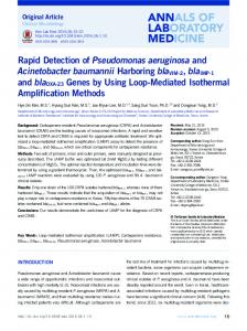
mean Ct used for a linear regression analysis. A slope of -3.33 (efficiency 99.7) and a regression R2 value of 0.99 indicated a good fitness for a real-time PCR assay (Fig. 1). A panel of P. avellanae strains and other bacterial species were tested to check the specificity. Results are summarized in Table 1. The assay detected all P. avellanae strains from various geographic locations used in this study (Fig. 2). The amplification plot showed that all isolates crossed the threshold-line between cycles 14 and 15, when the same amount of bacterial DNA (from 100 ng to 10 pg per reaction per strain) was tested. The other hazelnut pathogens (i.e. P. syringae pv. coryli, P. s. pv. syringae and X. a. pv. corylina), other phytopathogenic pseudomonad strains, and Pantoea agglomerans did not react (Fig. 3). Remarkably. P. s. pv. actinidiae strains originating from Italy showed a strong signal with our probe. P. s. pv. actinidiae NCPPB 3739, the type-strain of the pathovar, isolated in Japan, however, did not show amplification. No signal was detected for the negative controls included in the test. Sensitivity of the assay with samples of infected hazelnut twigs. Detection sensitivity was determined using three replicates of extracts of artificially inoculated hazelnut twigs. The assay was able to detect P. avellanae CRA-PAV 1265 artificially inoculated in low concentration (i.e. 8x10³ CFU/ml) on/in leaf scars up to nine days after inoculation. The addition of BSA proved essential to enable DNA amplification since no amplification was recorded when it was not added. No amplification signal was detected for the negative controls.
Performance and specificity of the assay. Parameters were determined by amplifying ten-fold dilutions of P. avellanae DNA (from 100 ng to 10 pg per reaction). Each dilution was amplified in three replicates and the
Fig. 1. Linear regression generated by ten-fold dilution of bacterial DNA (picograms) and the threshold cycle values as detected by the TaqMan real-time PCR assay. The amplification efficiency is 99.7 and the regression R2 is 0.99.
Fig. 2. Sensitivity of the TaqMan real-time PCR assay for the detection of Pseudomonas avellanae. Amplification plot from the Bio-Rad iCycler thermal cycler iQ5 detection system showing cycle threshold number (Ct) versus normalized fluorescent values of a dilution series of DNA from a panel of P. avellanae strains. Different panel of curves represent P. avellanae strains from Italy and the type-strain from Greece tested with the assay at decreasing doses. The reaction did not produce any detectable amplicon for the negative control samples.
Journal of Plant Pathology (2009), 91 (3), 561-566
Gervasi and Scortichini
565
to nine days from inoculation. Additional study is necessary to further investigate the sensitivity and specificity of the real-time PCR assay in for use in epidemiological studies with epiphytic and/or latent populations of the pathogen.
REFERENCES
Fig. 3. Specificity of the TaqMan real-time PCR assay for the detection of Pseudomonas avellanae, for a number of other bacterial hazelnut pathogens, other phytopathogenic pseudomonads and Pantoea agglomerans. Curves showing fluorescence increasing at cycle threshold number (Ct) between 14 and 15 correspond to DNA preparations from pure cultures of P. avellanae strains from Italy and the type-strain from Greece and an Italian strain of P. s. pv. actinidiae. The reaction did not produce any detectable amplicon (i.e. no increasing curves present at the bottom of the plot) for all the other species and pathovars reported in Table 1, including the type strain of P. s. pv. actinidiae from Japan.
DISCUSSION
The assay developed has potential for rapid detection of P. avellanae. Primers and probe targeted to the 16S rRNA gene of the bacterium enabled detection of a panel of P. avellanae strains from Greece and Italy. None of of the strains we tested of other bacterial pathogens inciting diseases on hazelnut, namely P. s. pv. coryli, P. s. pv. syringae and X. a. pv. arboricola, cross-reacted in the assay. Some Italian strains of P. s. pv. actinidiae, the causal agent of bacterial canker of kiwifruit (Actinidia deliciosa Liang et Ferguson) and yellow kiwifruit (A. chinensis Planchon) strains isolated in Italy (Scortichini, 1994; Ferrante and Scortichini, 2009), however, strongly cross-reacted with our probe. The P. s. pv. actinidiae type-strain, isolated in Japan (Takikawa et al., 1989), did not cross-react. These findings once more confirm the genetic relatedness of this pathovar with P. avellanae (Scortichini et al., 2002b) and point out that genetic variability exists within this pathovar. Similarly, Weller et al. (2007) found that their TaqMan real-time PCR assay, exploiting the pep (prolylendopeptidase) gene of Xanthomonas arboricola, detected not only X. a. pv. fragariae but also cross-reacted with some strains of X. a. pv. corylina and pv. pruni. For reliable use of the protocol, addition of BSA proved critical since, as previously noted (Scortichini and Marchesi, 2001), PCR-inhibiting compounds are present in macerated hazelnut twigs. For preliminary adjustment of the assay, we monitored the pathogen up
Ferrante P., Scortichini M., 2009. Identification of Pseudomonas syringae pv. actinidiae as causal agent of bacterial canker of yellow kiwifruit (Actinidia chinensis Planchon) in central Italy. Journal of Phytopathology 157 (in press). Kageyama K., Komatsu T., Suga H., 2003. Refined PCR protocol for detection of plant pathogens in soil. Journal of General Plant Pathology 69: 153-160. Kreader C.A., 1996. Relief of amplification inhibition in PCR with bovine serum albumin or T4 gene 32 protein. Applied and Environmental Microbiology 62: 1102-1106. Kutyavin I.V., Afonina I.A., Mills A., Gorn V.V., Lukhtanov E.A., Belousov E.S., Singer M.J., Walburger D.K., Lokhov S.G., Gall A.A., Dempcy R., Reed M.W., Meyer R.B., Hedgpeth J., 2000. 3’minor groove binder-DNA probes increase sequence specificity at PCR extension temperatures. Nucleic Acids Research 28: 655-661. Janse J.D., Rossi M.P., Angelucci L., Scortichini M., Derks J.H.J., Akkermans A.D.L., De Vrijer R., Psallidas P., 1996. Reclassification of Pseudomonas syringae pv. avellanae as Pseudomonas avellanae (spec. nov.), the bacterium causing canker of hazelnut (Corylus avellana L.). Systematic and Applied Microbiology 19: 589-595. Loreti S., Gallelli A., 2002. Rapid and specific detection of virulent Pseudomonas avellanae strains by PCR amplification. European Journal of Plant Pathology 108: 237-244. Palacio-Bielsa A., Cambra M.A., López M.M., 2009. PCR detection and identification of plant-pathogenic bacteria: updated review of protocols (1989-2007). Journal of Plant Pathology 91: 249-297. Pfaffl M.W., 2001. A new mathematical model for relative quantification in real-time RT PCR. Nucleic Acids Research 29: 2002-2007. Rasmussen R., 2001. Quantification in the lightcycler. In: Meuer S., Wittwer C., Nakagawara K. (eds). Rapid cycle real-time PCR. Methods and Applications, pp. 21-34. Springer, Heidelberg, Germany. Scortichini M., 1994. Occurrence of Pseudomonas syringae pv. actinidiae in Italy. Plant Pathology 43: 1035-1038. Scortichini M., Lazzari M., 1996. Systemic migration of Pseudomonas syringae pv. avellanae in twigs and young shoots of hazelnut and symptom development. Journal of Phytopathology 144: 215-219. Scortichini M., Marchesi U., 2001. Sensitive and specific detection of Pseudomonas avellanae using primers based on 16S rRNA gene sequences. Journal of Phytopathology 149: 527-532. Scortichini M., 2002. Bacterial canker and decline of European hazelnut. Plant Disease 86: 704-709. Scortichini M., Marchesi U., Rossi M.P., Di Prospero P., 2002a. Bacteria associated with hazelnut (Corylus avellana L.) decline are of two groups: Pseudomonas avellanae and
566
Detection of Ps. avellanae by real-time PCR
strains resembling P. syringae pv. syringae. Applied and Environmental Microbiology 68: 476-484. Scortichini M., Marchesi U., Di Prospero P., 2002b. Genetic relatedness among Pseudomonas avellanae, P. syringae pv. theae and P. s. pv. actinidiae, and their identification. European Journal of Plant Pathology 108: 269-278. Takikawa Y., Serizawa S., Ichikawa S., Tsuyumu S., Goto M., 1989. Pseudomonas syringae pv. actinidiae pv. nov., the
Received February 6, 2009 Accepted April 27, 2009
Journal of Plant Pathology (2009), 91 (3), 561-566 causal bacterium of canker of kiwifruit in Japan. Annals of the Phytopathological Society of Japan 54: 224-228. Weller S.A., Beresford-Jones N.J., Hall J., Thwaites R., Parkinson N., Elphinstone J.G., 2007. Detection of Xanthomonas fragariae and presumptive detection of Xanthomonas arboricola pv. fragariae, from strawberry leaves, by real-time PCR. Journal of Microbiological Methods 70: 379-383.