Detection of Salmonella spp. Using a Generic and Differential FRET-PCR Jilei Zhang1,2, Lanjing Wei1,2, Patrick Kelly3, Mark Freeman4, Kirsten Jaegerson3, Jiansen Gong2,5, Bu Xu2,5, Zhiming Pan2,6, Chuanling Xu1,2, Chengming Wang1,2* 1 Yangzhou University College of Veterinary Medicine, Yangzhou, Jiangsu, P. R. China, 2 Jiangsu Co-Innovation Center for the Prevention and Control of Important Animal Infectious Diseases and Zoonoses, Yangzhou, Jiangsu, P. R. China, 3 Ross University School of Veterinary Medicine, Basseterre, St. Kitts and Nevis, 4 Virginia-Maryland Regional College of Veterinary Medicine, Blacksburg, Virginia, United States of America, 5 Poultry Institute, Chinese Academy of Agricultural Sciences, Yangzhou, Jiangsu, P. R. China, 6 Yangzhou University College of Bioscience and Biotechnology, Yangzhou, Jiangsu, P. R. China
Abstract To facilitate the detection of Salmonella and to be able to rapidly and conveniently determine the species/subspecies present, we developed and tested a generic and differential FRET-PCR targeting their tetrathionate reductase response regulator gene. The differential pan-Salmonella FRET-PCR we developed successfully detected seven plasmids that contained partial sequences of S. bongori and the six S. enterica subspecies. The detection limit varied from ,5 copies of target gene/per PCR reaction for S. enterica enterica to ,200 for S. bongori. Melting curve analysis demonstrated a Tm of ,68uC for S. enterica enterica, ,62.5uC for S. enterica houtenae and S. enterica diarizonae, ,57uC for S. enterica indica, and ,54uC for S. bongori, S. enterica salamae and S. enterica arizonae. The differential pan-Salmonella FRET-PCR also detected and determined the subspecies of 4 reference strains and 47 Salmonella isolated from clinically ill birds or pigs. Finally, we found it could directly detect and differentiate Salmonella in feline (5/50 positive; 10%; one S. enterica salamae and 4 S. enterica enterica) and canine feces (15/114 positive; 13.2%; all S. enterica enterica). The differential pan-Salmonella FRET-PCR failed to react with 96 non-Salmonella bacterial strains. Our experiments show the differential pan-Salmonella FRET-PCR we developed is a rapid, sensitive and specific method to detect and differentiate Salmonella. Citation: Zhang J, Wei L, Kelly P, Freeman M, Jaegerson K, et al. (2013) Detection of Salmonella spp. Using a Generic and Differential FRET-PCR. PLoS ONE 8(10): e76053. doi:10.1371/journal.pone.0076053 Editor: Yung-Fu Chang, Cornell University, United States of America Received June 23, 2013; Accepted August 17, 2013; Published October 16, 2013 Copyright: ß 2013 Zhang et al. This is an open-access article distributed under the terms of the Creative Commons Attribution License, which permits unrestricted use, distribution, and reproduction in any medium, provided the original author and source are credited. Funding: This study was funded by the Ross University School of Veterinary Medicine, the Priority Academic Program Development of Jiangsu Higher Education Institutions of China (PAPD) and by Jiangsu Co-Innovation Center for the Prevention and Control of Important Animal Infectious Diseases and Zoonoses. The funders had no role in study design, data collection and analysis, decision to publish, or preparation of the manuscript. Competing Interests: The authors have declared that no competing interests exist. * E-mail:
[email protected]
The diagnosis of salmonellosis using conventional culture and serotyping is time consuming, laborious and requires large numbers of reagents. Effective nucleic acid-based methods could greatly simplify the procedure and a number of DNA-based techniques have been described to target specific Salmonella serovars [7–12]. Most commonly, they specifically identify S. enterica or only S. enterica subsp. enterica [13–19]. Although a multiplex PCR assay has been described to differentiate species and subspecies of Salmonella [20], the procedure requires organisms isolated in culture, is time consuming, and may be associated with decreased sensitivity and specificity due to complex interactions among five sets of primers. The FRET-PCR assay largely eliminates these problems and we therefore attempted to use this technique to design and test a differential pan-Salmonella FRETPCR to sensitively and specifically detect and differentiate Salmonella species and subspecies. The ability to respire tetrathionate is a characteristic of Salmonella and tetrathionate broth is used as a standard enrichment medium for Salmonella species. The tetrathionate reductase locus (ttr), including the tetrathionate reductase response regulator gene (ttrR), is located within the Salmonella pathogenicity island 2 and is highly conserved with minimal genetic polymorphism among Salmonella species/subspecies. The inclusivity and exclusivity of the PCR technology
Introduction Salmonella are important zoonotic pathogens which affect the health of people and animals worldwide. Advances in methods to detect Salmonella and a better understanding of their genomics have resulted in an improved classification system for the organisms. Currently, the genus Salmonella consists of only 2 species, S. bongori and S. enterica, with the latter containing 6 subspecies: enterica (I), salamae (II), arizonae (IIIa), diarizonae (IIIb), houtenae (IV), and indica (VI) [1,2]. Using the White-Kauffmann-Le Minor Scheme based on somatic, flagellar and capsular antigens, around 2600 serovars are recognized of which over 99.5% belong to the S. enterica species and more than half are S. enterica subspecies enterica [1]. The S. enterica enterica serotypes are primarily associated with warmblooded animals while S. bongori and the remaining five S. enterica subspecies are mainly found in cold-blooded animals and the environment [3]. While most human and animal infections are associated with S. enterica enterica, the growing popularity of reptiles as pets has led to an increasing number of infections with S. bongori and S. enterica subspecies other than S. enterica enterica [2,4,5,6]. A retrospective analysis of over 75,000 isolates collected between 1985–2009 showed that S. enterica subspecies salamae, arizonae, diarizonae and houtenae caused invasive disease in people significantly more frequently than did S. enterica enterica [2]. PLOS ONE | www.plosone.org
1
October 2013 | Volume 8 | Issue 10 | e76053
A Differential Pan-Salmonella PCR
targeting ttrR gene was found to be 100% while applying on 110 Salmonella strains and 87 non-Salmonella strains [16,18]. This makes the ttrR an ideal target for molecular probes. The results of our experiments using the ttrR to develop a FRET-PCR to differentiate Salmonella are described in this report.
Table 1. Salmonella strains used for selectivity of panSalmonella FRET-PCR.
Materials and Methods
Serotype
Time, host and location of Salmonella collection
Tm
S. Indiana
2012; Goose; Danyang, Jiangsu, China
67.5uC
Fecal samples
2010; Goose; Yangzhou, Jiangsu, China
The study was reviewed and approved by the Institutional Animal Care and Use Committee of the Ross University School of Veterinary Medicine (RUSVM). Between June and November of 2011, convenience free-catch fecal samples were obtained from dogs (n = 114) owned by consenting RUSVM veterinary students, and from stray cats (n = 50) involved in the RUSVM Small Animal Spay/Neuter Outreach Program. The animals from which feces were collected were clinically healthy and had no signs of diarrhea. Within 2 hours of collection into clean plastic fecal pots, around 1 g of the feces was transferred with a sterile spatula into a 2.0 ml sterile Eppendorf tube and frozen at 280uC until thawed at room temperature for DNA extraction.
2011, Chicken; Yangzhou, Jiangsu, China 2012; Chicken; Xinxiang, Henan, China S. Potsdam
2010; Goose; Yangzhou, Jiangsu, China
68.0uC
2012; Goose; Yancheng, Jiangsu, China 2012; Chicken; Yangzhou, Jiangsu, China 2009; Chicken; Yangzhou, Jiangsu, China S. Kottbus
2011; Duck; Tianchang, Anhui, China
68.0uC
S. Saintpaul
2010; Duck; Yangzhou, Jiangsu, China
68.0uC
2009; Chicken; Yangzhou, Jiangsu, China S. Typhimurium 2012; Pigeon; Xinxiang, Henan, China
68.0uC
2012; Goose; Yangzhou, Jiangsu, China
Salmonella strains
2008; Chicken; Taian, Shandong, China
To test the sensitivity and specificity of the differential panSalmonella FRET-PCR, we used 47 Salmonella strains (Table 1) comprising 20 serotypes isolated from clinically ill chickens, ducks, geese, pigeons and pigs in five provinces of China between 1999– 2013 [21], 4 reference strains [S. Typhimurium (ATCC 14028); S. Enteritidis (Gaertner) Castellani and Chalmers (ATCC 13076); S. Choleraesuis from Denmark (CMCC 50018); S. Gallinarum from China (CVCC 536)], and 96 non-Salmonella bacterial strains (84 Escherichia coli strains, 10 Campylobacter spp. strains, 1 Pseudomonas aeruginosa strain, and 1 Enterococcus faecalis strain). The Chinese samples from clinically ill birds and pigs had been grown on MacConkey agar, identified by biochemical characteristics (Biolog Inc., Hayward, California, USA) and serotyped using standard agglutination tests with O and H antisera (Tianrun, Ningbo, China).
2009; Chicken; Yangzhou, Jiangsu, China 2012; Pig; Nanjing, Jiangsu, China ATCC 14028 S. Heidelberg
68.0uC
2009; Chicken; Yangzhou, Jiangsu, China S. Reading
2009; Chicken; Yangzhou, Jiangsu, China
68.0uC
S. Derby
2009; Chicken; Yangzhou, Jiangsu, China
67.5uC
S. Enteritidis
2009; Chicken; Haian, Jiangsu, China
2012; Pig; Nanjing, Jiangsu, China 68.0uC
1999; Duck; Xinjin, Sichuan, China 2003; Duck; Mianyang, Sichuan, China 2007; Duck; Weifang, Shandong, China ATCC 13076
Artificially inoculated dog feces Two hundred ml of the ATCC strain S. Typhimurium 14028 with a concentration of 106 CFU/ml was thoroughly mixed for 1 minute with 1 gram of Salmonella culture-negative canine feces suspended in 0.8 ml of sterile PBS. An aliquot of 100 ml was streaked onto MacConkey agar (Remel, Lenexa, Kansas, 66215, USA) and cultured for 18-hour culture at 37uC. As described below, the DNA was extracted from a further aliquot of 200 ml and tested with the differential pan-Salmonella FRET-PCR.
S. Gullinarum
1956; Chicken, CVCC536 strain; Xian, Shanxi, China 68.0uC
S. Montevideo
2009; Chicken; Yangzhou, Jiangsu, China
S. Thompson
2009; Chicken; Yangzhou, Jiangsu, China
67.5uC
S. Bazenheid
2009; Chicken; Yangzhou, Jiangsu, China
68.0uC
68.5uC
S. Choleraesuis
CMCC 50018; Pig; Denmark
68.0uC
S. Kentucky
2009; Chicken; Yangzhou, Jiangsu, China
68.0uC
S. Blockley
2009; Chicken; Yangzhou, Jiangsu, China
68.5uC
S. Agona
2009; Chicken; Yangzhou, Jiangsu, China
66.5uC
S. Pullorum
2011–13; 10 strains from Chicken; Yangzhou, Jiangsu68.5uC
Extraction of DNA from cat and dog feces
2009; 2 strains from Chicken; Tianchang, Anhui, China
The QIAamp DNA Stool Mini Kit (Qiagen Inc., Valencia, CA, USA) was used to extract fecal DNA from cats and dogs following the manufacturer’s instructions. Briefly, the fecal sample (100 mg) was lysed for 2 h at 55uC with buffer ATL and proteinase K before the reaction was stopped with buffer AL at 70uC for 10 min. After centrifugation, anhydrous ethanol was added to the supernatant and the mixture passed through the QIAamp kit column. Following two washes with buffers AW1 and AW2, DNA was eluted in 50 mL of elution buffer and stored at 280uC until thawed at room temperature for testing.
S. Anatis
2012; Pig; Nanjing, Jiangsu, China
66.0uC
S. Newport
2012; Pig; Nanjing, Jiangsu, China
68.0uC
S. Meleagridis
2012; Pig; Nanjing, Jiangsu, China
66.5uC
doi:10.1371/journal.pone.0076053.t001
AM933172, S. Weltevreden FR775221, S. Paratyphi CP000857, S. Typhi LT2 AF282268, S. Dublin CP001144) and five other S. enterica subspecies (salamae, arizonae, diarizonae, houtenae, indica) (Table 2) were obtained from GenBank (www.ncbi.nlm.nih.gov). The Clustal Multiple Alignment Algorithm was used to identify a highly conserved region of the ttrR gene common to all the above Salmonella (Figure 1). It was also used to identify probe regions that
Differential pan-Salmonella FRET-PCR Primers and probes. The ttrR sequences of S. bongori, six S. enterica enterica serovars (S. Gallinarium AM933173, S. Enteritidis PLOS ONE | www.plosone.org
2012; Goose; Danyang, Jiangsu, China
2
October 2013 | Volume 8 | Issue 10 | e76053
A Differential Pan-Salmonella PCR
Table 2. Plasmids containing partial Salmonella ttrR gene used in this study.
Salmonella sequence contained in the plasmid
Gene accession number
Detection limit (copies/10 mL PCR reaction)
Melting temperature (Tm)
S. bongori
AY578066
,100
53uC
S. enterica diarizonae
AY578069
,50
62uC
S. enterica houtenae
AY578068
,50
62.5uC
S. enterica arizonae
CP000880
,100
53.5uC
S. enterica indica
AY578065
,100
57uC
S. enterica salamae
AY578070
,100
54uC
S. enterica enterica Typhimurium
AF282268
,5
68uC
doi:10.1371/journal.pone.0076053.t002
evaluated to determine the probe melting temperature (Tm). This setting is only possible on LightCycler 1.5 and 2.0 models which can accommodate very rapid temperature ramps because of their capillary tube reaction system. PCR products were verified using electrophoresis through 4% MetaPhor agarose gels and purified for automated DNA sequencing with a QIAquick PCR Purification Kit according to the manufacturer’s instructions (Qiagen, Valencia, CA, USA). Both strands of DNA of the PCR products were sequenced at the Genomic Sequencing Laboratory (Davis Sequencing, Davis, USA) using the forward and downstream primers (Figure 1).
were relatively conserved but had a low level of polymorphism that would enable us to differentiate species and subspecies of Salmonella by high-resolution melting curve analysis (Figure 1, Figure 2). The primers and probes were synthesized by Integrated DNA Technologies (Coralville, IA, USA). The differential pan-Salmonella FRET-PCR we developed amplifies a 216-bp target and the positions of primers and probes are shown in Figure 1: forward primer: 59-GATGTTYCTTAGCGCYTTACAGGC-39; downstream primer: 59-CCGACMGCGTAATATTTGGCTGAC-39; anchor probe: 59-ATACGCTTTCCGGCACGGCAAT-6-FAM39; reporter probe: 59-LCRED640-CGTCRGTGGATTWCCGTCGCCCT-Phosphate-39. The fluorescein probe was 39labeled with carboxyfluorescein (6-FAM) which acts as the FRET donor probe, excited by 488 nm light. The LCRed-640 probe was HPLC-purified and used as the FRET acceptor probe, emitting ,640 nm fluorescence following excitation by 6-FAM in close physical proximity. Plasmids. Seven plasmids (Integrated DNA Technologies, Coralville, IA, USA) containing portions of the ttrR gene of S. bongori, S. enterica enterica, S. enterica salamae, S. enterica arizonae, S. enterica diarizonae, S. enterica houtenae and S. enterica indica were used as the positive controls and for quantitative standards (Table 2). Seven nucleotide fragments representing partial ttrR regions of the above Salmonella were synthesized and inserted in the pIDTSMART cloning Vector (Integrated DNA Technologies, Coralville, IA, USA). The resulting plasmids were linearized with HindIII (Promega, Madison, WI, USA) and quantified by PicoGreenH DNA fluorescence assay (Molecular Probes, Eugene, OR, USA) for preparation of quantitative standards (104, 103, 102, 101, 100 copies of ttrR molecules/10 mL). Thermal cycling. Differential pan-Salmonella FRET-PCR was performed in a LightCyclerH 2.0 real-time PCR platform using a thermal protocol and PCR conditions as described [22]. Each reaction was performed in a 20 mL final volume containing 10 mL of extracted DNA. Thermal cycling consisted of 18 highstringency step-down cycles followed by 30 relaxed-stringency fluorescence acquisition cycles. The 18 high-stringency step-down thermal cycles were 660 sec @ 95uC, 12 sec @ 64uC, 8 sec @ 72uC; 960 sec @ 95uC, 12 sec @ 62uC, 8 sec @ 72uC; 360 sec @ 95uC, 12 sec @ 60uC, 8 sec @ 72uC. The relaxed-stringency fluorescence acquisition cycling consisted of 3060 sec @ 95uC, followed by fluorescence acquisition of 12 sec @ 56uC, and 10 sec @ 72uC. Following the completion of differential pan-Salmonella FRET-PCR, the melting curve analysis for probes annealing to the PCR products was determined by monitoring the fluorescence from 45uC to 80uC, and the first derivatives of F4/F1 were PLOS ONE | www.plosone.org
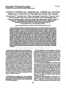
Results Development of the differential pan-Salmonella FRETPCR Our differential pan-Salmonella FRET-PCR detected each of the 7 plasmids containing partial ttrR sequences of S. bongori, the 6 S. enterica subspecies (Table 2). It also detected all 47 Salmonella isolates made from clinically ill birds and pigs in China and the four reference strains; the Tm for these isolates varied from 66.5uC to 68.5uC (Table 1). No reaction products were obtained when the differential pan-Salmonella FRET-PCR was performed on 96 strains of non-Salmonella bacteria. Using the Salmonella ttrR-containing plasmids as quantitative standards, we determined that the detection limit of the FRETPCR varied from ,5 copies of the S. enterica enterica ttrR gene per PCR reaction to ,200 copies per PCR reaction for S. bongori (Table 2). Melting curve analysis enabled a high level of differentiation of Salmonella with strains clustering in four groups based on their melting points: i) S. enterica subsp. enterica had a Tm of ,68uC; ii) ,62.5uC for S. enterica subsp. houtenae and S. enterica subsp. diarizonae; iii) ,57uC for S. enterica subsp. indica; iv) ,54uC for S. bongori, S. enterica subsp. salamae, and S. enterica subsp. arizonae (Figure 2). High-resolution melting analysis on samples containing mixtures of plasmids revealed multiple distinct melting peaks that enabled us to differentiate the Salmonella species/subspecies involved (data not shown).
Artificially inoculated dog feces Following incubation of the fecal sample typical black Salmonella colonies were observed on the inoculated MacConkey agar and confirmed to be S. Typhimurium by standard biochemical testing. Differential pan-Salmonella FRET-PCR of the fecal sample was positive with a Tm of 68uC which was consistent with S. Typhimurium.
3
October 2013 | Volume 8 | Issue 10 | e76053
A Differential Pan-Salmonella PCR
Direct detection of fecal Salmonella DNA by the differential pan-Salmonella FRET-PCR Of the 164 fecal samples studied, 10% of the feline specimens (5/50) and 13.2% of the canine specimens (15/114) were positive for Salmonella by the differential pan-Salmonella FRET-PCR. Of the positive specimens, 95% (19/20) were S. enterica enterica based on their Tm of ,68uC and this was confirmed by gel electrophoresis and sequencing (Figure-S1; Figure-S2). One fecal DNA from a cat had a Tm of 54uC which was consistent with S. enterica salamae and this was also confirmed by sequencing of the reaction products.
Discussion By systematically aligning the ttrR sequences of representative Salmonella species, subspecies and serovars, we were able to identify a region suitable for targeting in a differential pan-Salmonella FRET-PCR. The primers we designed proved to be highly conserved and enabled us to amplify all the Salmonella species and subspecies we tested, including four reference strains and 47 isolates made from clinically ill birds and pigs. We were unable, however, to amplify non-Salmonella bacteria which shows our test has a high specificity. The probes we designed were also conserved but had a low level of polymorphism with the different Salmonella species and subspecies which resulted in different melting points for the organisms. Based on their melting points we were able to rapidly (under 4 hours from DNA extraction to melting curve analysis) and clearly detect and differentiate S. enterica enterica which is the most important subspecies, and in addition the less common and important pathogen S. enterica indica. Although our test did not enable us to differentiate between S. enterica subsp. houtenae and S. enterica subsp. diarizonae, and also between S. bongori, S. enterica subsp. salamae, and S. enterica subsp. arizonae, these organisms are only isolated comparatively rarely in clinical samples [3]. In some areas, however, they are being recognized more frequently [2] and once the possible subspecies have been found in our FRET-PCR, differentiation between them would require sequencing of the reaction products. If biochemical tests are to be used to make the identification, knowledge of the potential subspecies gained from the FRET-PCR would enable more specific testing to be performed.
Figure 2. Melting curves of the pan-Salmonella FRET-PCR. Seven plasmids containing portions of ttrR gene of S. bongori and 6 S. enterica subspecies were used as the positive controls and for melting curve analysis of pan-Salmonella FRET-PCR. The difference in the numbers and types of nucleotide mismatches in the fluorescein and LCRed-640 probes we designed enabled us to identify 4 distinct groups of Salmonella based on their Tm: S. enterica subsp. enterica has a Tm of ,68uC (pink and solid line); ,62.5uC for S. enterica subsp. houtenae (blue and solid line) and S. enterica subsp. diarizonae (blue and dashed line); ,57uC for S. enterica subsp. indica (black and solid line); ,54uC for S. bongori (green and dashed line), S. enterica subsp. arizonae (red and solid line), and S. enterica subsp. salamae (red and dashed line). doi:10.1371/journal.pone.0076053.g002
While a multiplex PCR has been described which identifies all the Salmonella species and subspecies, the test requires cultures of organisms and uses 5 primers [20], the products of which need to be resolved on gels by electrophoresis. Our differential panSalmonella FRET-PCR requires only a single set of primers and one set of probes, and the products are revealed as the differential panSalmonella FRET-PCR assay is performed. This greatly simplifies the procedure and assures the sensitivity and specificity of Salmonella detection, and the rapidity of the test means it will be very useful in situations where screening of large numbers of
Figure 1. Alignment of primers and probes used in the differential pan-Salmonella FRET-PCR. Nucleotide sequences of the primers and probes are shown at the top of the figure and the Salmonella tested are shown down the side. Dots indicate that nucleotides are identical. The upstream primer and probes were used as shown while the downstream primer was used as a downstream oligonucleotide. The upstream and downstream primers and the LCRed 640 probe had 2, 1, and 2 degenerate nucleotides (shown in capital letters). The nucleotides between the primers and probes or between the probes are not shown. doi:10.1371/journal.pone.0076053.g001
PLOS ONE | www.plosone.org
4
October 2013 | Volume 8 | Issue 10 | e76053
A Differential Pan-Salmonella PCR
samples is required in a timely and convenient fashion. Compared to the classical gel-based PCR, our differential pan-Salmonella FRET-PCR might be more expensive but has the advantages of reducing labor costs, eliminating the need for electrophoresis equipment, gels and expensive biohazard disposal, and, most importantly, in minimizing the chances of carry-over contamination. Numerous PCR assays have been described for the detection of Salmonella DNA in a variety of samples, food in particular [7– 10,13–20]. As with routine cultures, however, these PCR methods described to date for Salmonella detection usually require a preenrichment step which delays testing by 6 to 24 hours [19,23], and make the tests unsuitable for rapid screening of large numbers of samples. Our differential pan-Salmonella FRET-PCR was effective in directly detecting Salmonella DNA in experimentally inoculated dog feces when we tested a sample containing approximately 105 organisms, at least some of which were viable and could be cultured. This experiment indicates the differential pan-Salmonella FRET-PCR is sensitive and capable of differentiating its target in a complex matrix containing a large variety of microbial flora and host genome. We were also able to use the differential panSalmonella FRET-PCR to detect Salmonella in dog and cat feces from apparently healthy animals. Although we did not culture the feces from the dogs and cats and were therefore unable to precisely determine the sensitivity and specificity of the test when performed directly on fecal samples, our findings are comparable with other studies using PCR which found Salmonella in 6% and 2% of dogs [24] and cats [25] with normal feces, respectively. Further studies are underway in our laboratory to determine the sensitivity of the PCR in detecting DNA of Salmonella directly in tissue, fecal and environmental samples. In conclusion, our study established a novel differential panSalmonella FRET-PCR with high sensitivity and specificity to quantitatively detect all Salmonella species and subspecies. Melting
curve analysis following amplification and real-time detection conclusively and conveniently differentiated Salmonella species and subspecies and enabled us to diagnose, for the first time, the presence of S. enterica salamae in the feces of a clinically healthy cat.
Supporting Information Figure S1 Representative melting curves of the panSalmonella FRET-PCR of canine fecal samples. DNA extracted from canine fecal samples was used for the pan-Salmonella FRET-PCR described in this study. The positive samples showed an identical Tm of 68uC while the melting curves of negative samples remained flat. (TIF) Figure S2 Gel electrophoresis (4.0% agarose) analysis of the pan-Salmonella FRET-PCR’s amplified products. Lane 1: Trans2K Plus DNA Marker (Beijing Transgen Biotech Co., Ltd.); Lanes 2–3: positive fecal samples from cats; Lane 4: positive fecal sample from dog; Lanes 5–11: 7 plasmids containing part of ttrR gene in the following order: S. enterica enterica, S. enterica houtenae, S. enterica diarizonae, S. enterica indica, S. enterica salamae, S. enterica Arizonae and S. bongori; Lanes 12–13: negative fecal samples from cat and dog; Lanes 14–17: DNAs extracted from Escherichia coli, Campylobacter spp., Pseudomonas aeruginosa, and Enterococcus faecalis, respectively. (TIF)
Author Contributions Conceived and designed the experiments: CW JZ. Performed the experiments: JZ LW PK MF KJ JG BX ZP CX CW. Analyzed the data: JZ PK CW. Contributed reagents/materials/analysis tools: MF JG BX ZP CX. Wrote the paper: JZ LW PK MF JG CW.
References 12. Zhou LQ, Pollard AJ (2010) A fast and highly sensitive blood culture PCR method for clinical detection of Salmonella enterica serovar Typhi. Ann Clin Microbiol and Antimicrob. 9:14. 13. Chen S, Yee A, Griffiths M, Larkin C, Yamashiro CT, et al. (1997) The evaluation of a fluorogenic polymerase chain reaction assay for the detection of Salmonella species in food commodities. Int J Food Microbiol. 35: 239–250. 14. Agarwal A, Makker A, Goel SK (2002) Application of the PCR technique for a rapid, specific and sensitive detection of Salmonella spp. in foods. Mol Cell Probes 16: 243–250. 15. Ellingson JL, Anderson JL, Carlson SA, Sharma VK (2004) Twelve hour realtime PCR technique for the sensitive and specific detection of Salmonella in raw and ready-to-eat meat products. Mol. Cell. Probes 18: 51–57. 16. Malorny B, Paccassoni E, Fach P, Bunge C, Martin A, et al. (2004) Diagnostic real-time PCR for detection of Salmonella in food. Appl Environ Microbiol 70: 7046–7052. 17. Hein I, Flekna G, Krassnig M, Wagner M (2006) Real-time PCR for the detection of Salmonella spp. in food: An alternative approach to a conventional PCR system suggested by the FOOD-PCR project. J Microbiol Methods 66: 538–547. 18. Malorny B, Ma¨de D, Teufel P, Berghof-Ja¨ger C, Huber I, et al. (2007) Multicenter validation study of two blockcycler- and one capillary-based realtime PCR methods for the detection of Salmonella in milk powder. Int J Food Microbiol 117: 211–218. 19. Josefsen MH, Krause M, Hansen F, Hoorfar J (2007) Optimization of a 12-hour TaqMan PCR-based method for detection of Salmonella bacteria in meat. Appl. Environ. Microbiol. 73: 3040–3048. 20. Lee K, Iwata T, Shimizu M, Taniguchi T, Nakadai A, et al. (2009) A novel multiplex PCR assay for Salmonella subspecies identification. J Appl Microbiol 107: 805–811. 21. Gong J, Xu M, Zhu C, Miao J, Liu X, et al. (2013) Antimicrobial resistance, presence of integrons and biofilm formation of Salmonella Pullorum isolates from Eastern China (1962–2010). Avian Pathol 42: 290–294. 22. Kelly PJ, Xu C, Lucas H, Loftis A, Abete J, et al. (2013) Ehrlichiosis, babesiosis, anaplasmosis and hepatozoonosis in dogs from St. Kitts, West Indies. PLoS One. 8:e53450.
1. Josefsen MH, Lofstrom C, Olsen KEP, Molbak K, Hoorfar J (2011) Salmonella. In: Liu D Molecular detection of human bacterial pathogens. Boca Raton: Taylor and Francis Group. pp. 1023–1035. 2. Abbott SL, Ni FC, Janda JM (2012) Increase in extraintestinal infections caused by Salmonella enterica subspecies II-IV. Emerg Infect Dis. 18:637–639. 3. Bopp CA, Brenner FW, Fields PI, Wells JG, Strockbine NA (2003) Escherichia, Shigella, and Salmonella. In: Murray PR, Baro EJ, Jorgensen JH, Pfaller MA, Yolken RH, Manual of clinical microbiology ed. Washington, DC: ASM Press. pp. 654–671. 4. Ma JS, Chen PY, Lau YJ, Chi CS (2003) Brain abscess caused by Salmonella enterica subspecies houtenae in a patient with chronic granulomatous disease. J Microbiol Immunol Infect 36:282–284. 5. Giammanco GM, Pignato S, Mammina C, Grimont F, Grimont PA, et al. (2002) Persistent endemicity of Salmonella bongori 48:z35:– in Southern Italy: molecular characterization of human, animal, and environmental isolates. J Clin Microbiol 40:3502–3505. 6. Pedersen K, Lassen-Nielsen AM, Nordentoft S, Hammer AS (2009) Serovars of Salmonella from captive reptiles. Zoonoses and Public Health 56:238–242. 7. Galan JE, Curtiss R 3rd (1991) Distribution of the invA, -B, -C, and –D genes of Salmonella typhimurium among other Salmonella serovars: invA mutants of Salmonella typhi are deficient for entry into mammalian cells. Infect. Immun. 59: 2901–2908. 8. Rahn K, De Grandis SA, Clarke RC, McEwen SA, Gala´n JE, et al. (1992). Amplification of an invA gene sequence of Salmonella Typhimurium by polymerase chain reaction as a specific method of detection of Salmonella. Mol. Cell. Probes. 6: 271–279. 9. Chaudhry R. Chandel DS, Pandey A Dhawan B, Gulati V, et al. (2005) Utility of PCR in diagnosing complicated cases of unusual clinical manifestations of Salmonella enterica var. Paratyphi A. Am J Med.118: 799–800. 10. Chart H. Cheasty T, de Pinna E, Siorvanes L,Wain J, et al. (2007) Serodiagnosis of Salmonella enterica serovar Typhi and S. enterica serovars Paratyphi A, B and C human infections. J Med Microbiol. 56, 1161–1166. 11. McCarthy N, Reen FJ, Buckley JF, Frye JG, Boyd EF, et al. (2009) Sensitive and rapid molecular detection assays for Salmonella enterica serovars Typhimurium and Heidelberg. J Food Prot 72:2350–2357.
PLOS ONE | www.plosone.org
5
October 2013 | Volume 8 | Issue 10 | e76053
A Differential Pan-Salmonella PCR
23. Gado´ I, Major P, Kira´ly M, Pla´veczky MG (2000) Rapid combined assay for Salmonella detection in food samples. Acta Microbiol Immunol Hung 47: 445– 456. 24. Tupler T, Levy JK, Sabshin SJ, Tucker SJ, Greiner EC, et al. (2012) Enteropathogens identified in dogs entering a Florida animal shelter with normal feces or diarrhea. J Am Vet Med Assoc 241: 338–343.
PLOS ONE | www.plosone.org
25. Sabshin SJ, Levy JK, Tupler T, Tucker SJ, Greiner EC, et al. (2012) Enteropathogens identified in cats entering a Florida animal shelter with normal feces or diarrhea. J Am Vet Med Assoc 241: 331–337.
6
October 2013 | Volume 8 | Issue 10 | e76053