2 (March-April 1991), pp. 45-47. Detection, signal .... figure shows the labelling enzymes (aldolase or ... right-hand side of the figure: the labelling enzyme ALP,.
Journal of Automatic Chemistry, Vol.
13, No. 2 (March-April 1991), pp. 45-47
Detection, signal processing, and calibration In lmmunoassay systems P. A. Bonini, G. Banff Istituto H.S.
Table 1. Activities
Raffaele, Dipart#nento di Medicina di Laboratorio, 20132 Milano,
1251
Italy
M. Pazzagli
of isotopic and nonisotopic labels. detectable events/s/7"5 x 106 labelled molecules detectable event/s/5"6 x 108 labelled molecules Determined by detectability of
H
Universita’ di Firenze, Istituto di Endocrinologia, Firenze, Italy
G. Messeri
Enzymes
Ospedale di Careggi, Laboratorio Analisi, Firenze, Italy
Chemiluminescent labels Fluorescent labels
reaction
and A. Roda Universiti di Messina, Istituto di Chimica, Messina, Italy
The new trends in immunochemistry related to the replacement of radioisotopic labels with non-radioactive labels are presented. Immunoenzymatic, fluorescent and chemiluminescent techniques are described in terms of their basic principles and their most common applications. The advantages of computer-controlled calibration are also discussed.
product
detectable event/s/labelled molecule Many detectable events/s/labelled molecule
labelling substances in contact between antigens and antibodies. Consequently, various isotope and nonisotope mediated immunochemical reactions show very wide ranges of sensitivity: table 2 shows the best detection limits reported in literature under a variety of conditions
[].
Introduction
Immunoenzymatic techniques
The problems of signal detection and elaboration in nonisotopic assays are clearly related to the kind of label used. Various physical properties (other than radioactivity) of the label itself, or, alternatively, of labelmediated products are measured to monitor immunochemical reactions.
Many enzymes
The signal can be detected either continuously or in a non-continuous way. An example of the first case is the measurement of the optical density of an enzyme mediated reaction; photon counting during fluorescent or chemiluminescent reactions is typical of non-continuous detection. Radioisotopes, which have been the most popular labels for immunochemical reactions in the last 30 years, are increasingly being replaced by nonisotopic labels; nevertheless, radioisotopes are the ’gold standards’ when comparing the performanceg (sensitivity and specificity) of new labels.
Specific activity and detection limits
are widely used as labels in immunochemical reactions: table 3 shows the characteristics of an ideal enzyme label [3]. Peroxidase (HRP) (EC 1.11.1.7), alkaline phosphatase (ALP) (EC 3.1.3.1), and betagalactosidase (Gal) (EC 3.2.1.23) are the enzymes most commonly used in immunochemistry; acetylcholinesterase (EC 3.1.1.7), glucose-6-phosphatedehydrogenase (EC 1.1.1.49), catalase (EC 1.11.1.6), and other enzymes are less frequently used. ALP usually splits paranitrophenylphosphate (pNPP) into phosphate and photometrically measurable paranitrophenol, but also methylumbelliferylphosphate can be used with production of fluorescent methylumbelliferone. Orthonitrophenylgalactopyranoside (oNPG), as well as methylumbellipherylgalactoside, can be used as substrates of Gal with a final photometric or fluorimetric reaction. The availability of different substrates for a single enzyme (for example for peroxidase) can result in the release of different enzymatic products with various absorbance
spectra: this can facilitate the automation of such The term ’specific activity’ was used in the past to indicate the number of radioactive disintegrations per unit of time per unit of weight of the isotope compound in radioisotopic labelled immunoassays. It is now used, in a wider sense, to indicate detectable events per unit of time per unit of weight of label material in nonisotopic mediated reactions. As shown in table 1, the specific activity of chemiluminescent and fluorescent labels is notably higher than with radioisotopes ]. This does not mean that chemiluminescent of fluorescent labels always result in more sensitive tests: in fact, many factors play an important role in determining sensitivity: the most important one is the possible steric hindrance due to some
reactions
[4].
Stopping enzymatic reactions by using various solutions (table 4) can be very useful in manual determinations, even if potentially harmful substances need to be used; in automatic systems this step can be avoided.
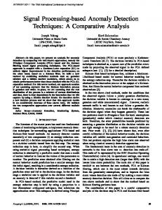
Enzymes are, per se, able to amplify the immunochemical reactions; their amplifying power can be further enhanced as shown in figure 1. The left-hand side of the figure shows the labelling enzymes (aldolase or phosphoglucoisomerase) acting as primary system enzymes in an amplifying system, and generating, 45
0142-0453/91 $3.00
t)
1991 Taylor & Francis Ltd.
P. A. Bonini et al. Immunoassay systems
Table 2. Detection limits of different labels used in immunoassays (reprinted by permission ’Thermochemiluminescence and its Application in Immunoassay’ [Drukkers, Groningen, 1988]).
of T.
Luider [Editor]
Type of label
Examples of labels
Detection limit
Radio-isotopes
3H 125I
Enzymes
Beta-galactosidase Horseradish peroxidase Alkaline phosphatase Firefly luciferin/luciferase
5 x 10-15 mole 5 x 10-17 mole 1.5 x 10⁻¹⁶ 3 X 10-- 16 mole 5 x 10-17 mole 1016 photons/min/mg 5"6 10-19 mole 10-19 mole 2 X 10 -17 mole 10-13 mole < 10-15 mole
Bioluminescence
Enzyme enhanced Firefly luciferin/luciferase Europium(III) ion
Fluorescence
Fluorescein
Enzymes/chemiluminescence
Peroxidase/luminol/enhancer
Glucose-6-phosphate dehydrogenase peroxidase/
10-18 mole
isoluminol
respectively, fructose-bisphosphate (FBP) from glyceraldehydetriphosphate (G3P) + dihydroacetonphosphate (DHAP) and fructose-6-phosphate (F6P) from glucose-6phosphate (G6P). Either FBP or F6P can act as substrates for the secondary system, driven by the enzymes phosphofructokinase and fructosebisphosphatase, with production of inorganic phosphate. Redox cycle is a secondary amplifying system as shown in the right-hand side of the figure: the labelling enzyme ALP, acting as primary system enzyme, generates the substrate nicotinamide adeninnucleotide (NAD) for the redox cycle driven by the enzymes alcoholdehydrogenase and lipoamidedehydrogenase; the cycle needs an excess of ethanol and generates formazan dye as product. A ten-fold increase in absorbance can be obtained in this way
[5].
Nonisotopic methods based on photon counting as signal measurement include fluorescence and chemiluminescence.
Fluorescent and chemiluminescent techniques
In fluorescence a molecule (fluorophor), excited by light, moves to an excited state and then returns to the natural state. Energy is emitted during this process as photons and then measured without chemical modification. a chemical
Conversely, chemiluminescence is based on Table 3. Characteristics
of an ideal enzyme label.
High enzyme activity at low substrate concentration Enzyme stable at pH required for good antibody-antigen binding Presence of reactive groups through which enzymes can be covalently linked to antibody, antigen or hapten with minimum loss of enzyme or immune activities Availability of soluble, purified enzyme at low cost Absence of health hazards attributable to enzyme, substrates and cofactors Enzyme labelled conjugates stable under routine storage and assay conditions
46
from
transformation of a substance into another one. In such a chemical reaction, energy is emitted and then measured. In the field of fluorescent immunoassays (FIA), various methods have been implemented and are routinely used; the most popular are described below.
Fluorescence polarization immunoassay (FPIA) is characterized by a fluorogenic label being excited by polarized light and emitting a partially polarized fluorescence at a right angle to the incident beam.
Front surface fluorescence is characterized by a fluorescence mediated immunoreaction taking place on a solid-phase device, reflecting the generated fluorescence in the same direction as the exciting light.
In
time resolved fluorescent immunoassays (TRF) immunoreagents are labelled with a special class of fluorogens (lanthanide ions, usually Europium), characterized by emission peaks at 614 nm, very narrow and well separated from the scattering caused by excitation (at 340 nm) and from interfering fluorescence of serum (from 400 to 600 nm) (see figure 2). In addition, the specificity of this technology is further enhanced by the decay time of these substances, which is much longer (10-1000 s) than that of the background (1-20 ns); thus, specific fluorescence can be detected once the interfering one has fully decayed
[6]. Luminescence is based on the property of some natural (bioluminescence) or non-natural (chemiluminescence) chemical compounds to emit energy (photons) when
Table 4. Stopping solutions currently used in immunochemistry.
HRP
TMB ALP Beta gal
--
OPD sulphuric acid 2 mol/l ABTS--* sodium azide 10 mmol/1 oDIA hydrochloric acid 5 mol/1
sulphuric acid
mol/1
pNPP sodium carbonate 0"5 mol/1 oNPG-- sodium carbonate 0"5 mol/1
P. A. Bonini et al. Immunoassay systems
NADP
NAD
F6P
"
"(
I
ADP
Ethanol Acetaldehyde
NADH
FBP
G3P- DHAP Figure 1. Amplification of enzyme immunoassays. enzymatically oxidized. The most commonly used molecules in chemiluminescence are isoluminol and acridinium esters. Some chemical compounds (typically phenol and naphthols) are used to enhance chemiluminescence--this results in a raised intensity of the emitted light, as well as in its transformation from a flash to a constant glow. A 1000 times amplification and a stabilization up to 20 minutes of the signal is obtained in this way, thus allowing an optimization of the reaction
[7].
Calibration Calibration is an important step in an immunochemical reaction: it is classically performed by using various standards, containing different, well-defined amounts of the analyte. This calibration, typical of the manual procedures, has been applied to semi-automated and automated instruments. The introduction of automation to immunochemistry has meant that the calibration points in the daily work have been reduced to one or two points, while the complete multipoint standard curve is stored in the computer. The advantages of such procedure are obvious.
A very simple and inexpensive calibration system is represented by so-called ’master curve’: the ’true’ calibration in these cases is performed by the manufacturer and memorized by the computer which is then able to retrieve the calibration data following an instruction from the operator (usually represented by a barcode connected to a single lot of reagents). References
Figure 2. Time resolvedJtuoroimmunoassays. From left to right: excitation spectrum of Europium, emission spectrum of serum, excitation and emission spectra of Jtuorescein, and absorption spectrum of Europium.
1. EKINS, R., CI-m, F. and MICALEFF, J., In Alternative Immunoassays (John Wiley and Sons, New York, 1985), 64-66. 2. LUIDF, R, T. (Ed.), Thermochemiluminescence and its Application in Immunoassay (Drukkers, Groningen, 1988), 23-24. 3. BLAKE, C. and GOULD, B. J., Analyst, 109 (1984), 533. 4. ALKAISSI, .g. and MOSTRATOS, A., Journal of Immunological Methods, 58 (1983), 127. 5. BATFS, L. D., Annales de Biologie Clinique, 47 (1989), 527. 6. SOINI, g. and HEMMILA, I., Clinical Chemistry, 25 (1979), 353. 7. PAZZAGLI, M. and MESSWRI, G., In Non Radiometric Assays: Technology and Application in Polypeptide and Steroid Hormone Detection (Alan R. Liss, New York, 1988), 61-77.
47