Detection without deflection in hair cells
385
Detection without deflection? A hypothesis for direct sensing of sound pressure by hair cells ANDREW BELL Research School of Biological Sciences, The Australian National University, Canberra, ACT 0200, Australia (Fax, 61-2-6125-3808; Email,
[email protected]) It is widely thought that organisms detect sound by sensing the deflection of hair-like projections, the stereocilia, at the apex of hair cells. In the case of mammals, the standard interpretation is that hair cells in the cochlea respond to deflection of stereocilia induced by motion generated by a hydrodynamic travelling wave. But in the light of persistent anomalies, an alternative hypothesis seems to have some merit: that sensing cells (in particular the outer hair cells) may, at least at low intensities, be reacting to a different stimulus – the rapid pressure wave that sweeps through the cochlear fluids at the speed of sound in water. This would explain why fast responses are sometimes seen before the peak of the travelling wave. Yet how could cells directly sense fluid pressure? Here, a model is constructed of the outer hair cell as a pressure vessel able to sense pressure variations across its cuticular pore, and this ‘fontanelle’ model, based on the sensing action of the basal body at this compliant spot, could explain the observed anomalies. Moreover, the fontanelle model can be applied to a wide range of other organisms, suggesting that direct pressure detection is a general mode of sensing complementary to stereociliar displacement. [Bell A 2007 Detection without deflection? A hypothesis for direct sensing of sound pressure by hair cells; J. Biosci. 32 385–404]
1.
Introduction
Because hair cells bear prominent hair-like projections called stereocilia, it is only natural to assume that they react to bending of the stereocilia induced by particle displacement. But this paper suggests it may be worthwhile to critically reexamine this long-standing assumption. Is it always the case that a hair cell’s receptor potential arises if, and only if, its stereocilia are deflected? Particle displacement and pressure are complementary acoustic variables (Morse 1991, pp 221 ff.), so it makes just as much sense for an organism to detect sound using a pressure sensor as a displacement sensor – just as some microphones are pressure detectors while others detect particle velocity. What would a cell look like if it were designed to be a pressure detector, and can we find any likely candidates? This paper considers these questions seriously and concludes that the outer hair cells from the mammalian cochlea (figure 1) may well be pressure sensors. If so, this would explain a collection of anomalies that have
Keywords.
accumulated in hearing science over the years and which have now becoming pressing. The end point of the investigation is that it is possible to see the outer hair cell as a dual detector: agreed, it can detect displacement via its stereocilia, but, crucially, it can also sense pressure. This wide-ranging review sets out a circumstantial case for this unconventional supposition, and looks at some of the wider implications for auditory detection in general. There is no doubt that deflection of stereocilia stimulates a hair cell (Hudspeth and Corey 1977; Hudspeth 2000), and this idea is not under challenge. The case to be assembled here is that some types of hair cell – mammalian outer hair cells in particular – are sensitive to a second, simultaneously applied, input: compression of their cell body. This can occur, it is supposed, by pressure stimulation of the cuticular pore (aptly named the fontanelle by Hawkins 1976), a hole on top of the rigidly constructed cell which is covered only by a compliant cell membrane. The hole is a remnant of where the cell’s kinocilium used to be before it disappeared during development. At this spot still resides the cellular
Basal body; cochlea; fontanelle; kinocilium; outer hair cell; pressure
http://www.ias.ac.in/jbiosci
J. Biosci. 32(2), March 2007, 385–404, © Indian J. Academy of Sciences 385 Biosci. 32(2), March 2007
Andrew Bell
386 Sensory hairs (Stereocilia) Rudimentary kinocilium
Cuticular plate
Apical cistern Mitochondrion
Subplasma membrane
Hensen's body
Outer nuclear membrane Subsurface cistern Nucleus
Subsynaptic cistern Synaptic bar
Deiters' cell
Efferent nerve ending Afferent nerve ending
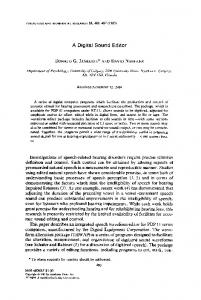
Figure 1. Structure of an outer hair cell, showing the hole at the top of the cell where the “rudimentary kinocilium” or basal body is well placed to sense pressure across it. Note the unusual spherical body (Hensens body) which could be a source of compressibility. [From Lim (1986) with permission of Elsevier Science.]
machinery, the basal body, that once drove this motile organelle and which now works in reverse as a sensor of intracochlear fluid pressure. In other words, hair cells are dual sensors, able to respond to both displacement via their stereocilia and pressure via their fontanelle. This dual scheme enables them to more efficiently sense their environment, a proposal set out as part of the author’s PhD thesis (Bell 2005). According to the thesis, outer hair cell stereocilia are, at low sound intensities, essential feedback devices – not primary sensors – although they do function as standard sensors at high sound levels. It J. Biosci. 32(2), March 2007
is suggested that, at least in the human case, the pressuresensing function is more sensitive than the displacementsensing one, so that below about 60 dB sound pressure level, the pressure component of a sound signal is more readily detected; only at higher levels does the conventional travelling wave (Békésy 1960) come to have appreciable effects. This dual arrangement can explain a number of puzzling anomalies in cochlear mechanics – especially how cells can sense the fast pressure wave that reaches them well before the peak of displacement associated with the travelling wave. In terms of signal processing across frequencies, there are considerable advantages in sensing a fast, cochlea-wide signal – there are virtually no delays in the system that need to be compensated for neurally. However, rather than dwell on the details of cochlear mechanics (which are addressed in Bell 2005), this paper brings together observations from the literature which suggest that pressure sensing by cells is a widespread phenomenon throughout the animal kingdom. Motile machinery, once used to drive kinocilia, has been evolutionarily adapted to act as a sensor by working in reverse, at least among insects, fish, and other aquatic animals. Instead of acting electromechanically, like a rotary motor, in these cases the basal body operates mechanoelectrically, like a dynamo. There is reason to think such a scheme could operate successfully in mammals as well. If confirmed, such a perspective would have wide ramifications and provide a major shift in our understanding of the hearing process. 2. Two signals in the cochlea A basic starting point is to realise there are two different, although related, signals in the cochlea. Figure 2 shows how they arise. The first is the usual acoustic pressure wave that, following back-and-forth vibration of the stapes, is communicated to the cochlear fluids at the speed of sound in water (1500 m/s). This wave creates, nearly instantaneously, a hydraulic pressure field, the magnitude of which depends crucially on the stiffness of the round window (which is the major point of pressure relief) since the rest of the cochlea, mostly water, is nearly incompressible. This hydraulic pressure, p+, is sometimes called common-mode pressure, for it occurs, in phase, on both sides of the sensory partition. The second signal is the differential pressure, p–, caused by the presence of the partition: it is the difference between pv, the pressure in the upper gallery (scala vestibuli) and pt, the pressure in the lower gallery (scala tympani). Thus, the common mode pressure p+ is given by p+ = (pv + pt)/2, whereas the differential pressure p– = (pv – pt)/ 2. The former has traditionally been assumed to have no direct effect on cochlear mechanics whereas the latter
Detection without deflection in hair cells
387
stapes
pv
oval window
partition (basilar membrane) round window
helicotrema
pt
Figure 2. In response to vibration of the stapes, a pressure wave fills the nearly incompressible cochlear fluids virtually instantly. The pressure in the upper gallery is taken to be pv, while that in the lower is pt. The differential pressure across the partition p– is (pv – pt)/2, and this is the origin of the conventional travelling wave that deflects the stereocilia of hair cells on the partition. The common-mode pressure p+ (common to both galleries) is (pv + pt)/2, and is usually thought to have no sensible effect. But if the hair cells contained a compressible element, sensitivity to pressure could arise.
gives rise to a slowly propagating travelling wave, a wave of displacement on the basilar membrane that propagates from base to apex and is presumed to stimulate hair cells by bending their stereocilia. It is the striking appearance of these stereocilia which, not unreasonably, leads us to think that common-mode pressure has no sensible effect on hair cells. But if outer hair cells were able to sense the compressional wave (as proposed in Bell 2003), this would explain a number of anomalies in hearing science, particularly those where the auditory nerve appears to give a response before the travelling wave can reach them (Guinan et al 2005; Ren and Nuttall 2006; Ren 2004). At low sound pressure levels, it is possible that the pressure stimulus may in fact be the primary one, and this raises the possibility that the cochlea may be actively operating along resonance lines rather than relying exclusively on passive travelling wave principles (Helmholtz 1875; Bell 2004, 2005). 3.
Cochlear sensitivity to pressure
The idea that hearing might result from direct detection of sound has had a long history. In the 20th century the idea was considered by Pohlman (1933) who reviewed various opposing theories at the time and was not averse to the idea that “the auditory cells react directly to the vibrations in the liquid in which they are immersed” [p. 193] rather than to transverse vibrations in the cochlear duct. The advantage of this “piezo-electrical” concept, he said, is that it simplifies
matters enormously and does not confuse the issue by employing mechanical structures extrinsic to the auditory cells. Guild (1937) was the first to suggest, albeit diffidently, that the outer hair cells may be directly stimulated by sound. He made the leap (p. 370) from the purely anatomical observation that outer hair cells are surrounded by large fluid-filled spaces – the spaces of Nuel – to the physical interpretation that this is an ideal arrangement for detecting pressure changes in the cochlear fluids. In recent times, the peculiarity of the spaces of Nuel has also been noted, and in a popular review (Brownell 1998) we are reminded that in no other organ of the body does one find cells surrounded on all sides by large fluid spaces. Other cells, inner hair cells included, are surrounded by supporting cells. Davis et al (1934) addressed the pressure-sensitivity question when considering the origin of the cochlear microphonic. They suggested that the cochlear microphonic arose from the body of hair cells being squeezed. “We venture the hypothesis that the electrical potential arises from the sensory cells themselves as a result of mechanical deformation … [and that] the difference of potential is developed between the upper and lower ends of these cells.” [p. 329]. Crucially, though, they thought that the out-ofphase responses of the oval and round windows “definitely rules out the possibility that the potential is generated merely by increased or diminished pressure within the cavity of the inner ear” [loc. cit.]. The idea that the cochlea senses pressure is important because, as foreshadowed, such a facility could explain J. Biosci. 32(2), March 2007
Andrew Bell
388
behaviour that the travelling wave theory cannot. For example, cochlear echoes show a similar waveform as input signal strength is raised. Active travelling wave models have not yet replicated this behaviour and one pertinent paper (Shera 2001) stated in its abstract that such a characteristic “contradicts many, if not most, cochlear models”. De Boer and Nuttall (2003) noted the difficulty of formulating a satisfactory time-domain model and suggested that “noncausal” factors must be at work. In addition, people with blocked round windows can still hear, as can those who have lost middle ears to disease – observations difficult to square with a travelling wave model (Bell 2005). If a pressure wave is the exciting stimulus in these cases, it raises the possibility of parallel excitation of a resonant system (Bell 2004). In such a scheme, it is possible to set up a bank of resonant elements using standing waves (based on slow-velocity, high-dispersion “squirting waves”) between the rows of outer hair cells (Bell and Fletcher 2004) so that the cochlear partition could be tuned over the whole auditory range using wave-mediated positive feedback between one row and another. Evidence supporting the idea is set out in Bell (2005), where it is shown that if the ear were to possess a small degree of compressibility it could detect common-mode pressure. A range of experiments are considered in that work, and they all leave the question unresolved, mostly because they were conducted either on dead specimens, on single isolated cells, or at high sound pressure levels, conditions under which the cochlear amplifier would not function. Detection of pressure is therefore a live option, and it is worth considering how, at least at low sound intensities, the cochlea might detect common-mode pressure. The underlying assumption here is that a live cochlea is likely to function very differently to a dead one, and this circumstance should not surprise us. 4.
Detecting sound underwater
The problem of detecting pressure in the cochlear liquids can be seen in a similar light to the problem confronting some marine creatures in detecting underwater sound. The answer, as found by a number of aquatic animals, is to use compressible materials in order to transform pressure signals into displacements. This analogy leads us to suspect that mammalian outer hair cells also use this strategy. The hypothesis is therefore made that the cells’ characteristic Hensen bodies (and perhaps the accompanying cisternal system – see figure 1) enclose compressible material, most likely air, just like cells in the macula neglecta of sharks appear to do. Later, a proposal will be made of how that compression leads to cellular excitation. The idea is that the kinocilium (and its degenerate form, the cuticular pore) are important elements in this process, as they contain the actual sensory apparatus: the basal body. J. Biosci. 32(2), March 2007
In the light of this model, some confirmatory evidence is presented below. Let us first look at some basic principles. Immersed as they are in liquid, outer hair cells can be considered to act like mini-hydrophones, detecting sound propagating through water. In this context, underwater acoustics, and the approaches that marine creatures use to detect sound in water, is informative. We should also remember that our hearing sense first evolved in water and only later became adapted to life in air. A creature living in water faces a major problem in detecting sound in its environment. As pointed out by Pumphrey (1950), its tissues are largely composed of water, and so it will have nearly the same compressibility and density as the medium in which it lives. In other words, sound will pass straight through it without interacting; effectively, the animal is acoustically transparent. The way around this is to introduce an impedance discontinuity, but first we need to be clear in our terminology. A basic point is that all plane propagating waves carry two different, but intimately related properties: particle displacement (or its time derivative, particle velocity) and sound pressure (Wartzok and Ketten 1999). Thus, a plane wave moving through a medium of density ρ at phase velocity c will have an rms particle velocity u and rms sound pressure p. The characteristic impedance of the medium, p/u, is given by p/u = ρc. In air the acoustic impedance is close to 415 Pa s/m (rayls), whereas in water the corresponding figure is 1.5 × 10⁶, about 3600 times greater. Because the sound intensity, I (the acoustic power per unit area) is given by the product of pressure and particle velocity (I = p × p/ρ c = p2/ρc), this means that for the same acoustic power the pressure in water is 60 times greater than it is in air and the particle velocity 60 times less. Rephrased, in detecting sound underwater, sensing the pressure component of a wave is easier than extracting its displacement component – provided, of course, that one has a way of introducing a compressible element to detect the pressure. This situation, however, only applies to plane waves, far from the source. As described by van Bergeijk (1964), when we are close to the source, as well as the propagated compressional wave there is an important near-field displacement effect to take into account. Van Bergeijk considers as source the case of an oscillating air bubble. As the bubble expands and contracts, it displaces water away from, and towards, itself. Because water is virtually incompressible, this radial displacement is passed on from one concentric layer to the next. The displacement amplitude falls off as 1/r2, where r is the distance from the source. If water were completely incompressible, this displacement would be, van Bergeijk observes, the only detectable phenomenon. But due to water’s small compressibility, the
Detection without deflection in hair cells vibrating bubble will also generate an acoustic wave, the familiar compressional wave propagating at 1500 m/s. This wave decreases in amplitude as 1/r. Therefore, close to the source, in the near-field, the displacement amplitudes are greater than expected from a plane wave; in the far-field, the displacement amplitudes are virtually all contributed by the compressional wave. The boundary between the two regimes, where the two displacement amplitudes are about equal, is generally taken to be one-sixth of a wavelength (Rossing and Fletcher 1995, §6.2). In water at 1 kHz, this is about 25 cm. A radially oscillating bubble gives the strongest near-field effect. Other moving bodies, such as solid spheres vibrating side to side, will also produce near-field effects, but the displacements will fall off faster (1/r3 for the sphere) and there will be directional effects as well. A wriggling worm will only make its presence felt very close by (Rossing and Fletcher 1995, §7.1). The major conclusion to this discussion is to see that in detecting sound underwater we are probably better off, in the near field, with using a displacement-sensitive detector, but in the far field both pressure and displacement detectors will work, although pressure detection is far easier than it is in air.
A
5.
389
Detecting pressure and displacement
The classic displacement detector is the hair cell (figure 3a), and it is usually considered that all hair cells of the acousticolateralis system – from which mammalian ears evolved – are displacement (or velocity) detectors. On the other hand, if we are to detect pressure, an impedance discontinuity is needed, and a common implementation is a diaphragm across an enclosed space so as to detect the force generated across it, as shown in figure 3b. The distinction is spelt out in Ewing (1989, pp 58 ff.). Fish use both these mechanisms. Firstly, the hair cells in the lateral line organ respond to movement of fluid as the fish swims, giving important information about its nearby environment and relative motion. Secondly, most fish also detect compressional waves – they hear – by using an impedance discontinuity. One way is to introduce a material with density greater than water, such as calcium carbonate; this is the option taken by cod which have calcite otoliths sitting on top of hair cells (Fletcher 1992, Ch. 4). The second way is to use a light or compressible material, and many fish species use a gas bladder filled with air (Fletcher 1992, §9.2). The bladder is useful for buoyancy, but it also oscillates in volume and displacement as compressional
B u
p
Figure 3. Underwater detection of displacement or pressure. A hair on a hair cell (A) responds to water displacement u by bending; a sensor mechanism (striped) responds neurally. To respond to a pressure wave, p, in the water, a diaphragm across an impedance discontinuity is required (B); the sensor is set to detect movement of the diaphragm. The space is filled with a compressible material like air. [Adapted from Ewing (1989).] J. Biosci. 32(2), March 2007
Andrew Bell
390
waves pass through. By coupling the surface of the bladder to the ear via special bones (the Weberian ossicles), the fish can hear long-distance sound waves (van Bergeijk 1964). The following demonstration from Békésy (1967) well illustrates how a compressible volume can be very efficient in allowing vibration to be detected within nearly incompressible water. He placed his finger inside a cavity formed out of a heavy block of lead and filled the cavity with paraffin oil (figure 4). On one face of the cavity was a flexible membrane driven by a vibrator, in this way supplying oscillating pressure to the oil. When he placed a small piece of foam rubber inside the cavity, he reported that touching the foam rubber “produced the sensation of strong vibrations” [p. 424]. His experiment was done in the context of a discussion of whether we hear by sensing pressure or displacement. Anatomy seems to suggest that with the mammalian cochlea containing hair cells, as in figure 3a, it is designed to respond to fluid displacements. If that is so, then the middle ear can be regarded as a device, like the swim bladder, for converting sound pressures into volume displacements. Such an account accords with the travelling wave theory – in which displacement of the basilar membrane is the focal stimulus. But I want to look closer at anatomy and point out that there appears to be another pressure-to-displacement converter, Békésy’s piece of foam rubber, strategically located inside the hair cell itself – an ideal arrangement for detecting underwater sound directly which so far appears to have been overlooked. The material to follow therefore presents a novel extension of the underwater acoustics story. It describes a situation that
lead block paraffin oil
vibrator
membrane foam rubber
Figure 4. Békésy’s demonstration of an efficient way to hear underwater. Touching the foam rubber produced “the sensation of strong vibrations”. J. Biosci. 32(2), March 2007
neatly reflects Békésy’s revealing experiment. We note that figure 4 is an arrangement in which a compressible material is completely enveloped by incompressible surrounds to create a pressure-to-displacement converter. In the ear the otic capsule is made of material even harder than a lead block – ivory-like bone, the hardest in the human body – and is filled with nearly incompressible water. Helmholtz elaborates on this arrangement by noting that “an incompressible fluid... contained between solid walls is distinguishable from a compressible one in this: that every impulse [however minute] which reaches any part of its surface communicates itself immediately throughout the whole fluid…” (Helmholtz 1874, p. 106). Now, if we introduce compressible hair cells into the system, this permits efficient detection of vibration. This possibility is explored by first considering sharks and other creatures who, without swim bladders, hear remarkably well underwater. 6. The revealing case of shark hearing How sharks hear has been a long-standing puzzle. As with all elasmobranchs – sharks, rays and skates – they lack bone and make do with cartilage instead. Unlike the bony fish, they also lack swim bladders. While some of the cells in their labyrinth bear otoconia, others, particularly those most sensitive to vibration, do not. The standard conclusion (Corwin 1981a,b; Kalmijn 1988a; Myrberg 2001) is that sharks must rely on detecting water displacement directly. The problem is that these motions are extremely minute far from the source, and amount to molecular dimensions. Some authors have even doubted that sharks could hear over large distances, and that they converge on prey through the use of smell, but Corwin’s review mentions observed acoustic responses over 250 m in some sharks. At such distances, particle displacements, must be extremely small and physiological thresholds have been measured as low as 5 × 10⁻¹⁰ m. If shark hearing is comparable to that of fish, then another perspective on the problem (Rogers and Cox 1988) suggests that the displacements are about 2 × 10–12 m, about 1/100th the diameter of the hydrogen atom. But the real problem is not the minute size of these motions but that they are theoretically undetectable because any small sensor will move in step with its surroundings as an acoustic pressure wave passes through (Pumphrey 1950; Myrberg 2001). There is no relative motion to sense, a situation aggravated by wavelengths underwater being so long. In order to hear the shark must somehow introduce an impedance discontinuity so that the sound wave will produce differential forces. The shark has two populations of cells in its labyrinth that are involved in vibration responses: the sacculus, covered in otoliths, and the well-named macula (or papilla) neglecta (Lewis et al 1985, pp. 58–62; Myrberg 2001) which is covered only with a gel.
Detection without deflection in hair cells The otoliths, with a density some 3 times higher than water, will undergo a relative displacement of about foursevenths that of the surrounding water, and perhaps this motion is detected. The outstanding problem relates to the neglecta and its 200 000 or so cells, which are highly sensitive to vibration (Corwin 1981; Lewis et al 1985). There should be no relative difference between the motion of the water and the motion of the covering gel (which is mainly water). One suggestion (van den Berg and Schuijf 1983) has been that this sensory patch of cells may react to some pressure-to-displacement conversion in the labyrinth, but such a process seems physically unrealistic and has been criticized (Kalmijn 1988a). The answer lies, I think, in a closer study of the anatomy of the macula neglecta. The microscopic study of Corwin (1977) showed that the cells of this sensory region contained both ‘dark’ and ‘light’ types, and that the light cells contained ‘many vacuities’ of unknown function. I suggest that if these vacuities were filled with air, then, finger-like, the detection problem would be solved. One review has claimed it ‘highly unlikely’ that, in fish at least, the inner ear could act as a pressure transducer independent of the swimbladder (Tavolga 1981, p. 581), but in the years since it was written the sensitivity and resolution of experimental apparatus has improved enormously. Also of relevance to the vacuity question, Wever (1978) points out that “the papilla neglecta is responsive to vibratory stimuli… [although] … it does not lie in a path of vibratory fluid flow and can be expected to be insensitive to aerial and aquatic sounds” (p. 974). A logical inference is that the cells are sensitive to pressure. 7.
Bubbles and underwater acoustics
Pictorially, a clear example of what I have in mind is the mechanosensor of a primitive marine polyp (Tardent and Schmid 1972) which reacts to vibration near its tentacles by shooting out spikes (nematocysts) at high speed. Microscopic anatomy of the sensor reveals that it comprises a kinocilium, surrounded at its base by a bell-shaped basal body, sitting directly on top of a large clear vesicle (figure 5). The function of the vesicle is unknown, and its contents could not be ascertained, but the authors say it may have functional significance. Given that the purpose of the cell is detection of vibration, it is reasonable to think that the vesicle is an example of a pressure-to-displacement converter – a piece of foam rubber – and that its contents are compressible. The material might be compressible lipid (Mouritsen 2005) able to change volume under pressure, or it could even be air. There is a distinct advantage in having an ‘on-the-spot’ pressure-to-displacement converter. First, it is a simple scheme, not requiring complicated anatomical structures such as the Weberian ossicles. It is also direct and robust, and each cell operates independently of the others. The
391
Figure 5. A vibration sensor on a marine polyp. A kinocilium (SC), surrounded at its base with a basal body (black wavy vertical lines), sits on top of a vesicle (V). The contents of the vesicle are not known, but it makes sense to presume it contains air or other compressible material. [Reproduced from Tardent and Schmid (1972) with permission of Elsevier Science.]
problem with the swim bladder is that there’s only one, an arrangement that compromises direction-finding, as each ear senses the same signal; some researchers have therefore doubted that bony fish can localize sound sources (van Bergeijk 1964, p. 290), a proposition which, if true, would have enormous evolutionary disadvantages. The suggestion made here is that many marine creatures – polyps, at least some species of fish, and sharks – have learnt to use air bubbles enclosed within hair cells as detectors of underwater sound. Air bubbles have dramatic effects on underwater acoustics, and recognition of this opens the door to a fastgrowing literature (Leighton 2004). Although we do not have the space to consider the details, we note that “gas bubbles are the most potent naturally-occurring entities that influence the acoustic environment in liquids” (Leighton 2004, p. 3267) and there is “exceptionally efficient coupling between bubbles and acoustic waves” [p. 3272], properties that nature may have exploited in more ways than currently appreciated. Ultrasound technologists makes use of the phenomenon to increase the contrast of the human bloodstream by injecting saline in which microscopic air bubbles are suspended (Stewart 2003). Whales and dolphins J. Biosci. 32(2), March 2007
Andrew Bell
392
use it by surrounding schools of fish with cylindrical “bubble nets” into which they project intense sounds, generating a reverberating wall of sound through which the fish cannot or will not swim (Leighton 2004, pp. 3284–3289); once corralled, the cetaceans enter for the feed. When bubbles interact with an underwater sound field they can undergo strong resonances, or “ringing”, with the stiffness coming from the bubble’s compliance and the inertia from the surrounding liquid. The Q varies between about 5 and 30, depending on whether the bubble’s radius is micrometres or millimetres (Leighton 2004, p. 3278) and attenuations can exceed 200 dB/m (Leighton 2004, p. 3291). The resonance frequency, fr, depends approximately on the inverse of the bubble radius, r, so that (Leighton 2004, Eq. 1): fr =
1 2σ 2σ 4η2 ( 3κ ( p0 − pν + )− + pν − 2 )1/ 2 r r ρr 2π r ρ
where ρ is the density of the liquid, κ is an index between 1 and 1.4 depending on the thermodynamic properties of the gas, p0 is the static pressure surrounding the bubble, pv is the vapour pressure inside the bubble, σ is the surface tension, and η is the viscosity. This equation holds for free-field conditions, and does not include thermal losses, acoustic radiation losses, or bubble–bubble interactions, which appreciably lower the natural frequency. This equation was first derived in simplified form by Minnaert in 1933 by equating the kinetic and potential energies in a linear oscillating system. Then fr = (1/2πr)·(3γp0/ρ)1/2, and we see that spherical bubbles will resonate in the audio range when of millimetre dimensions and in the ultrasonic range when of micrometre size. Of course, as Leighton points out, real bubbles are non-linear and the system is difficult to treat analytically. Nevertheless, the point is the strong interaction between sound and bubbles, whether by resonant or nonresonant means, and this can provide the basis of a sensitive acoustic detector. 8.
Cells containing air
When cells contain air bubbles, microscopically identifying the fact is difficult. Tiny bubbles tend to dissolve rapidly in water, and fixation techniques involving various stages of dehydration are bound to give unpredictable results. Examining cells that most definitely contain air bubbles therefore gives some insight. We mentioned the swim bladders of fish, which convert pressure into displacement. Fish also use their gas bladders to achieve neutral buoyancy, saving them constant swimming effort. The bladder is filled by a gas gland, which takes molecules of gas, mostly oxygen, out of solution from the blood. The process sounds simple, but J. Biosci. 32(2), March 2007
it is physically remarkable and incompletely understood (Phleger 1991, pp 209–247). Somehow a metabolic process concentrates gas against an increasing tendency for it to dissolve in water as the pressure increases (Henry’s law). Since even chemically inert gases like argon are concentrated, some physical process must be harnessed. The partial pressure of gas dissolved in sea water stays constant at about the level present at the surface (that is, about 0.2 atmosphere for oxygen). A fish living at 100 m will therefore need to concentrate oxygen from 0.2 atmospheres in the blood to 2 atmospheres in the swim bladder (Wittenberg 1958). Even some fish that live at a depth of more than 7000 m are able to fill their swim bladders with gas (Nielsen and Munk 1964). At that depth, the pressure is 630 atmospheres and the density of air is close to that of water, suggesting that the function of the bladder is for hearing (using its contents’ compressibility) rather than for buoyancy. On the other hand, many fish species – such as lantern fish, which migrate daily over a depth of 500 m – have their swim bladders filled with wax esters (Nevenzel et al 1966; Nevenzel et al 1969), which provides buoyancy independent of depth (a drawback of gas is that its buoyancy is a function of depth, which is not ideal for fish ranging hundreds of metres vertically). Other fish species have a swim bladder filled with gas while they are juvenile, but progressively fill it with lipids as they mature until it is completely full. In both these last two cases, the standard interpretation would be that these fish have compromised their hearing – an unlikely situation from an evolutionary standpoint; alternatively, if they were to have other sites of compressibility, such as in the body of their hearing cells, the problem would not exist. At this point it is appropriate to draw attention to one remarkable facet of fish hearing: in contrast to the early literature that gives the upper frequency limit of fish hearing at a few kilohertz, recent research shows that a number of species – notably those in a family that include the herring – can hear well into the ultrasonic range, up to 180 kHz (Higgs 2004). The probable reason is to enable detection, and avoidance, of predatory whales and dolphins which emit strong ultrasound signals for echolocation. The observed sensitivity is sufficient to detect cetaceans at a distance of more than 1 km. But the facility raises the question of how these fish do it, as mechanically their auditory system appears ill-suited to high-frequency operation. A simple answer may come from noting that a 10-µm air bubble possesses a resonance frequency in water of about 170 kHz, and as we saw earlier, this resonance can increase the amplitude of vibration by orders of magnitude as the mass of the water interacts with the springiness of the air. The prediction is that the hair cells of these fish, like sharks, contain microscopic air bubbles. In terms of the resonance model, it is also noteworthy that the utricle of
Detection without deflection in hair cells herring-like fish contains “bands of hair cells of alternating polarity” (Higgs 2004, p. 180), just like the supposed pattern of outer hair cells in the mammalian cochlea. Another reason for introducing a discussion of swim bladders is to look more closely at the gas gland itself. Somehow cells of the gland, typically 10–100 µm in diameter, must create tiny gas bubbles and release them into the swim bladder. Bubble formation is a tricky process in that it has to overcome surface tension between the gas and the liquid in which it forms. Because the pressure inside a gas bubble depends directly on the surface tension and inversely on the radius (Laplace’s law), a minute bubble faces immediate extinction because the pressure will be so high as to dissolve it away again (Vogel 2003, pp. 110–111). For a 0.1 µm diameter air bubble in water, the pressure due to surface tension will be nearly 30 atmospheres. Nevertheless, the plain fact is that bubbles do appear out of liquids (beer, for example) and it seems that indents in a surface, such as scratches in a glass, offer some protection from collapsing pressures. Another way of assisting bubble formation is to modify the surface tension, and so it seems no coincidence that gas glands are associated with various kinds of oily lipids. An oil will allow gas to spread on its surface at the
393
same time as repelling water, which explains why lipid droplets are often pearly white (as are the lipid droplets secreted by Hensens cells in the cochlea). In the same way, the swim bladder wall of freshly captured specimens is often observed to be brilliantly white (Phleger 1991, p. 217) and reflective due to a lipid coating. Microscopic examination of gas gland cells confirms this picture. A general feature is the presence of vacuoles and lipid droplets (Pelster 1995, p. 217). Somehow the hydrophobic lipids allow the cell to generate gas bubbles. A difficulty for the microscopist is that it is hard to tell whether the observed vacuoles contain gas or lipid (Fänge 1953, p. 34; Fänge 1966, p. 315), due to the fact that bubbles quickly dissolve once the cell dies, and preparation for microscopy induces further changes. Nevertheless, inspection of published gas gland micrographs (figure 6) shows such a profusion of vacuoles that, given the cell’s function, there is little doubt some of the vacuoles must be air bubbles (Dorn 1961). Coincidentally, in Dorn’s work he identifies two types of neighbouring cells – “helle” (light, left of figure 6) and “dunkle” (dark, right), the same terminology that Corwin uses to describe the two types of interleaving cells in the neglecta of the shark.
Figure 6. Two types of gas gland cell, which take dissolved oxygen and other gases from the blood and pump them into the swim bladder (here, the cavity below the thick line). The cells contain many vacuoles and it is unclear whether, in vivo, they are filled with gas or lipid, since preparation for microscopy will compromise their natural appearance. Given the cells’ function, it is hard to resist the conclusion that some of these vacuoles are air bubbles. [From Dorn (1961) and used with the permission of Springer-Verlag.] J. Biosci. 32(2), March 2007
Andrew Bell
394
Before concluding, it is worth noting a review (Macdonald and Fraser 1999) of how marine animals, including those without an obvious gas phase, are able to accurately sense depth. The field is full of uncertainties and few definite conclusions can be reached, prompting the authors to suggest that such organisms “may still contain a small gas pocket which would… [enable] stretch receptors to transduce micro-hydrostatic pressure changes” (Macdonald and Fraser 1999, p. 27). The conclusion must be that, despite obstacles, it is possible for living cells to produce, and sustain, tiny gas bubbles within. 9. An air bubble in outer hair cells? If outer hair cells are pressure detectors, there must be some compressible material within. There are non-aqueous materials inside cells that may have compressibility higher than that of water (45 Mbar–1), but the obvious candidates – conventional lipids and proteins (Kharakoz 2000) – have compressibility coefficients about the same as water (22–130 and 10–25 Mbar–1, respectively). When seeking responses to micropascal pressures, it is hard to escape the conclusion that the compressible material is a gas, and air, or a component of air, is the logical choice, as consideration of §7, and our underwater heritage, suggests. Following Boyle’s law, a gas will halve its volume for doubling of pressure, so that a threshold pressure elevation of 0.5 mPa in the cochlear liquids (that is, a 20 µPa threshold pressure in air × 25 middle ear gain) will cause the volume of a bubble at atmospheric pressure to decrease by a factor of 1 part in 100 kPa/0.5 mPa = 2 × 10⁸. Below I outline a scheme by which it appears physically possible to detect such a small change. Outer hair cells contain two distinctive multilayered structures that appear unique to these cells: Hensens body and subsurface cisternas (figure 1). These features, of largely unknown function, are closely related anatomically, and it makes sense to think that Hensens body may be composed of a multi-lamellar lipid (Mouritsen 2005, Ch. 5) associated with gas generation in the same way as these lamellar bodies are associated with gas adsorption in the lung (Mouritsen 2005, Ch. 10). These bodies, and possibly the connected subsurface cisterns, may therefore contain gas, making the cell contents compressible. These features are now examined. As described in a review by Lim (1986), Hensens body is a spherical whorl of endoplasmic reticulum just below the cuticular plate connected to an elaborate cisternal system residing largely on the walls of the cell. Mitochondria lie nearby. Both organelles show a multilayered but fenestrated structure. The subsynaptic portion of the cisterns is close to efferent terminals, suggesting a susceptibility to efferent J. Biosci. 32(2), March 2007
control. The number of layers and abundance of discrete bodies increases after acoustic stimulation and toxic treatments; distinct vacuoles also become more common (Leonova and Raphael 1998). Aspirin is one agent particularly effective in causing blistering and vacuolization of the cisterns (and of course in reducing hearing sensitivity). The body is named after Hensen, who first described it in 1863; it appeared to him to have a spiral arrangement (Engström and Wersäll 1958). The appearance of Hensens body varies from worker to worker, but clear renderings of the layered structure are seen in a TEM by Engström and Ades (1973) and a freeze-fracture micrograph by Mammano et al (1999, figure 6B). Some revealing micrographs can also be found in Harada et al (1990, figures 5–7). Using a different staining method, Spicer et al (1998) found that the bodies generally looked like a cluster of vesicles, although there was a remarkably diverse appearance. An ultrastructural study of the cisternal system of guinea pigs (Saito 1983) showed that each cell usually had between two and four Hensens bodies. Their concentric layers were connected to a cisternal system that typically had 4–7 parallel stacks, but sometimes up to 12. The lumens of the bodies and the cisterns were found to be filled with an electron opaque material, suggestive of neither water nor air; on the other hand, empty areas (caveolae) were found next to bulging and dilated cisterns (their figures 8 and 9), and these look like remnants of aggregated air bubbles. The figures arouse a suspicion that in vivo a thin layer of air may exist next to the electron-dense generating apparatus. Again the association between layered lipids and gas exchange (Mouritsen 2005) is a factor to keep in mind. Studies of the subcisternal layers with certain vital fluorescent dyes are also revealing. When the lipophilic dyes CTC and DiOC6 are applied (Forge et al 1993; Ikeda and Takasaka 1993; Pollice and Brownell 1993), the whole cisternal system lights up. It appears to occupy an appreciable fraction of the cell’s contents, particularly in the region of Hensens body, but also below the nucleus. A feature revealed by these dyes is that the cisternal system is lipophilic, implying some associated lipids; in turn this suggests a role for the lipids in separating a gas from its aqueous substrate via surface tension effects, as they do in the swim bladder (Pelster 1995, p. 104). The lipids could originate from within the cell itself (outer hair cells are able to synthesize lipids: Schacht and Zenner 1987) or from the nearby lipid-rich Hensen cells. Another fluorescent lipophilic dye, FM1-43, is indicative of cell membrane turnover, and Meyer et al (2001) found that it strongly stained Hensens body in a guinea pig outer hair cell. The dye cannot penetrate passive cell membranes, but when turnover (endocytosis) of membrane occurs, FM143 can be readily taken through into the interior of the cell. In an OHC, dye molecules enter the cell through its apical
Detection without deflection in hair cells end, suggesting that the dye penetrates the cuticular pore (shown as the ‘rudimentary kinocilium’ in figure 1) and is carried to Hensens body. This fits in with the authors’ finding that the dye does not penetrate through the other possible route – the MET or mechanoelectric transducer channels of the stereocilia (at least when the MET channels are blocked). The work by Meyer et al therefore reveals a close association between Hensens body and the cuticular pore, a relationship that will be detailed further below. An important finding is that the cisternal system is essential for electromotility, and hence hearing. When the system is disrupted by high doses of salicylate (Dieler et al 1991), motility disappears, along with hearing sensitivity, only to return when the drug is rinsed away. Salicylate enlarged the distances between the cisternal layers, and led to increased numbers of vesicles next to them. A number of authors have noted the importance of the cisternal system and speculated on its function. Engström (1955) noted the distinctive regular layers, or lamellæ, beneath the outer membrane (and in Hensens body) and suggested that polarized molecules aligned within them might produce potentials when distorted, in this way explaining the origin of the cochlear microphonic. More recently, Brownell (1986) and Brownell and Popel (1998) have tried to relate the ultrastructure of the cisterns to the necessary expansions and contractions of the cell wall, and mentioned electrostatic and electro-osmotic mechanisms. While these ideas have merit in understanding electromotility, they are secondary to the main point under discussion here: what is the key initial (or ‘adequate’) stimulus – bending of stereocilia or compression of a bubble within the cell body? If it is the latter, then the outstanding role of the cisternal system is to provide compressibility. Let us calculate the volume of the cisternal system. A single Hensens body, with a diameter of 3 µm, has a volume of 1.5 × 10⁻¹⁷ m3, and there may be a handful of such organelles, but probably amounting to no more than 10–16 m3. Cisterns have a typical thickness of 0.5 µm, so the volume they occupy in a cell 10 µm in diameter and 50 µm long is about 5 × 10–16 m3. Take the volume of enclosed air to be half that, about 2 × 10–16 m. If threshold sound pressure causes a reduction in volume of 2 × 10⁻⁸, as calculated above, then this will produce a change in the volume of the cell of 10–24 m3 (which, in terms of cell-sized units, is 10–6 µm3 or a cube with edges of 1/100th of micrometre – small, but not vanishingly so). Do outer hair cells show compressibility? The common assumption is that they possess no compressibility (Steele 1990; Iwasa and Chadwick 1992), a move predicated on the idea that the cells are filled with water. When the cells expand and contract at acoustic frequencies, there is not enough time for water to pass in and out across the cell membrane, so the shape change must be isovolumetric – when the cell
395
lengthens, its diameter narrows accordingly. This can be expressed in terms of the Poisson ratio, σ, the ratio of the radius strain to the length strain. For an incompressible material, σ = 0.5 (so that for a cylinder the length must change twice as much as the radius), and we would expect measurements on individual OHCs to return such a value. Actual measurements (Iwasa and Chadwick 1992) on an OHC gave a value of σ between 1.85 and 2.3, implying that the incompressibility assumption is wrong (Allen 2001, §3.2.2) and that length changes must be accompanied by appreciable changes in axial and circumferential stresses and internal pressure). In actual fact, not too much credence can be put on reported measurements since the in vivo changes we are looking for are, as calculated above, in the region of parts in 108, way below the levels detectable by standard measurement techniques. How could an OHC detect an internal volume change of 10–24 m3? There are two important features. (i) The first is that the outer hair cell, test-tube like, is constructed so as to resist pressure deformations. Brownell aptly describes the OHC as a pressure vessel (Brownell 1990; Brownell and Shehata 1990) or cylindrical hydrostat (Brownell and Popel 1998), capped by a solid plate (the cuticular plate) and encircled by strong helically wound fibres that cross clockwise and anticlockwise like a reinforced garden hose. The actin fibres are set at an angle of 9–15° to the circumference, forming a cytosketetal spring that, together with a rippled outside plasma membrane, makes it possible for the cell to undergo length changes (Brownell and Popel 1998). To increase rigidity, the cells are inflated to a hydrostatic pressure (turgor pressure) of about 1 kPa (Ratnanather et al 1993). (ii) The second is the presence of a small compliant spot on top of the cell, a hole in the cuticular plate anatomically associated with sensory capabilities. I suggest that this strategically placed organelle – the cuticular pore or fontanelle – is the pressure sensor.
10. The fontanelle as a pressure sensor Whereas figure 1 shows the cuticular pore (the rudimentary kinocilium) in vertical section, a cross-section through the cuticular plate shows it as a tiny circular hole. The cylindrical pore was first observed by Held early in the 20th century, and has been consistently seen by others (Engström et al 1962; Flock et al 1962; Hawkins 1965; Wersäll and Lundquist 1966; Sobkowicz et al 1995), but its function is unclear. An image from Flock et al (1962), traced in figure 7 below, shows the familiar array of stereocilia and a distinct hole, about 0.1 µm in diameter, near the vertex of the V. Flock et al (1962) call it a cuticular pore; Hawkins (1976) calls it, rather aptly, a fonticulus or fontanelle. J. Biosci. 32(2), March 2007
396
Andrew Bell
Flock and colleagues identify it as a basal body through its obvious similarity at high magnification (their figure 3a and shown below in figure 8a here) to the characteristic 9-fold symmetry of that familiar organelle, the basal body, at the base of kinocilia. Clear views of the basal
Figure 7. Stereocilia and cuticular pore (arrowed) of an OHC (guinea pig) traced from figure 1 of Flock et al (1962).
body in vertical section are depicted in figures 21 and 24 of Engström and Ades (1973). It is significant that most mammals, humans included, have a kinocilium during gestation, but it disappears at or shortly after birth, the standard interpretation being that it has become functionless. As its name implies, the kinocilium is a true cilium, with the well-known 9 + 2 arrangement of 9 filaments arranged around 2 central ones. (Many researchers have noted that stereocilia are inappropriately named, as they are not cilia at all but modified microvilli, but the name seems too well established.) Kinocilia have a clear motile function in certain animals in certain places – allowing unicellular animals to move about and allowing wax-laden dirt to be transported out of the ear canal, for example – but their association with stereocilia is unclear and in any event generally considered unimportant. Engström et al (1962) appear to have been among the few researchers to have attributed prime functional significance to the cuticular pore. They point out its connection with the basal bodies of kinocilia, which in turn derive from the distinctive centrioles that all animal cells – from amœba to human – display. Because of the major organizing influence of centrioles in the growth and maintenance of cells, they suggest that the basal body “should be regarded as the essential excitable structure of the hair cell”. (Engström et al 1962, p. 1363). Indeed, since they thought that stereociliar deflection was the adequate stimulus, the stereocilia must therefore not bend but act as stiff levers, transmitting force
Figure 8. Left: the cuticular pore, showing its 9-fold rotational symmetry; its inside diameter is about 0.1 µm. Right: image of a centriole, of similar size, enhanced to show detail. The hooks are dynein arms. [Cuticular pore from figure 3a of Flock et al (1962) and reproduced with permission of the Acoustical Society of America; centriole from frontispiece of Wheatley (1982) and used with permission.] J. Biosci. 32(2), March 2007
Detection without deflection in hair cells to the cuticular plate and thereby exciting the basal body. The idea received support in a review by Fex (1974): “For evolutionary reasons, it would seem more likely that the site of the mechano-transducer of mammalian cochlear hair cells would be in the membrane of the cuticle free region of the hair cell top, or very close to this membrane, rather than anywhere else” (p. 596). Engström and colleagues also pointed out that the pore is covered only by the plasma membrane of the cell and tends to bulge out, sometimes forming balloon-like protuberances during fixation (Lavigne-Rebillard and Pujol 1986). This indicates “a high degree of compliance”, and, if not artefactual, “must represent structures of great physiological importance”. The basal body is surrounded by a radiating pattern of many organelles – mitochondria, small vesicles, granules and vacuoles – suggestive of a close functional and metabolic relationship; immunolabelling for tubulin, the major component of microtubules, gives an intense fluorescent spot at this point (Steyger et al 1989). Hillman (1969) noted the pliability of the membrane at the base of the kinocilium, and suggested that tilting of the kinocilium excited the cell by its plunger-like action at this point. My proposal follows Engström and colleagues in accepting the essential importance of the basal body, but rather than having it stimulated by bending of stereocilia, I suggest it could be stimulated directly by intracochlear fluid pressure. Although the kinocilium may be absent in humans after birth, it seems reasonable to suppose that all the machinery for operating it is still in place. That is, the cell has remodelled and refined an existing structure, not wantonly thrown one away. Specifically, I suggest this machinery has been adapted to operate in reverse, allowing it to act as a sensor. A subsequent literature search indicated a close approach to this idea, perhaps even the same: Hillman, pursuing the plunger analogy, concluded a 1971 study (with Lewis) of kinocilia movement at the cell surface of the frog labyrinth (Hillman and Lewis 1971) by referring to the mammalian auditory system and speculating (ibid., p. 418) that the “basal kinociliary remnant” could be a “diaphragm-like, pressure-sensitive spot”. The authors do not specify exactly what the cause of such a deformation might be, and the idea was not taken further. A subsequent major review by one of the pair (Lewis et al 1985) gives only passing reference to this work and fails to even mention the speculation. Let us now revisit the figure of 10–24 m3 for the volume change inside an OHC at threshold. We see in figure 8 that the bore of the fontanelle has a diameter of 0.1 µm, which means its area is about 10–14 m2. At threshold pressure, therefore, a volume change of 10–24 m3 inside the cell will be accomplished by fluid flowing through the channel a distance of 10–10 m. Detecting such a threshold displacement seems relatively easy, as mammalian stereocilia are called on
397
to sense a deflection of 10–10 to 10–11 m at threshold (Dallos 1996), and, even in crickets (Thurm et al 1983), nerve impulses are produced when their kinocilia are deflected at their base by 10–12 m. The reference to crickets provides a good example of how kinocilia can act as sensors as well as motors. To understand how this can happen, and to give a clearer insight into the mechanism proposed here, we need to take a general survey of kinocilia and the basal bodies at their base. 11.
Kinocilia, basal bodies, and centrioles
The starting point here is a wide-ranging review by Flock (1971) in which he discusses sensory transduction in hair cells with a focus on the inner ear. Both stereocilia and kinocilia are given treatment. Stereocilia are really modified microvilli and emerge in distinct arrays from the hair cell’s solid cuticular plate. Each hair cell usually has a single kinocilium that is strategically placed next to the stereocilia. The kinocilium is a true cilium, with a complex internal structure which in some cases allows it to move (hence the name). Both OHCs and IHCs bear them. The kinocilium is a complex, distinct structure that, rather than sitting on the cuticular plate, emerges from a neighbouring cuticle-free zone. Flock considers what the adequate stimulus may be, and comes to the conclusion that the hair cell, in all organs, is basically a directionally sensitive displacement detector. Static pressure is considered as a stimulus at one point (Flock 1971, p. 400), but with no direct evidence in its favour, is put to one side. Engström’s lever-action of stiff hairs, and excitation at the basal body, are mentioned as possibilities but a decision on the validity of these ideas must await improved knowledge of cellular mechanics since “the final mechanical transformer is probably of molecular dimensions” (p. 408). The 9-fold symmetry of the kinocilium and its basal body is set out, and its strange disappearance in adult mammals stated. The key question of the role of the kinocilia in transduction is tackled, and among some possibilities the best answer, to my mind, provided (p. 424): that a kinocilium can act as a motile cilium in reverse. This idea was first proposed in 1958 after study of the tympanic organ of a locust (Gray and Pumphrey 1958). A micrograph of its sensory unit shows the distinctive 9 + 2 pattern seen in kinocilia, and Gray and Pumphrey suggested that a kinocilium can, through a reversed sense, play the part of a receptor. Lowenstein and Wersäll echoed the reverse transduction idea after examining the arrangement of kinocilia in the labyrinths of guinea pigs and rays (Lowenstein and Wersäll 1959). If cellular electricity can drive a motor, then all the parts exist for motion of the motor to generate an electrical signal (Lewis et al 1985, p. 126; Wiederhold 1976). J. Biosci. 32(2), March 2007
398
Andrew Bell
Experiments leave no doubt that kinocilia can act as sensors, although this work has largely been confined to insects and molluscs. Thurm et al (1983) describe how by slight modification of the ciliary shaft – removing dynein arms and adding some extra components – the cell can become sensory. In this way a stimulus can modulate the receptor current and depolarize the cell (producing excitation), or hyperpolarize it (producing inhibition). The outstanding common feature of this work is that the final stimulus is not bending of the hair, per se, but pressure (or force) exerted at the base of the hair, in the region of the basal body. Thus, if a cilium is made completely pliable, by application of a drug (chloral hydrate), they collapse and lie flat; nevertheless, pressure on the cell surface still produces a generator potential (Alkon 1983a,b). Conversely, making the cilium completely rigid – so it is unable to bend – still allows potentials to be generated. Another relevant observation is that paramoecium, which swims, and senses, using motile cilia, still shows depolarizing and hyperpolarizing responses after removal of its cilia. The clearest instance of the basal body acting as a pressure sensor can be seen in a study of the sensory spines on the legs of cockroaches, in which vertically directed pressure on the spine triggers recordable action potentials
Figure 9. A pressure sensor in the tactile spine of a cockroach. Pushing on the modified cilium transmits pressure to the basal body and generates action potentials. [Reproduced from Moran and Varela 1971, with permission.] J. Biosci. 32(2), March 2007
(Moran and Varela 1971). Significantly, the spine contains a modified cilium that makes a 9 + 0 connection to a basal body, as shown in figure 9. In the same way, a naked basal body, without the connecting cilium, might well directly sense the hydrostatic pressure surrounding the cell, and this is precisely how the outer hair cell is constructed (figure 1). The outer hair cell’s basal body senses the hydraulic pressure generated in the incompressible fluid of the cochlea, a pressure that varies in accordance with the inward and outward motion of the stapes induced by sound waves striking the ear drum (figures 1 and 2). Because the cell’s contents are compressible, while the extracellular fluid is not, a pressure difference appears directly across the fontanelle, and this is what stimulates the cell. 12.
How can basal bodies detect pressure?
The focus has now shifted from the kinocilium to the basal body. How can this organelle make the test-tube-like entity shown in figure 1 into a pressure transducer? All cilia have at their base a characteristic 9-fold structure, the basal body, from which they grow. Unique among cell organelles, they have a constant size, shape, and ultrastructure (Afzelius 1983). The mystery comes from their close similarity, if not identity, to the other vital but enigmatic cell organelle, the centriole (Wheatley 1982). Centrioles, which come in matched pairs usually at right angles to each other, spring from the nucleus and play a major part in organization, structure, polarity, growth, division and death of cells. They are ubiquitous throughout the animal kingdom, indicating they have some vital function to perform. A suggestive image is that, like a spider in a web, centrioles sit at microtubular centres of the cell, surveying activity (Schliwa 1992). Both sensory and non-sensory cells have centrioles, but in a sensory cell one centriole will migrate to its surface and grow a cilium, its partner usually nearby (Wersäll et al 1965). Theories about centriole function, and the significance of the 9 arms, abound (Albrecht-Buehler 1992); Wheatley (1982) highlights one scientist as saying “Biologists have long been haunted by the possibility that the primary significance of centrioles has escaped them” (p. 185). Nevertheless, here I will briefly set out one model that provides a cogent picture of what the sensory fontanelle could be doing. According to Brinkley and Stubblefield (1970), the entity we see in figure 8b is a motor with rotating blades. The blades are angled from one end of the centriole to the other, to give the simplified turbine-like structure seen in figure 10. In this figure, the inner dynein arms (visible in figure 8b) that presumably drive the turbine have been omitted. An attractive feature of the turbine model is the straightforward way by which flow through the core of the system might cause the blades to rotate.
Detection without deflection in hair cells
Figure 10. Simplified structure of a centriole or basal body showing the turbine-like arrangement of the blades. [From Albrecht-Buehler (1992) and used with the permission of Elsevier Science.]
A slightly different formulation was put forward by Bornens (1979), who considered that the centriole may act more like a stepper motor so that portions of the device either (i) rotated or (ii) oscillated backwards and forwards a number of degrees in an electrical field between the triplets. Either way, a torsional oscillator of fixed vibrational frequency might result, allowing the centriole to act as a pacemaker for the rest of the cell. Any slight change in the device’s mechanical environment could affect its oscillation frequency and be detected by comparison with the frequency of the companion centriole. In this connection, we may even consider quantum mechanical effects, as in paired detectors set at right angles which sense electron spin (Stern–Gerlach effect). The unexpected finding of silicon in centrioles may be coincidental but it highlights the possibility of electrical interactions.
399
The idea that the basal body of outer hair cells could be an electrical motor or, acting in reverse, a rotational sensor is given credence by consideration of a comparable structure in motile bacteria. These microorganisms propel themselves using a rotating flagellum, and ultrastructural studies show it is a miniature electric motor, complete with stator, rotor, drive shaft, bushing, universal joint and helical propeller (Namba and Vonderviszt 1997; Thomas et al 1999). Notably, the motor can rotate well into the auditory range, at up to 1700 revolutions per second (Magariyama et al 1994). Unifying the picture we have been constructing, the rotary motor originates from the cell’s basal body. A micrograph and diagram of such a rotational motor are shown in figure 11. If we identify the basal body of the outer hair cell as a flagellum-less version of this motor, configured to act as a sensor, then the forces acting on it are minute. If the intracochlear pressure at threshold is 0.5 mPa, as before, this would produce a force on the sensory pore of 5 × 10⁻⁶ pN. In comparison, the force required for activation of a typical mechanosensitive ion channel is 10–20 pN (Morris 2001), so in these terms the oscillator system must operate with orders of magnitude greater sensitivity. Is this realistic? A legitimate doubt may be raised here, although we point out that there are possibilities for resonance – outer hair cells are graded in length from base to apex (Pujol et al 1992), suggesting something like a Helmholtz resonator – and there are also two positive feedback mechanisms that might be expected to improve sensitivity. The first is mechanical, and relates to the observation that the Poisson ratio for the OHC is greater than 0.5. This means that as the electromotility mechanism is engaged and the cell changes length, the volume will tend to change accordingly. For a hyperpolarization (say) induced by initial increase in pressure on the sensory pore, the effect is towards increasing the volume – which will decrease the internal pressure and cause an increased pressure difference across the sensory pore; the result will be increased hyperpolarization – a positive feedback effect. Depolarization (induced by a transient decrease in intracochlear pressure) will lead to an opposite sequence, but producing positive feedback once again. The positive feedback loop acts so as to make the cell’s contents appear more compressible than under static conditions. In effect, the cell contains not air but supercompressible or rarefied air, a conclusion that the ancient Greeks would find agreeable (Bell 2004). The second positive feedback loop is electrical. Electromotility causes OHCs to change length in accordance with imposed electrical fields (Brownell 2002); in the same way, the cochlear microphonic – the electrical potential generated by collective outer hair cell activity – might induce further length changes in those cells. The essential role of the cochlear microphonic in affecting the responses J. Biosci. 32(2), March 2007
Andrew Bell
400
Figure 11. A rotary motor, 45 nm in diameter, located at the basal body of a bacterium’s flagellum. At left is a micrograph of the basal body in cross-section, showing the multiple rings of the stepper motor. At right is a schematic diagram (labels omitted) illustrating the major molecular components. A similar motor, minus the flagellum, could occur at the basal body of an outer hair cell and might operate in reverse as a sensor of pressure: fluid flow through the pore might induce rotation. [From Thomas et al (1999) and Namba and Vonderviszt (1997), with permission of the National Academy of Sciences USA and Cambridge University Press.]
of hair cells (that is, it is not just an ineffectual by-product of cochlear action, an epiphenomenon) was first put forward by Davis et al (1934), and has since been taken up by a number of others. Here, the electrical voltages generated by sensory transduction in the hair cell would feed back in a way that led to additional length changes and voltages – a positive feedback loop. Sharks and other electrosensitive fish can detect voltage gradients as low as 0.5 µV/m, presumably by using this mechanism (Kalmijn 1988b, 2000; Zakon 1988). These animals have specialized hair cells modified for electrical sensing; interestingly, these cells lack stereocilia, but retain a kinocilium. Whether these refinements to the proposed scheme are sufficient to improve, to detectable levels, its sensitivity to direct pressure stimuli is a complex matter and beyond resolution here. Nevertheless, taken together, a wide literature points towards the pressure-detection scheme being physically possible; I hope the case made here is sufficient for this somewhat unorthodox possibility to be seriously entertained. 13.
Conclusion
This paper reinforces the idea that the outer hair cell is a pressure vessel, a good starting point from which to make a J. Biosci. 32(2), March 2007
pressure sensor. At the same time, although the cell wall is stiff and can resist internal (and external) pressure changes, its structure allows it – like a garden hose – to change length, permitting electromotility and subsequent tuning processes to occur (Bell 2006, 2007). The other property required of a pressure sensor is a compliant spot that allows movement to be registered. This, one presumes, is the function of the fontanelle and is the spot where motion is registered by the basal body. The final ingredient is internal compressibility, and here we conjecture that Hensens body plays a role in either generating compressible lipids or, perhaps via special physical and biochemical effects, air. One of the notable features of the model is that it gives a central role to the cilium, and in particular the cellular machinery that drives it. Despite progress, knowledge of this process is still incomplete (Sowa et al 2005; Wheatley 2005), and the present integrating hypothesis is designed to steer us towards a better comprehension of how this remarkable machinery works. The hypothesis makes a number of different conjectures that can be put to experimental test. The model integrates a wide range of peculiar cellular structures, notably fontanelles, basal bodies, and Hensens bodies, and was developed as an attempt to enlarge and unify our understanding of how
Detection without deflection in hair cells hearing happens (Hudspeth 1997). If it allows auditory science to make some progress on that front, its speculative outlook has been worthwhile. Acknowledgements This work originated from the author’s PhD studies, which were supported by an ANU PhD scholarship, partial funding from the University of Tübingen (through Prof. A.W. Gummer), and an RSBS Biotechnology Research Centre grant. The ethical treatment of animals is a matter of concern to the author (http://eprints.anu.edu.au/archive/00001532) and he hopes that experiments which might arise from this paper can be done non-invasively and without causing harm. Useful comments from T. Maddess, N. H. Fletcher and the reviewers are much appreciated. References Afzelius B A 1983 Basal bodies in the immotile-cilia syndrome; J. Submicrosc. Cytol. 15 111–114 Albrecht-Buehler G 1992 Function and formation of centrioles and basal bodies; in The centrosome (ed) V I Kalnins (San Diego: Academic Press) pp 69–102 Alkon D L 1983a The role of statocyst sensory cilia in mechanotransduction; J. Submicrosc. Cytol. 15 145–150 Alkon D L 1983b Sensory function of cilia; J. Submicrosc. Cytol. 15 173–176 Allen J B 2001 Nonlinear cochlear signal processing; in: Physiology of the ear (eds) A F Jahn and J Santos-Sacchi (San Diego, CA: Singular Thomson Learning) (2nd edition), pp 393–442 Békésy G v 1960 Experiments in hearing (New York: McGrawHill) Békésy G v 1967 Some similarities in sensory perception of fish and man; in Lateral line detectors (ed.) P H Cahn (Bloomington: Indiana University Press) pp 417–435 Bell A 2003 Are outer hair cells pressure sensors? Basis of a SAW model of the cochlear amplifier; in Biophysics of the cochlea: from molecules to models (ed.) A W Gummer (Singapore: World Scientific) pp 429–431 Bell A 2004 Hearing: travelling wave or resonance? PLoS Biol. 2 e337 Bell A 2004 Resonance theories of hearing: a history and a fresh approach; Acoustics Australia 32 108–113 Bell A 2005 The underwater piano: a resonance theory of cochlear mechanics, Ph.D. thesis, Australian National University, Canberra, Australia Bell A 2006 Sensors, motors, and tuning in the cochlea: interacting cells could form a surface acoustic wave resonator; Bioinsp. Biomim. 1 96–101 Bell A 2007 Tuning the cochlea: wave-mediated positive feedback between cells; Biol. Cybern. DOI 10.1007/s00422-006-0134-0 Bell A and Fletcher N H 2004 The cochlear amplifier as a standing wave: “squirting” waves between rows of outer hair cells? J. Acoust. Soc. Am. 116 1016–1024
401
Bornens M 1979 The centriole as a gyroscopic oscillator. Implications for cell organization and some other consequences; Biol. Cellulaire 35 115–132 Brinkley B R and Stubblefield E 1970 Ultrastructure and interaction of the kinetochore and centriole in mitosis and meiosis; Adv. Cell Biol. 1 119–185 Brownell W E 1986 Outer hair cell motility and cochlear frequency selectivity; in Auditory frequency selectivity (eds) B C J Moore and R D Patterson (New York: Plenum) Brownell W E 1990 Outer hair cell electromotility and otoacoustic emissions; Ear Hear. 11 82–92 Brownell W E 1998 How the ear works: Nature’s solutions for listening; Volta Rev. 99 9–28 Brownell W E 2002 On the origins of the outer hair cell electromotility; in Hair cell micromechanics and otoacoustic emission (eds) C I Berlin, L J Hood and A Ricci (Clifton Park, NY: Delmar Learning / Singular Publishing) pp 25–47 Brownell W E and Popel A S 1998 Electrical and mechanical anatomy of the outer hair cell; in Psychophysical and physiological advances in hearing (eds) A R Palmer, A Rees, A Q Summerfield and R Meddis (London: Whurr) pp 89–96 Brownell W E and Shehata W E 1990 The effect of cytoplasmic turgor pressure on the static and dynamic mechanical properties of outer hair cells; in Mechanics and biophysics of hearing (eds) P Dallos, C D Geisler, J W Matthews, M A Ruggero and C R Steele (Berlin: Springer) pp 52–59 Corwin J T 1977 Morphology of the macula neglecta in sharks of the genus Carcharhinus; J. Morphol. 152 341–362 Corwin J T 1981a Audition in elasmobranchs; in Hearing and sound communication in fishes (eds) W N Tavolga, A N Popper and R R Fay (New York: Springer) pp 81–105 Corwin J T 1981b Peripheral auditory physiology in the lemon shark: evidence of parallel otolithic and non-otolithic sound detection; J. Comp. Physiol. 142 379–390 Dallos P 1996 Overview: cochlear neurobiology; in The cochlea (eds) P Dallos, A N Popper and R R Fay (New York: Springer), 1−43 Davis H, Derbyshire A J, Lurie M H and Saul L J 1934 The electric response of the cochlea; Am. J. Physiol. 107 311–332 de Boer E and Nuttall A L 2003 Properties of amplifying elements in the cochlea; in Biophysics of the cochlea: from molecules to models (ed.) A W Gummer (Singapore: World Scientific) pp 331–342 Dieler R, Shehata-Dieler W E and Brownell W E 1991 Concomitant salicylate-induced alterations of outer hair cell subsurface cisternae and electromotility; J. Neurocytol. 20 637–653 Dorn E 1961 Über den Feinbau der Swimmblase von Anguilla vulgaris L; Z. Zellforsch. 55 849–912 Engström H 1955 Morphological studies on the possible origin of cochlear microphonics; Rev. Laryngol. Otol. Rhinol. (Bord). 76 808–816 Engström H and Ades H W 1973 The ultrastructure of the organ of Corti; in The ultrastructure of sensory organs (ed.) I Friedman (Amsterdam: North-Holland) pp 83–151 Engström H, Ades H W and Hawkins J E 1962 Structure and functions of the sensory hairs of the inner ear; J. Acoust. Soc. Am. 34 1356–1363 J. Biosci. 32(2), March 2007
402
Andrew Bell
Engström H and Wersäll J 1958 The ultrastructural organization of the organ of Corti and of the vestibular sensory epithelia; Exp. Cell Res. Suppl. 5 460–492 Ewing A W 1989 Arthropod bioacoustics: neurobiology and behaviour (Edinburgh: Edinburgh University Press) Fänge R 1953 The mechanisms of gas transport in the euphysoclist swimbladder; Acta Physiol. Scand. Suppl. 110 1–133 Fänge R 1966 Physiology of the swimbladder; Physiol. Rev. 46 299–322 Fex J 1974 Neural excitatory processes of the inner ear; in Handbook of sensory physiology (eds) W D Keidel and W D Neff (Berlin: Springer) vol 5.1 pp 585–646 Fletcher N H 1992 Acoustic systems in biology (New York: Oxford University Press) Flock Å 1971 Sensory transduction in hair cells; in Handbook of sensory physiology (ed) W R Loewenstein (Berlin: Springer) vol. 5.1, pp 396–441 Flock Å, Kimura R S, Lundquist P-G and Wersäll J 1962 Morphological basis of directional sensitivity of the outer hair cells in the organ of Corti; J. Acoust. Soc. Am. 34 1351–1355 Forge A, Zajic G, Li L, Nevill G and Schacht J 1993 Structural variability of the sub-surface cisternae in intact, isolated outer hair cells shown by fluorescent labelling of intracellular membranes and freeze-fracture; Hear. Res. 64 175–183 Gray E G and Pumphrey R J 1958 Ultra-structure of the insect ear; Nature (London) 181 618 Guild S R 1937 Comments on the physiology of hearing and the anatomy of the inner ear; Laryngoscope 47 365–372 Guinan J J, Lin T and Cheng H 2005 Medial-olivocochlear-efferent inhibition of the first peak of auditory-nerve responses: evidence for a new motion within the cochlea; J. Acoust. Soc. Am. 118 2421–2433 Harada Y, Tagashira N, Takunida M, Suzuki M, Nagasawa A and Sakai T 1990 Three-dimensional ultrastructure of cochlea: a review; in Inner ear pathobiology (Adv. Otorhinolaryngol. v. 45) (eds) M Ciges and A Campos (Basel: Karger) pp 49–68 Hawkins J E 1965 Cytoarchitectural basis of the cochlear transducer; Symp. Quant. Biol. 30 147–157 Hawkins J E 1976 Drug ototoxicity; in Handbook of sensory physiology (eds) W D Keidel and W D Neff (Berlin: Springer) vol. 5.3 pp 707–748 Helmholtz H L F v 1874 The mechanism of the ossicles and the membrana tympani (translated by J Hinton) (London: New Sydenham Society) Helmholtz H L F v 1875 On the sensations of tone as a physiological basis for the theory of music (London: Longmans, Green) Higgs D M 2004 Neuroethology and sensory ecology of teleost ultrasound detection; in The senses of fish (eds) G von der Emde, J Mogdans and B G Kapoor (Boston, MA: Kluwer) pp 173–188 Hillman D E 1969 New ultrastructural findings regarding a vestibular ciliary apparatus and its possible functional significance; Brain Res. 13 407–412 Hillman D E and Lewis E R 1971 Morphological basis for a mechanical linkage in otolithic receptor transduction in the frog; Science 174 416–419 Hudspeth A J 1997 How hearing happens; Neuron 19 947–950 J. Biosci. 32(2), March 2007
Hudspeth A J 2000 Hearing; in Principles of neural science (eds) E R Kandel, J H Schwartz and T M Jessell (New York: McGrawHill) 4th edition, pp 590–613 Hudspeth A J and Corey D P 1977 Sensitivity, polarity, and conductance change in the response of vertebrate hair cells to controlled mechanical stimuli; Proc. Natl. Acad. Sci. USA 74 2407–2411 Ikeda K and Takasaka T 1993 Confocal laser microscopical images of calcium distribution and intracellular organelles in the outer hair cell isolated from the guinea pig cochlea; Hear. Res. 66 169–176 Iwasa K H and Chadwick R S 1992 Elasticity and active force generation of cochlear outer hair cells; J. Acoust. Soc. Am. 92 3169–3173 Kalmijn A J 1988a Hydrodynamic and acoustic field detection; in Sensory biology of aquatic animals (eds) J Atema, R R Fay, A N Popper and W N Tavolga (New York: Springer) pp 83–130 Kalmijn A J 1988b Detection of weak electric fields; in Sensory biology of aquatic animals (eds) J Atema, R R Fay, A N Popper and W N Tavolga (New York: Springer) pp 151–186 Kalmijn A J 2000 Detection and processing of electromagnetic and near-field acoustic stimuli in elasmobranch fishes; Philos. Trans. R. Soc. London B355 1135–1141 Kharakoz D P 2000 Protein compressibility, dynamics, and pressure; Biophys. J. 79 511–525 Lavigne-Rebillard M and Pujol R 1986 Development of the auditory hair cell surface in human fetuses; Anat. Embryol. 174 369–377 Leighton T G 2004 From seas to surgeries, from babbling brooks to baby scans: the acoustics of gas bubbles in liquids; Int. J. Modern Phys. B18 3267–3314 Leonova E V and Y Raphael 1998 Alteration of membranous structures in ototoxically damaged outer hair cells of the organ of Corti (Midwinter Meeting, Florida, Association for Research in Otolaryngology) Lewis E R, Leverenz E L and Bialek W S 1985 The vertebrate inner ear (Boca Raton, FL: CRC Press) Lim D J 1986 Functional structure of the organ of Corti: a review; Hear. Res. 22 117–146 Lowenstein O and Wersäll J 1959 A functional interpretation of the electron-microscopic structure of the sensory hairs in the cristæ of the elasmobranch Raja clavata in terms of directional sensitivity; Nature (London) 184 1807–1808 Macdonald A G and Fraser P J 1999 The transduction of very small hydrostatic pressures; Comp. Biochem. Physiol. A122 13–36 Magariyama Y, Sugiyama S, Muramoto K, et al 1994 Very fast flagellar rotation; Nature (London) 371 752 Mammano F, Frolenkov G I, Lagostena L, et al 1999 ATP-induced Ca2+ release in cochlear outer hair cells: localization of an isositol triphosphate-gated Ca2+ store to the base of the sensory hair bundle; J. Neurosci. 19 6918–6929 Meyer J, Mack A F and Gummer A W 2001 Pronounced infracuticular endocytosis in mammalian outer hair cells; Hear. Res. 161 10–22 Moran D T and Varela F G 1971 Microtubules and sensory transduction; Proc. Natl. Acad. Sci. USA 68 757–760 Morris C E 2001 Mechanosensitive ion channels in eukaryotic cells; in Cell physiology sourcebook: a molecular approach
Detection without deflection in hair cells (ed.) N Sperelakis (New York: Academic Press) 3rd edition pp 745–760 Morse P M 1991 Vibration and sound (New York: Acoustical Society of America) Mouritsen O G 2005 Life: as a matter of fat (Berlin: Springer) Myrberg A A 2001 The acoustical biology of elasmobranchs; Environ. Biol. Fishes 60 31–45 Namba K and Vonderviszt F 1997 Molecular architecture of bacterial flagellum; Q. Rev. Biophys. 30 1–65 Nevenzel J C, Rodegker W, Mead J F and Gordon M S 1966 Lipids of the living coelacanth, Latimeria chalumnae; Science 152 1753–1755 Nevenzel J C, Rodegker W, Robinson J S and Kayama M 1969 The lipids of some lantern fishes (family Myctophidae); Comp. Biochem. Physiol. 31 25–36 Nielsen J G and Munk O 1964 A hadal fish (Bassogigas profundissimus) with a functional swimbladder; Nature (London) 204 594–595 Pelster B 1995 Metabolism of the swimbladder tissue; in Biochemistry and molecular biology of fishes (eds) P W Hochachka and T P Mommsen (Amsterdam: Elsevier) vol. 4 pp 101–118 Phleger C F 1991 Biochemical aspects of buoyancy in fishes; in Biochemistry and molecular biology of fishes (eds) P W Hochachka and T P Mommsen (Amsterdam: Elsevier) vol. 1 pp 209–247 Pohlman A G 1933 A reconsideration of the mechanics of the auditory apparatus; J. Laryngol. Otol. 48 156–195 Pollice P A and W E Brownell 1993 Characterization of the outer hair cell’s lateral wall membrane; Hear. Res. 70 187–196 Pujol R, Lenoir M, Ladrech S, Tribillac F and Rebillard G 1992 Correlation between the length of outer hair cells and the frequency coding of the cochlea; in Auditory physiology and perception (Advances in the biosciences volume 83) (eds) Y Cazals, L Demany and K Horner (Oxford: Pergamon) pp 45–51 Pumphrey R J 1950 Hearing; Symp. Soc. Exp. Biol. 4 3–18 Ratnanather J T, Brownell W E and Popel A S 1993 Mechanical properties of the outer hair cell; in Biophysics of hair cell sensory systems (eds) H Duifhuis, J W Horst, P van Dijk and S M van Netten (Singapore: World Scientific) pp 199–206 Ren T 2004 Reverse propagation of sound in the gerbil cochlea; Nat. Neurosci. 7 333–334 Ren T and Nuttall A L 2006 Cochlear compression wave: an implication of the Allen–Fahey experiment; J. Acoust. Soc. Am. 119 1940–1942 Rogers P H and Cox M 1988 Underwater sound as a biological stimulus; in Sensory biology of aquatic animals (eds) J Atema, R R Fay, A N Popper and W N Tavolga (New York: Springer) pp 131–149 Rossing T D and Fletcher N H 1995 Principles of vibration and sound (New York: Springer) Saito K 1983 Fine structure of the sensory epithelium of guinea-pig organ of Corti: subsurface cisternae and lamellar bodies in the outer hair cells; Cell Tissue Res. 229 467–481 Schacht J and Zenner H P 1987 Evidence that phosphoinositides mediate motility in cochlear outer hair cells; Hear. Res. 31 155–160
403
Schliwa M 1992 Cell polarity and centrosomes; in The centrosome (ed.) V I Kalnins (San Diego: Academic) pp 331–351 Shera C A 2001 Intensity-invariance of fine-structure in basilarmembrane click responses: implications for cochlear mechanics; J. Acoust. Soc. Am. 110 332–348 Sobkowicz H M, Slapnick S M and August B K 1995 The kinocilium of auditory hair cells and evidence for its morphogenetic role during the regeneration of stereocilia and cuticular plates; J. Neurocytol. 24 633–653 Sowa Y, Rowe A D, Leake M C, et al 2005 Direct observation of steps in rotation of the bacterial flagellar motor; Nature (London) 437 916–919 Spicer S S, Thomopoulos G N and Schulte B A 1998 Cytologic evidence for mechanisms of K+ transport and genesis of Hensen bodies and subsurface cisternae in outer hair cells; Anat. Rec. 251 97–113 Steele C R 1990 Elastic behaviour of the outer hair cell wall; in Mechanics and biophysics of hearing (eds) P Dallos, C D Geisler, J W Matthews, M A Ruggero and C R Steele (Berlin: Springer) pp 76–83 Stewart M J 2003 Contrast echocardiography; Heart 89 342–348 Steyger P S, Furness D N, Hackney C M and Richardson G P 1989 Tubulin and microtubules in cochlear hair cells: comparative immunocytochemistry and ultrastructure; Hear. Res. 42 1–16 Tardent P and Schmid V 1972 Ultrastructure of mechanoreceptors of the polyp Coryne pintneri (Hydrozoa, Athecata); Exp. Cell Res. 72 265–275 Tavolga W N 1981 Retrospect and prospect: listening through a wet filter; in Hearing and sound communication in fishes (eds) W N Tavolga, A N Popper and R R Fay (New York: Springer) pp 573–587 Thomas D R, Morgan D G and Derosier D J 1999 Rotational symmetry of the C ring and a mechanism for the flagellar rotary motor; Proc. Natl. Acad. Sci. USA 96 10134–10139 Thurm U, Erler G, Godde J, Kastrup H, Keil T, Volker W and Vohwinkel B 1983 Cilia specialized for mechanoreception; J. Submicrosc. Cytol. 15 151–155 van Bergeijk W A 1964 Directional and nondirectional hearing in fish; in Marine bio-acoustics (ed) W N Tavolga (Oxford: Pergamon), vol. 1, pp 281–299 van den Berg A V and Schuijf A 1983 Discrimination of sounds based on the phase difference between particle motion and acoustic pressure in the shark Chiloscyllium griseum; Proc. Roy. Soc. London B 218 127–134 Vogel S 2003 Comparative biomechanics (Princeton: Princeton University Press) Wartzok D and Ketten D R 1999 Marine mammal sensory systems; in Biology of marine mammals (eds) J E Reynolds and S A Rommel (Washington: Smithsonian) pp 117–175 Wersäll J, Flock Å and Lundquist P-G 1965 Structural basis for directional sensitivity in cochlear and vestibular sensory receptors; Symp. Quant. Biol. 30 115–132 Wersäll J and Lundquist P-G 1966 Morphological polarization of the mechanoreceptors of the vestibular and acoustic systems; Second symposium on the role of the vestibular organs in space exploration (Washington, DC: NASA), SP-115, pp 57–72 Wever E G 1978 The reptile ear (Princeton: Princeton University Press) J. Biosci. 32(2), March 2007
Andrew Bell
404
Wheatley D N 1982 The centriole: a central enigma of cell biology (Amsterdam: Elsevier) Wheatley D N 2005 Landmarks in the first hundred years of primary (9+0) cilium research; Cell Biol. Int. 29 333–339 Wiederhold M L 1976 Mechanosensory transduction in “sensory” and “motile” cilia; Annu. Rev. Biophys. Bioeng. 5 39–62
Wittenberg J B 1958 The secretion of inert gas into the swimbladder of fish; J. Gen. Physiol. 41 783–804 Zakon H H 1988 The electroreceptors: diversity in structure and function; in Sensory biology of aquatic animals (eds) J Atema, R R Fay, A N Popper and W N Tavolga (New York: Springer) pp 813–850
MS received 14 July 2006; accepted 4 December 2006 ePublication: 4 January 2007 Corresponding editor: VIDYANAND NANJUNDIAH
J. Biosci. 32(2), March 2007