J. Braz. Chem. Soc., Vol. 10, No. 4, 281-286, 1999. Printed in Brazil.
© 1999 Soc. Bras. Química 0103 – 5053 $6.00 + 0.00 Article
Determination of Correlation Times from Selective and Non-Selective Spin-Lattice Relaxation Rates and their Use in Drug-Drug and Drug-Albumin Interaction Studies Luzineide Wanderley Tinoco, and José Daniel Figueroa-Villar* Departamento de Química do Instituto Militar de Engenharia, Praça General Tibúrcio, 80 - Urca, Rio de Janeiro - RJ, Brazil Os efeitos da variação da concentração da amostra nos valores de deslocamento químico e nas velocidades de relaxação spin-rede seletiva (R1S) e não seletiva (R1NS) foram medidos em solução para os três isômeros das guanil hidrazonas derivadas do nitrobenzaldeído (NBGH) puros e com a albumina do soro bovino (BSA). Os resultados foram usados para determinar o tempo de correlação (τc), mostrando que o grau de interação intermolecular droga-droga varia com a posição do grupo nitro no anel aromático e que este grau de associação interfere na interação destas drogas com a BSA. Os resultados sugerem que o grau de interação droga-droga e droga-BSA estão relacionados com a atividade in vitro destas drogas contra o Trypanosoma cruzi. The effects of the changes in sample concentration on the NMR chemical shifts and on the selective and non-selective spin-lattice relaxation rates (R1S and R1NS) of the three isomers of nitrobenzaldeyde guanyl hydrazone (NBGH) pure and with bovine serum albumin (BSA) were measured in solution. The results were used to determine the correlation times (τc), showing that the degree of intermolecular drug-drug association varies with the nitro group position on the ring and that this degree of association interferes with the interaction of these drugs with BSA. The results suggest that the degree of drug-drug and drug-BSA association are related to the in vitro anti-Trypanosoma cruzi activity of these compounds.
Keywords: intermolecular interactions, relaxation rates, correlation times, guanyl hydrazones
Introduction Aromatic guanyl hydrazones are a new family of cationic compounds which we have shown to be potential candidates for the chemotherapy of Chagas disease1. This disease is third in the ranking of importance for the World Health Organization, and is responsible for about 45.000 deaths per year in South America. The transfusion of blood contaminated with T. cruzi is the second most important way of infection of this disease in endemic areas, and the only way of contagion in countries outside the endemic zone2,3. The probability of receiving a blood transfusion with blood contaminated with this protozoon can be as high as 2.19% in certain regions of South America4. Some guanyl hydrazones are able to eliminate the trypomastigote forms of Trypanosoma cruzi from contaminated blood in concentrations as low as 17 µM, making them good candidates for the prophylaxis of blood in blood banks in ene-mail
[email protected]
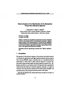
demic areas1. The mechanism of action of these cationic antibiotics is still unknown, but it is believed that it may be related to their capacity to interact with either the cellular membrane or the DNA of the parasite5. In any case, it is clear that this interaction occurs through the cationic side chain. In order to determine if the guanyl hydrazones can be safely used in blood prophylaxis it necessary, among other studies, to determine the type and degree of their interaction with plasmatic proteins. In our previous studies we showed, using non-selective T1 studies, that the guanyl hydrazones derived from nitrobenzaldeyde (NBGH - Fig. 1) interact with bovine serum albumin (BSA) with different affinities6. These compounds showed different activity depending on the isomer, for 2-nitrobenzaldehyde (2NBGH, ID50 87.5 µM), 3-nitrobenzaldehyde (3NBGH, ID50 182.6 µM) and 4-nitrobenzaldehyde (4NBGH, ID50 55.9 µM)1. Our initial hypothesis to explain this difference in activity was based on the possible
282
Tinoco & Figueroa-Villar
6
H7
5
N
4 3
N
NO2
H7
6
H NH2 +
-
Cl
5
N
4
J. Braz. Chem. Soc.
H N
+
NH2
2
NH2
5
-
O2N
3NBGH
7
N
Cl
NO 2 2NBGH
H
6
NH2
2
H N
NH2 +
NH2
-
Cl
3
4NBGH
Figure 1. Nitrobenzaldeyde Guanyl Hydrazones (NBGH).
different degrees of drug-drug association, which could affect the availability of the cationic side chain of the drug for interaction with the parasite. This hypothesis was also supported by molecular modeling studies6. In this work, selective and non selective spin-lattice relaxation rates measurements were used to study the intermolecular interactions between the NBGH and BSA and the importance of the self-association of the nitroguanyl hydrazones on their interaction with BSA and their in vitro activity. The measurement of selective and non selective spinlattice relaxation rates was suggested by Freeman et al to ascertain whether the relaxation mechanism is dipolar or other, such as chemical shift anisotropy or spin rotation7. The use of mono-, bi- and non selective spin-lattice relaxation rates have been applied to conformational studies of amino acids and peptides and in studies of interactions between small molecules with macromolecules8-14. To study systems were there is fast conversion between the bound and free states, as is usually the case for the drug-macromolecule complexes, it have been demonstrated that selective spin-lattice relaxation rates (R1S) are more sensitive to the process than the non selective spinlattice relaxation rates (R1NS)8-14. In a multi-spin system the relaxation rate can be approximated by a sum of pairwise 1H-1H dipolar interactions, yielding an initial rate constant for the recovery of the Iz magnetization given by Eq. 112-15. RNS = ∑ ρij + ∑ σij 1 i≠j
(1)
i≠j
In this equation ρij is the direct relaxation rate and σij is the cross relaxation term for a ij proton pair. Selective excitation inverts only the magnetization of spin i while not perturbing all other spins (j ≠ i). Consequently, the cross-relaxation rates do not contribute to the initial recovery constant, which is now given by Eq. 212-15: RS1 = ∑ ρij
(2)
i≠j
Some researchers have shown that R1 can be measured in the initial rate approximation with the 180° - τ - 90° sequence provided that the 180° pulse is selective and that,
in the extreme narrowing limit (ωτc 4NBGH. This result, which shows the lowest molecular mobility or higher tendency for drug-drug interaction for 3NBGH, is in agreement with the results obtained with the relaxation measurements at the lowest concentration (13 mM). HowTable 3.
R1NS/R1S ratios for
ever, in this case, it seems that the ortho and para isomers do not display a similar behavior, with 4NBGH being the least likely to form molecular aggregates. In the study of drug-BSA intermolecular interaction the analysis of selective and non-selective relaxation rates was carried out as for the pure drugs. The values of the R1NS / R1S ratio were evaluated for each NBGH isomer in 13 mM, 25 mM, 50 mM and 75 mM solutions in the presence of BSA (7.25 x 10⁻⁵ M). The results are shown in Tables 4, 5 and 6. Initially, the analysis was carried out using only the average R1NS / R1S ratio for all the isomers at the 13 mM solutions. Then, the same procedure was carried out using the overall average values for all the hydrogens at all the different concentrations, as it was done for the pure drugs. For each case, the correlation times were determined from Fig. 2, and the results are summarized in Table 7. For the 13 mM 2NBGH solution, the average value of the relaxation rate ratio in the presence of BSA is 0.6, which indicates that τc corresponds to 1.7 x 10⁻⁹ s. This τc value is significantly longer than the τc value for the pure drug (3.7 x 10⁻¹⁰ s). In fact it is possible to say that the molecule now presents long correlation times values, and that it is on the ωτc >> 1 region. This result strongly suggests that 2NBGH is forming a complex with BSA. It is well known that BSA presents a correlation time of 3,8 x 10⁻⁸ s and that the small molecules present values of τc in the range of 10-10 s to 10-12 s.18 Clearly, because of the association with BSA, 2NBGH is showing a τc value proximal to the τc value of BSA. An identical conclusion is reached if the analysis is carried out using the overall average values of the R1NS / R1S ratio for 2NBGH. In this case, the data from Tables 4 and 7 show that the average of the values of R1NS / R1S ratio is 0.6 for the 13 mM and 25 mM 2NBGH solutions, and 0.1 for the 50 mM and 75 mM 2NBGH solutions. Both results, when evaluated on the plot of Fig. 2, show that the 2NBGH molecules in the presence of BSA are on the ωτc >> 1 region, which is indicative of complex formation with BSA. These results also show that the increase in 2NBGH Table 4. R1NS/R1S ratios for 2NBGH with BSAa. 6
a
pure 4NBGH .
5
6 5
O2N
7
N
H N
+
NH2
2
-
Cl
13 mM
25 mM
50 mM
1.3
1.5
1.2
NH2
25 mM
50 mM
75 mM
H3
0.6
0.6
0.1
0.1
H4
0.7
0.4
0.1
0.1
1.3
H5
0.7
0.6
0.1
0.1
75 mM
3
H2,6
H N
13 mM
3
NH2
H7 N
4
H
J. Braz. Chem. Soc.
NO2
+
NH2
-
Cl
H3,5
1.3
1.2
1.2
1.2
H6
0.5
0.6
0.1
0.1
H7
1.4
1.2
1.2
1.7
H7
0.5
0.7
0.1
0.1
0.6
0.6
0.1
0.1
NS
Average R1 /R1
S
1.3
1.3
1.2
1.4
a- The absolute uncertainty for all results is equal to or less than ± 0.1.
NS
average R1 /R1
S
a- The absolute uncertainty for all results is equal to or less than ± 0.04.
Vol. 10, No. 4, 1999
Selective and Non-Selective Spin-Lattice Relaxation Rates
concentration facilitates the drug-BSA intermolecular interaction. The results for 3NBGH in the presence of BSA, are summarized in Table 5. The analysis of the data obtained with the 13 mM solution affords an average value for the R1NS / R1S ratio of 0.7, which corresponds to τc = 1.4 x 10⁻⁹ s, which is slightly greater than τc for the pure drug (1.1 x 10⁻⁹ s). This value is indicative of a weaker drug-BSA interaction for 3NBGH. When the analysis is carried out for all the data, it is observed that the average R1NS / R1S ratio is 0.7 for all the concentrations. The value of the average overall R1NS/R1S ratio for the pure 3NBGH solutions is 1.1 (τc = 5.3 x 10⁻¹⁰), indicating that the increase in tc due to the presence of BSA for 3NBGH (0.9 x 10⁻⁹ s) is only about half of what it was observed for 2NBGH (2.0 x 10⁻⁹ s). With these results it is possible to conclude that 3NBGH interacts with BSA, but with lower affinity than 2NBGH. The values of the R1NS/R1S ratio for 4NBGH with BSA are summarized in Table 6. It is possible to observe that the average ratio is 0.5 for the 13 to 50 mM solutions and 0.3 for the 75 mM solution. This values correspond to the ωτc >> 1 region in Fig. 2. The average R1NS / R1S ratio values for the 13 mM solution, as well as for all other solutions is 0.5, which is significantly smaller than the value for the pure drug (1.3). The difference between the correlation time for pure 4NBGH (3.7 x x10-10 s) and the correlation time for the drug with BSA (2.3 x 10⁻⁹ s) is 1.9 Table 5. R1NS/R1S ratios for 3NBGH with BSAa. 6
H7
5
N
4
H N
NH2 +
NH2
2
Cl-
13 mM
25 mM
50 mM
75 mM
x 10-9 s, showing that 4NBGH interacts with BSA with a similar affinity as 2NBGH. Since the correlation time presents an inverse correlation with the degree of molecular mobility, it is possible to say that the intensity of drug-BSA intermolecular interaction for the nitro guanyl hydrazones decrease in the order 2NBGH > 4NBGH >> 3NBGH, suggesting that the 2NBGH have, for a short margin over 4NBGH, the greater affinity for BSA. However, if we calculate the τc (PURE)/ τc (BSA) ratio for each drug (Table 7), thus considering the effect of drug-drug intermolecular interaction, it can be observed that the lower τc (PURE)/ τc (BSA) ratio corresponds to 4NBGH, indicating that this drug has the lowest propensity for drug-drug interaction and the highest tendency for drug-BSA interaction. In this case, it is possible to say that the degree of drug-BSA intermolecular interactions decrease in the order 4NBGH > 2NBGH >> 3NBGH. Interestingly, this last result suggests that there is a correlation between the τc PURE/τc BSA ratio and the in vitro ID50 values, which follow the order 4NBGH > 2NBGH > 3NBGH.
Conclusion Selective T1 measurements are very sensitive and convenient for the investigation of the binding of small molecules to macromolecules, allowing the calculation of the correlation times. Our results indicate that all the NBGH are able to establish intermolecular interactions with BSA, and that of the three isomers 4NBGH and 2NBGH present the highest affinity for this protein, with 4NBGH being the most affine. The meta isomer, 3NBGH, on the other hand Table 6. R1NS/R1S ratios for 4NBGH with BSAa.
NO2
H
6 5
H2
0.7
0.8
0.7
285
N
0.6 O2N
7
H N
NH2 +
NH2
2
-
Cl
13 mM
25 mM
50 mM
75 mM
H4
0.7
0.7
0.7
0.6
H5
0.6
0.7
0.8
0.8
H2,6
0.5
0.5
0.5
0.3
H6
0.6
0.9
0.7
0.7
H3,5
0.5
0.6
0.5
0.4
1.0
0.6
0.7
1.0
H7
0.6
0.6
0.5
0.2
0.5
0.6
0.5
0.3
H7 NS
S
average R1 /R1
0.7
0.7
0.7
3
NS
0.7
S
average R1 /R1
a- The absolute uncertainty for all results is equal to or less than ± 0.05.
a- The absolute uncertainty for all results is equal to or less than ± 0.05.
Table 7. R1NS/R1S ratio and correlation times for the NBGH.
NBGH 2NBGH
R1ns /R1s pure
τc pure (s, x 1010)
R1ns /R1s BSA
τc BSA (s, x 109)
τc pure/ τc BSA
a
b
a
b
a
b
a
b
a
b
ID50 (µM)
1.3
1.2
3.7
4.8
0.6
0.4
1.7
2.5
0.22
0.19
87.5
3NBGH
0.8
1.1
11.0
5.3
0.7
0.7
1.4
1.4
0.79
0.38
182.6
4NBGH
1.3
1.3
3.7
3.7
0.5
0.5
2.3
2.3
0.16
0.16
55.9
a- Values calculated for the 13 mM solutions only. b- Values calculated using the average of all the hydrogens at all the studied concentrations.
286
Tinoco & Figueroa-Villar
is the drug with the highest tendency to drug-drug association, and the less affine to BSA. The analysis of the values of τc PURE/τc BSA ratio shows that as the self association tendency of the drugs increases, the tendency for drug-BSA association and the in vitro activity decreases. These results strongly favor the hypothesis that the mechanism of interaction of these compounds with T. cruzi is of electrostatic nature, and that it occurs through the guanidinium moiety. In this way, the different tendencies for self association in solution, which disfavors the interaction of the drugs with the parasite, would explain the in vitro activity differences between the three isomers.
Acknowledgment We gratefully acknowledge the financial support given by PADCT/CNPq, the scholarship awarded by CNPq (J.D. Figueroa-Villar) and studentship awarded by CAPES (L.W. Tinoco).
References 1. Messeder, J.C.; Tinoco, L.W.; Figueroa-Villar, J.D.; Souza, E.M.; Santa Rita, R.; De Castro, S.L. Bioorg. Med. Chem. Lett. 1995, 5, 24, 3079. 2. Chmunis, G. A. Transfusion 1991, 31, 547. 3. Hamerschlak, N.; Pasternak, J.; Amato Neto, V.; Carvalho, M.B.; Guerra, C.S.; Coscina, A.L. Rev. Soc. Bras. Med. Trop. 1997, 30, 205. 4. Center for Disease Control (CDC). Emerging Infectious Diseases 1998, 4, 1, 5.
J. Braz. Chem. Soc.
5. Santos Filho, O.A.; Figueroa-Villar, J.D. Bioorg. Med. Chem. Lett. 1997, 7, 13, 1797. 6. Tinoco, L.W.; Ph.D. Thesis 1998, Instituto Militar de Engenharia, RJ, Brazil. 7. Freeman, R. ; Hill, H.D.W.; Tomlinson, B.L.; Hall, L.D. J. Chem. Phys. 1974, 61, 466. 8. Niccolai, N.; Miles, M.P.L.; Hehir, S.P.; Gibbons, W.A. J. Am. Chem. Soc. 1978, 100, 20, 6528. 9. Gaggelli, E.; Gaggelli, N.; Maccotta, A.; Valensin, G. J. Magn. Reson. 1994, B 104, 9. 10. Gaggelli, E.; Gaggelli, N.; Maccotta, A.; Valensin, G. Arch. Biochem. Biophys. 1994, 308, 1, 48. 11. Gaggelli, E.; Valensin, G.; Kushinir, T.; Navon, G. Magn. Reson. Chem. 1992, 30, 61. 12. Camparini, B.; Gaggelli, E.; Marchettini, N.; Valensin, G. Biophys. J. 1985, 48, 247. 13. Valensin, G.; Kushinir, T.; Navon, G. J. Magn. Reson. 1982, 23. 14. Bonechi, C.; Donati, A.; Picchi, M.P.; Rossi, C.; Tiezzi, E. Colloids and Surfaces: A 1996, 115, 89. 15. Hall, L.D.; Hill, H.D.W. J. Am. Chem. Soc. 1976, 98, 5, 1269. 16. Bodenhausen, G.; Freeman, R.; Morris, G.A. J. Magn. Reson. 1976, 23, 171. 17. Harris, C.D. Quantitative Chemical Analysis; W. H. Freeman and Company; New York, 1996. 18. Wallach, D. J. Chem. Phys. 1967, 47, 5258. Received: March 29, 1999