Dec 4, 2017 - Carolina Rojas, Daysi Diaz, Dr. Juan Beltran, Sergio Mendez, Roberto Masso, Fernando J. ... from her side and in her way to make me receive the Ph. D. degree by taking .... Multifunctional Nanomaterials, San Juan, PR, U.S.A., December .... growth, 1 sccm of 10% CH4/H2 was flowed for 80 minutes at the.
DEVELOPING A NOVEL APPROACH FOR LAYER CONTROLLED GRAPHENE SYNTHESIS AND TAILORING THE PROPERTIES FOR APPLICATIONS
A DISSERTATION SUBMITTED IN PARTIAL FULFILLMENT OF THE REQUIREMENTS FOR THE DEGREE OF DOCTOR OF PHILOSOPHY IN CHEMICAL PHYSICS
By Tej Bahadur Limbu Department of Physics University of Puerto Rico, Rio Piedras Campus San Juan, Puerto Rico, United States
December 4, 2017
DEVELOPING A NOVEL APPROACH FOR LAYER CONTROLLED GRAPHENE SYNTHESIS AND TAILORING THE PROPERTIES FOR APPLICATIONS
Accepted by the Faculty of the Doctoral Program in Chemical Physics of the University of Puerto Rico in partial fulfillment of the requirements for the degree of Doctor of Philosophy
_________________________________ Dr. Gerardo Morell Professor of the Department of Physics Thesis Advisor
_________________________________ Dr. Brad R. Weiner Professor of the Department of Chemistry Thesis Advisor
_________________________________ Dr. Ram S. Katiyar Professor of the Department of Physics Thesis Committee Member
December 4, 2017
Dedicated to My mother Chandramati Nembang
Abstract Graphene, a one atom thick two-dimensional crystal made up of sp2 bonded carbons has been attracting great interest because of its distinctive band structure and exceptional physical properties. There are several reports which demonstrate large area synthesis of high quality graphene on different substrates including the most common and inexpensive one, copper. However, layer controlled growth of graphene on copper is a challenging task as graphene growth on this substrate is understood as surface mediated self-limiting process which readily confines the growth to monolayer. In this dissertation work, we report that graphene can be grown in hot filament chemical vapor deposition reactor (HFCVD) in a layer controlled fashion that is not governed solely by the surface mediated self-limiting growth. We found that active carbon species generated by the dissociation of methane molecules at the hot filaments play a crucial role in the formation of biliayer and fewlayer graphene. Copper vapor produced during the graphene deposition appear to assist in the adlayer graphene formation. The results indicate that graphene adlayers grow on top of the previously grown layer, and show that HFCVD growth of graphene overcomes the difficulties in growing layer controlled graphene on copper. The work also demonstrates the grain size controlled growth of polycrystalline graphene with the largest grain size of ~16 µm. The high quality of grown graphene is confirmed by Raman spectroscopy, high resolution transmission electron microscopy, and electrical measurements. The extracted field effect hole mobilities of the largest grained monolayer, and bilayer graphene, and the thickest fewlayer graphene extracted are 4310 ± 348, 2745 ± 276, and 2472 ± 185 cm2V-1s-1, respectively, and the corresponding electron mobilities for monolayer, and bilayer graphene are 3610 ± 304, and 2250 ± 165 cm2V-1s-1, respectively, which are comparable to the high quality polycrystalline graphene reported in literature. The facile and inexpensive growth of high quality graphene in the HFCVD reactor, and its versatility for layer and grain size controllability show that this technique can be introduced for industrial scale production of graphene for device applications. We further report the optical and electrical properties of chemically-doped bilayer graphene stack by tetracyanoethylene, a strong electron acceptor. The tetracyanoethylene doping on the bilayer graphene via charge transfer was confirmed by Raman spectroscopy and Fourier transform infrared spectroscopy. Doped graphene shows a significant increase i
in the sheet carrier concentration of up to 1.520×1013 cm-2 with a concomitant reduction of the sheet resistance down to 414.1 Ω/sq. The high optical transmittance (ca. 84%) in the visible region in combination with the low sheet resistance of the tetracyanoethylene-doped bilayer graphene stack open up the possibility of making transparent conducting electrodes for practical applications. We also investigated the room temperature thermal conductivity of polycrystalline twisted bilayer graphene (tBLG) as a function of grain size measured by employing a noncontact optical technique based on micro-Raman spectroscopy. Polycrystalline tBLG sheets of different grain sizes were synthesized on copper by hot filament chemical vapor deposition. The thermal conductivity values are 1305 122 , 971 73 , and 657 42
Wm 1 K 1 for polycrystalline tBLG with average grain sizes of 54, 21, and 8 nm, respectively. Based on these thermal conductivity values, we also estimated the grain 10 2 1 boundary conductance, 14 .43 1.21 10 Wm K , and the thermal conductivity for single 1 1 crystal tBLG, 1510 103 Wm K . Our results show that the degradation of thermal
conductivity due to grain boundaries is smaller in bilayer than in monolayer graphene. Molecular dynamics simulations indicate that interlayer interactions play an important role in the heat conductivity of polycrystalline bilayer graphene. The quantitative study of the grain size dependent thermal conductivity of polycrystalline bilayer graphene is valuable in technological applications as well as for fundamental scientific understanding.
ii
Acknowledgements
My Ph. D. period has been incredibly rewarding both personally and professionally, and I am thankful to many people for this. I would like to express my sincere and deepest gratitude to my doctoral advisors Prof. Gerardo Morell and Prof. Brad R. Weiner for their constant support, encouragements, and supervision on my research. Their support and encouragements for my attendance at several meetings, conferences, and workshops are incredible, and are of highest values. I believe that without this experience, my graduate study would not have been meaningful and successful. I feel glad and lucky to have an opportunity to work in their research group. I would like to thank Prof. Ram S. Katiyar for agreeing to be one of the thesis committee members, and for his precious time to review this thesis. I am grateful to him for providing me an opportunity to use scientific equipment from his laboratory for my experiments. I would like to express my sincere thanks to all of my research collaborators Prof. Ram S. Katiyar, Prof. Benji Maruyama, Dr. Vladimir I. Makarov, Dr. Frank Mendoza, Dr. Konstanze R. Hahn, Dr. Satyaprakash Sahoo, Dr. Jennifer Carpena, Dr. Rajesh Katiyar, Dr. Danilo Barrionuevo, Mr. Joshua James Razink, and Mr. Jean C. Hernandez without the help of whom my Ph. D. research would not be a complete story. I am highly indebted to Dr. Vladimir I. Makarov for his collaboration and guidance in accomplishing a research work on molecular spectroscopy that does not make a part of this dissertation but adds a lot to the knowledge for my research career. My sincere thanks go to Dr. Frank Mendoza for his valuable collaboration, supports, and readiness for discussion on any research problems. I would like to thank Dr. Ratnakar Palai for giving me an access to use photolithography installed in his laboratory. I would like to thank Mr. Jose Guzman, our laboratory manager for all his helps through the supply of lab materials that helped to maintain the speed of my work. Mr. William Perez remained always very co-operative to help me in the Raman spectroscopy facility. Many thanks to him for that. I am very grateful to all my friends, colleagues, and laboratory group members for their company, helps, and encouragements: Bibek Thapa, Mohan Bhattarai, Sita Dugu, Dr. Kiran Dasari, Dr. Dhiren K. Pradhan, Dr. Shalini Kumari, Rafael Velazquez, Maried Rios Crespo, Ernesto Espada, Carolina Rojas, Daysi Diaz, Dr. Juan Beltran, Sergio Mendez, Roberto Masso, Fernando J. iii
Aponte Rivera, Samuel Escobar, Valerio Dorvilien, Monica Lopez, Dr. Javier Palomino, Dr. K. K. Mishra, Abelardo Colon, Angela Luis-Matos, Sandra Rodriguez Villanueva, Muhammad Shehzad, Dr. Geetika Khurana, Dr. Nitu Kumar, Alvaro Instan and Jose Alberto Hernandez-Perez. I would like to express my heartfelt gratitude to my Master’s thesis supervisors Prof. Sheshkanta Aryal and Dr. Kanchan Adhikari from Nepal whose guidance, encouragements, and supports made me obtain this opportunity to pursue Ph. D. study. My words are not enough to express my gratitude towards my mother Chandramati Nembang who does not understand much the meaning of education and degree but, provided endless love, care, and support to me to become a Ph. D. graduate. I would like to remember my late father Aita Bahadur Limbu (Nembang) whose untimely demise was a big gap in my family but his absence became a strength to move forward. Of course, ‘Thanks’ is not enough for my best friend, and beloved wife Januka Rai who always put her best efforts from her side and in her way to make me receive the Ph. D. degree by taking care of mine, and our beloved sons Misam Limbu and Suyen Limbu, and by taking a full responsibility of home management. I would like to remember and thank my sister Prem Kumari Ghimire and all family members for their unconditional love and support. Of course, this research would not have been possible without the financial supports. I would like to thank the funding agencies; NASA EPSCOR Puerto Rico for providing me research assistantship, and Institute for Functional Nanomaterials Puerto Rico for providing me research fellowship to support this research. Thanks also to those all if I have forgotten to mention. Thanks.
iv
Publications and Presentations Thesis Related Publications 1. Tej B. Limbu, Jean C. Hernandez, Frank Mendoza, Rajesh Katiyar, Joshua James Razink, Vladimir Makarov, Brad R. Weiner, Gerardo Morell, A novel approach to the Layer number-Controlled and grain size-controlled growth of high quality graphene for nanoelectronics (Under review). 2. T. B. Limbu, F. Mendoza, D. Barrionuevo, J. Carpena, B. Maruyama, R. S. Katiyar, B. Weiner and G. Morell, Study on the optical and electrical properties of tetracyanoethylene doped bilayer graphene stack for transparent conducting electrodes, AIP Advances 6 (2016) 035319. 3. Tej B. Limbu, Konstanze R. Hahn, Frank Mendoza, Satyaprakash Sahoo, Joshua James Razink, Ram S. Katiyar, Brad R. Weiner, Gerardo Morell, Grain size dependent thermal conductivity of polycrystalline twisted bilayer graphene, Carbon 117 (2017) 367-375. Other Publications 1. Tej B. Limbu, Kenneth J. Pérez Quintero, Arturo Hidalgo, Vladimir I. Makarov, Dachun Huang, Gerardo Morell, Brad R. Weiner, C2H radical detection by REMPI in the hot filament chemical vapor deposition of diamond, (Under review). 2. Tej B. Limbu, Kenneth J. Pérez Quintero, Arturo Hidalgo, Vladimir I. Makarov, Dachun Huang, Gerardo Morell, Brad R. Weiner, Observation of the C2H radical using
~
~
2 2 (1+2) REMPI via the B A' X transition, Chemical Physics 479 (2016) 91-98.
3. Frank Mendoza, T. B. Limbu, B. Weiner and G. Morell, Hot Filament Chemical Vapor Deposition: Enabling the scalable Synthesis of Bilayer Graphene and other Carbon Materials, InTech, Book Chapter, August-2016. 4. Surbhi Gupta, Rohit Medwal, Tej B. Limbu, Rajesh K. Katiyar, Shojan P. Pavunny, Monika Tomar, G. Morell, Vinay Gupta, and R. S. Katiyar, Graphene/semiconductor silicon modified BiFeO3/indium tin oxide ferroelectric photovoltaic device for transparent self-powered windows, Applied Physics Letters 107 (2015) 062902.
v
5. Frank Mendoza, T. B. Limbu, Brad Weiner and G. Morell, Large-area bilayer graphene synthesis in the hot filament chemical vapor deposition reactor, Diamond and Related Materials 51 (2015) 34-38.
Presentations 1. Layer number controlled synthesis of high quality and large area graphene of large grain size by hot filament chemical vapor deposition, Tej B. Limbu, Jean C. Hernandez, Frank Mendoza, Rajesh Katiyar, Joshua James Razink, Vladimir Makarov, Brad R. Weiner, Gerardo Morell, TechConnect World Innovation Conference & Expo, Washington, DC, U.S.A., May 14 to 17, 2017. (Oral) 2. Grain size dependent thermal conductivity of polycrystalline twisted bilayer graphene, Tej B. Limbu, Konstanze R. Hahn, Frank Mendoza, Satyaprakash Sahoo, Joshua James Razink, Ram S. Katiyar, Brad R. Weiner, Gerardo Morell, APS March meeting 2017, New Orleans, Louisiana, U.S.A., March 13-17, 2017. (Oral) 3. Defect density dependent thermal conductivity of misoriented bilayer graphene synthesized by hot filament chemical vapor deposition, Tej B. Limbu, Frank Mendoza, Jennifer Carpena, Satyaprakash Sahoo, Benji Maruyama, Ram S. Katiyar, Brad R. Weiner, Gerardo Morell, MRS fall meeting 2015, Boston, Massachusetts, U.S.A., November 29 to December 4, 2015. (Oral) 4. Controlled growth of high quality polycrystalline graphene of large crystallite size by hot filament chemical vapor deposition, Tej B. Limbu, Jean C. Hernandez, Frank Mendoza, Rajesh Katiyar, Joshua James Razink, Brad R. Weiner, Gerardo Morell, MRS fall meeting 2016, Boston, Massachusetts, U.S.A., November 27 to December 2, 2016. (Poster) 5. Synthesis of graphene nano-islands on germanium in hot filament chemical vapor deposition reactor for cold field emission, Tej B. Limbu, Frank Mendoza, Brad R. Weiner, Gerardo Morell, MRS spring meeting 2016, Phoenix, Arizona, U.S.A., March 27 to April 1, 2016. (Poster) 6. Study on the optical and electrical properties of tetracyanoethylene doped bilayer graphene stack for transparent conducting electrodes, T. B. Limbu, F. Mendoza, D. Barrionuevo, J. Carpena, B. Maruyama, R. S. Katiyar, B. Weiner and G. Morell, MRS vi
fall meeting 2015, Boston, Massachusetts, U.S.A., November 29 to December 4, 2015. (Poster) 7. Large area bilayer graphene synthesis by HFCVD for transparent conductive electrodes, Tej B. Limbu, Frank Mendoza, Brad R. Weiner, Gerardo Morell, Workshop on Multifunctional Nanomaterials, San Juan, PR, U.S.A., December 15-18, 2015. (Poster) 8. Physical properties of HFCVD bilayer graphene, Tej B. Limbu, Frank Mendoza, Kenneth J. Perez Quintero, Oscar Resto, Jean C. Hernandez, Brad R. Weiner, Gerardo Morell, MRS fall meeting 2014, Boston, Massachusetts, U.S.A., November 30 to December 5, 2014. (Poster) 9. Study on the C2H radical in diamond chemical vapor deposition: Experiment and modeling, Tej B. Limbu, Kenneth J. Pérez Quintero, Arturo Hidalgo, Vladimir I. Makarov, Dachun Huang, Gerardo Morell, Brad R. Weiner, MRS fall meeting 2014, Boston, Massachusetts, U.S.A., November 30 to December 5, 2014. (Poster) 10. Dynamics of C2H radical in HFCVD reactor during diamond thin film deposition, Tej B. Limbu, Kenneth J. Pérez Quintero, Arturo Hidalgo, Vladimir I. Makarov, Dachun Huang, Gerardo Morell, Brad R. Weiner, XIII International Materials Research Congress 2014, Cancun, Mexico, August 17-21, 2014. (Poster) 11. Spatial profile of radicals in the HFCVD reactor during diamond synthesis, Tej B. Limbu, Kenneth J. Pérez Quintero, Arturo Hidalgo, Vladimir I. Makarov, Dachun Huang, Gerardo Morell, Brad R. Weiner, New Diamonds and Nano Carbons (NDNC) Conference 2014, Chicago, U.S.A., May 25-29, 2014. (Poster)
vii
TABLE OF CONTENTS Abstract
i
Acknowledgements
iii
Publications and Presentations
v
List of Contents
viii
List of Figures
xi
List of Tables
xviii
List of Abbreviations
xviii
List of Contents Chapter 1. Introduction 1.1 Background
1
1.2 Exceptional Properties of graphene
3
1.2.1
Electrical Properties
3
1.2.2
Optical Properties
4
1.2.3
Thermal Properties
6
1.3 Layer Dependent Properties of Graphene
7
1.4 Fabrication of Large Area Graphene
8
1.4.1
Mechanical Exfoliation of Graphite
1.4.2
Epitaxial Growth on SiC
10
1.4.3
Chemical Vapor Deposition
11
1.4.3.1 Thermal Chemical Vapor Deposition
12
1.4.3.2 Plasma Enhanced Chemical Vapor Deposition
13
1.4.3.3 Hot Filament Chemical Vapor Deposition
15
1.5 Graphene Transfer
9
17
1.5.1
PMMA assisted wet transfer method
18
1.5.2
Thermal release tape (TRT) assisted dry transfer Method
20
1.6 Statement of the Problem
22 viii
1.7 Objectives
25
1.8 References
27
Chapter 2. Experimental and Analytical Techniques 2.1 Overview
33
2.2 Hot Filament Chemical Vapor Deposition (HFCVD)
34
2.2.1
Reactor System Overview
34
2.2.2
Sample Preparation
35
2.2.3
Graphene Deposition
36
2.3 Graphene Transfer
37
2.4 Characterization Techniques
38
2.4.1
Raman Spectroscopy
38
2.4.2
UV-visible Spectroscopy
40
2.4.3
Fourier Transform Infrared Spectroscopy
40
2.4.4
Optical Microscopy
41
2.4.5
Scanning Electron Microscopy
42
2.4.6
Transmission Electron Microscopy
43
2.4.7
Atomic Force Microscopy
44
2.4.8
Electrical Measurements
45
2.5 Fabrication of Graphene based field effect transistor
48
2.6 Molecular Dynamics Simulation
50
2.7 References
53
Chapter 3. Controlled Growth of High-Quality Graphene on Copper by Hot Filament Chemical Vapor Deposition 3.1 Introduction
54
3.2 Experimental Details
56
3.2.1
Substrate Cleaning Process
56
3.2.2
Graphene Growth Process
56
3.2.3
Graphene Transfer Process
57
3.2.4
Characterization Techniques
58
3.3 Results and Discussion 3.3.1
58
Raman Analysis
58 ix
3.3.2
Transmission Electron Microscopic Analysis
61
3.3.3
Estimation of Grain Size
63
3.3.4
Electronic Quality of HFCVD Graphene
66
3.3.5
Parametric Studies for understanding Graphene Growth
69
3.3.6
Graphene Growth Mechanism
73
3.3.6.1 Studies on graphene adlayer growth
73
3.3.6.2 Copper vapor assisted growth of graphene on SiO2/Si
76
3.3.6.3 Role of copper vapor on the layer-controlled graphene Growth
79
3.4 Conclusions
83
3.5 References
85
Chapter 4. Optical and Electrical Properties of Chemically Doped Bilayer Graphene Stack 4.1 Introduction
89
4.2 Experimental Details
90
4.2.1
Growth of Bilayer Graphene by HFCVD
90
4.2.2
Graphene Transfer onto Glass Substrate
91
4.2.3
Chemical Doping of Bilayer Graphene
91
4.2.4
Characterization Techniques
92
4.3 Results and Discussion
92
4.3.1
Characterization of Bilayer Graphene
92
4.3.2
Optical Properties of Chemically Doped Bilayer Graphene
93
4.3.3
Electrical Properties of Chemically Doped Bilayer Graphene
100
4.3.4
Estimation of Fermi Level Shift due to Chemical Doping
102
4.3.5
Doped Bilayer Graphene Stack as a Transparent Conducting Electrode
103
4.4 Conclusions
106
4.5 References
107
Chapter 5. Effect of Grain size on the Thermal Conductivity of Polycrystalline Twisted Bilayer Graphene 5.1 Introduction
111 x
5.2 Experimental Details
113
5.2.1
Graphene Growth by HFCVD
113
5.2.2
Graphene Transfer onto TEM Grid
113
5.2.3
Characterization of Graphene
115
5.2.4
Experimental Measurement of Thermal Conductivity
115
5.2.5
Thermal Conductivity Calculation by Molecular Dynamics Simulations
117
5.3 Results and Discussion
118
5.3.1
Graphene Growth and Characterization
118
5.3.2
Temperature Dependent Raman Studies
121
5.3.3
Laser Power Dependent Raman Studies and Thermal Conductivity Measurement
125
5.3.4
Estimation of Grain Boundary Conductance
128
5.3.5
MD Simulations and Comparison of Normalized Thermal Conductivity
131
5.4 Conclusions
135
5.5 References
137
Chapter 6. Summary and Future Directions 6.1 Summary
140
6.2 Future Directions
142
6.3 References
145
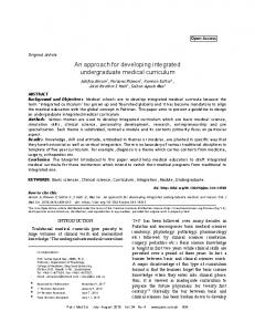
List of Figures Figure 1.1
(a) Structure of graphite formed by a stack of graphene layers. (b) Molecular structure of graphene showing sp2 bonded carbons.
Figure 1.2
(a) Electronic dispersion in the honeycomb lattice with enlarged Dirac cone. (b) Brillouin zone of the graphene lattice.
Figure 1.3
2
(a) Excitation processes responsible for absorption of light in graphene. (b) Optical photograph of a 50 µm aperture partially
xi
4
covered by graphene and its bilayer. The line scan profile shows the intensity of transmitted white light along the yellow line. Figure 1.4
5
Optical image of exfoliated graphene layers on 290 nm thick SiO2 on Si. Single and few-layer graphene regions are clearly distinguishable. 9
Figure 1.5
An illustration of epitaxial growth of graphene on a SiC wafer by thermal decomposition of SiC, together with the structural model of bilayer graphene on SiC. Blue broken line is the buffer layer.
Figure 1.6
10
A schematic diagram of TCVD system for graphene growth on copper.
12
Figure 1.7
Schematic illustration of basic PECVD set-up.
14
Figure 1.8
A schematic diagram of hot HFCVD reactor.
16
Figure 1.9
A schematic illustration of PMMA assisted wet transfer of graphene.
18
Figure 1.10
A schematic illustration of TRT assisted dry transfer of graphene.
20
Figure 1.11
A schematic illustration of the RtoR transfer of large area graphene grown on Cu onto a flexible PET substrate.
Figure 2.1
21
(a) Schematic of the HFCVD reactor. A photograph of (b) HFCVD system. (c) heater and filaments when they are cold. (d) reactor during the graphene deposition.
Figure 2.2
Schematic of the sequential steps followed in graphene deposition in the HFCVD reactor.
Figure 2.3
37
A simplified diagram of energy transitions for Rayleigh and Raman scattering.
Figure 2.4
34
39
Different configurations used for in van der Pauw method for measuring the electrical resistivity.
45
Figure 2.5
A simulated transfer characteristics of graphene based FET device.
47
Figure 2.6
A schematic of the different steps for the fabrication of graphene based FET device by photolithography.
Figure 3.1
(a) Raman spectra of monolayer (black), 25o twisted bilayer (red) and few-layer (blue) graphene grown controllably in the HFCVD. xii
49
The insets show the optical images of graphene transferred onto SiO2/Si. The scale bar is 1 cm. (b) Raman spectra collected from different grains of tBLG on SiO2/Si. The twist angles have been assigned to the Raman spectra by comparing their features with those of previous reports [23-25]. Raman maps for the ratio of 2D to G band intensities measured on (c) monolayer (d) twisted bilayer, and (e) few-layer graphene. The mapping area is 70 ×110 𝜇𝑚2 . (f) Optical transmittance spectra of monolayer, bilayer, and few-layer graphene. The inset shows the optical images of monolayer, bilayer, and few-layer graphene transferred onto glass substrates. The scale bar is 2 cm. Figure 3.2
59
HRTEM images of (a) monolayer graphene, (b) few-layer graphene, and (c) tBLG. The insets show the fast Fourier transform (FFT) of the respective images. The FFT of monolayer graphene shows a single set of six-fold reflection spots, bilayer graphene consists of two sets, and few-layer graphene consists of many sets depending upon the number of layers. The corresponding SAED patterns of (d) monolayer, (e) few-layer, and (f) twisted bilayer graphene. The measured twist angles are shown for each of the most frequently observed tBLG grains. (g) distribution of twist angles determined by SAED patterns and HRTEM images. Inset is the low magnification TEM image of a bilayer graphene suspended over TEM grid holes.
Figure 3.3
62
(a) Optical images of the bilayer graphene/Cu, and (b) monolayer graphene/Cu after oxidation process. Graphene grain boundaries can be seen due to the formation of copper oxide layer underneath the grain boundaries. Four different bilayer graphene/Cu samples with different grain size were grown and their grain sizes were estimated. (c) A plot of grain size vs methane flow rate during the growth. The
xiii
black solid diamonds and red solid circles correspond to monolayer and to bilayer graphene, respectively. Figure 3.4
64
(a) Optical image of bilayer graphene based FET device. The graphene channel has dimensions of 22 μm × 20 μm. The inset shows the schematic of a back-gated FET device based on graphene/SiO2/Si. (b) I-V characteristics of the graphene. (c) Transfer characteristics of monolayer, bilayer, and few-layer graphene FET devices. (Ids vs Vg at Vds = 0.1 Volt) (d) Charge carrier mobilities of HFCVD grown graphene as a function of number of layers. Some literature values are presented for comparison.
Figure 3.5
HRTEM images of (a) monolayer, and (b) twisted bilayer graphene with well-stitched grain boundaries.
Figure 3.6
70
D band evolution as a function of substrate temperature for (a) monolayer, (b) bilayer (250 twist angle), and (c) few-layer graphene.
Figure 3.8
68
Optical images of graphene on SiO2/Si systematically grown at different conditions.
Figure 3.7
67
(a) Schematic representation of few-layer graphene growth on monolayer graphene/Cu as a substrate. For few-layer graphene growth, 1 sccm of 10% CH4/H2 was flowed for 80 minutes at the filament temperature of 1800 oC, substrate temperature of 975 oC, and chamber pressure of 35 Torr. (b) Raman spectra collected from the areas with different number of layers marked as 1, 2, and 3 on the few-layer graphene (AB stacked) flake in the inset. The ID/IG ratio is smaller with increase in the number of graphene layers. (c), (d), (e), and (f) are schematics of the copper vapor assisted graphene growth mechanism in the HFCVD reactor. (c) Monolayer graphene growth with a marginal effect of copper vapors. (d) Bilayer graphene growth with a significant effect of copper vapors. Oval shaped structures connected by a curved arrow indicate the catalytic dissociation of methane and carbon radicals at the copper clusters. (e) Few-layer graphene growth with a precursor saturation in xiv
73
boundary layer. (f) Copper cluster assisted nucleation of adlayer graphene on pristine graphene surface. Figure 3.9
75
(a) Schematic of the graphene growth on SiO2/Si in the HFCVD reactor. (b) Raman spectrum collected from graphene grown on SiO2/Si, showing D, G and 2D bands. The inset shows a photograph of graphene/SiO2/Si. (c) Optical image of graphene/SiO2/Si. (d) EDS collected from graphene/SiO2/Si. There was no gas flow after the deposition ended. (e) EDS collected from graphene/SiO2/Si. 100 sccm of N2 gas was flowed after the deposition ended. (f) A high magnification SEM image of graphene/SiO2/Si from the process mentioned in (d), showing copper nanoparticles.
Figure 4.1
77
(a) Representative tapping mode AFM image of bilayer graphene on SiO2/Si showing a folding region and wrinkles. Inset shows the height profile of the graphene film along the white line. (b) HRTEM image of the bilayer graphene. The FFT of the image in the inset shows that it is a twisted bilayer graphene with a twist angle of 30o.
Figure 4.2
93
Raman spectra of the graphene transferred onto pyrex showing the blue shifting of G peak and red shifting of 2D peak positions on charge transfer doping. A slight decrease in the intensity of 2D peak with respect to G peak is also visible. The inset is the schematic diagram of the TCNE-doped stack of three graphene bilayers. Each bilayer graphene is shown with a combination of two monolayers separated by a small space indicating van der Waals bonding.
Figure 4.3
95
(a) FTIR spectra of neat TCNE (black), one bilayer graphene (red), two bilayers (blue), two bilayers with TCNE intercalation between them (pink) and three bilayers with TCNE intercalation between them (green). (b) Magnified portion of the FTIR spectra in Figure 3a in the region of C≡N stretch bands.
Figure 4.4
Optical transmittance of single bilayer graphene (black), undoped stack of two bilayers (red), undoped stack of three bilayers (green), xv
96
doped stack of two bilayers (blue), and doped stack of three bilayers (pink). Figure 4.5
99
Sheet resistance, charge carrier concentration and hole mobility values of the undoped and doped graphene. Note: BG stands for bilayer graphene.
Figure 5.1
101
Schematic illustration of the transfer process employed for bilayer graphene.
Figure 5.2
114
Raman spectra of tBLG materials: black color for low defect density, red for moderate defect density, and blue for high defect density.
Figure 5.3
119
Fit of D, G and D’ bands of the Raman spectra collected from each of the samples with average grain sizes of (a) 54, (b) 21, and (c) 8 nm respectively. The D and G bands were fitted with damped harmonic oscillator function (phonon model) whereas D’ band was fitted with Fano line shape.
Figure 5.4
120
(a) Tapping mode AFM image of bilayer graphene on SiO2/Si. Inset shows the height profile of the graphene film along the white line, showing a step height of 1.1 nm; (b) Field emission SEM image of suspended
nanocrystalline
tBLGs
on
bare
copper
grid;
(c) Representative HRTEM image of nanocrystalline tBLG for grain sizes of ~21 and ~54 nm showing a Moiré pattern; (d) Representative HRTEM image of nanocrystalline tBLG for a grain size of ~8 nm showing a Moiré pattern in each grain. Insets are the FFT of the images, showing two sets of six-fold reflection spots corresponding to the two graphene monolayers of the bilayer system rotated with respect to each other with a twist angle of ~21o. Figure 5.5
121
Raman G peak of the graphene with grain size of 54 nm recorded at different temperatures showing its gradual redshift on increasing temperature.
Figure 5.6
122
(a) A single linear plot of Raman G peak position vs temperature for suspended tBLG with different grain sizes. The experiments were xvi
performed at ~0.5 mW of laser power. (b) Decay rate of the G-mode optical phonons in nanocrystalline tBLG with different grain sizes. The experimental data (symbols) are fit (thick solid lines) to Equation (2). Figure 5.7
123
(a) A schematic diagram of laser probing on graphene/Cu grid for laser power- dependent Raman measurement. (b) Plots for the G peak shift vs absorbed laser power for different grain sizes. The data are fitted with black (54 nm average grain size), red (21 nm average grain size), and blue (8 nm average grain size) lines.
Figure 5.8
126
Grain size dependence of thermal conductivity in BLG. The filled squares are the experimental points. The thick curve is the guide to the eye for showing the non-linear trend of the data points.
Figure 5.9
129
Inverse of thermal conductivity versus the inverse of grain size ( d ). Red line is the linear fit, the slope of which gives the value of boundary conductance, 14 .43 1.21 1010 Wm 2 K 1 , and the yintercept gives the value of the thermal conductivity for single 1 1 crystalline tBLG, 1510 103 Wm K .
Figure 5.10
131
Constructed nanocrystalline graphene sheet with average grain size of 5 nm for (a) monolayer, and (b) tBLG sheets. (c) HRTEM image of a nanocrystalline tBLG sample of average grain size of 8 nm with different twist angles. Different Moire patterns are observable in (b) and (c) showing a variety of twist angles can exist in a polycrystalline tBLG.
Figure 5.11
132
Normalized thermal conductivity K K ref as a function of grain size
(d ) . The thermal conductivity of the HFCVD grown nanocrystalline tBLG (blue filled inverted triangles) have been normalized to K ref K P . The thermal conductivity for monolayer (green filled
diamonds) and twisted bilayer (red filled triangles) graphene
xvii
obtained from MD simulations have been normalized to the corresponding values of K ref .
134
List of Tables Table 4.1
Values of sheet resistance, optical transparency and corresponding Figure of Merit (FoM) of the TCEs reported in the references cited above.
Table 5.1
105
The values of the fit parameters and phonon decay rates using Equation (2) to describe the experimental temperature dependence of the anharmonic decay rate of the G-mode phonons for nanocrystalline tBLG of different grain sizes.
124
List of Abbreviations AEMD
Approach-to-equilibrium Molecular Dynamics
AFM APCVD
Atomic Force Microscopy Atmospheric Pressure Chemical Vapor Deposition
APS
Ammonium Persulfate
BLG
Bilayer Graphene
CCD
Charge Coupled Device
CMOS
Complementary Metal Oxide Semiconductor
CVD
Chemical Vapor Deposition
CYTOP
Cyclic Transparent Optical Polymer
FESEM
Field Emission Scanning Electron Microscopy
FFT
Fast Fourier Transform
FWHM
Full Width at Half Maximum
DI
Deionized
DOS
Density of States
FET
Field Effect Transistor
FoM
Figure of Merit
FTIR
Fourier Transform Infrared
FTO
Fluorine-doped Tin Oxide xviii
HFCVD HFTCVD
Hot Filament Chemical Vapor Deposition Hot Filament Thermal Chemical Vapor Deposition
HOPG HRTEM
Highly Oriented Pyrolytic Graphite High Resolution Transmission Electron Microscopy
IR
Infrared
ITO
Indium Tin Oxide
LA LAMMPS
Longitudinal Acoustic Large-scale Atomic/Molecular Massively Parallel Simulator
LOR
Lift off Resist
LPCVD
Low Pressure Chemical Vapor Deposition
MD
Molecular Dynamics
MFC
Mass Flow Controller
PET
Polyethylene Terephthalate
PECVD
Plasma Enhanced Chemical Vapor Deposition
PMMA
Poly(methyl methacrylate)
PR
Photoresist
REBO
Reactive Empirical Bond Order
RtoR
Roll-to-roll
SAED
Selected Area Electron Diffraction
SEM
Scanning Electron Microscopy
SWNT
Single Walled Carbon Nanotube
TBLG
Twisted Bilayer Graphene
TCE
Transparent Conducting Electrode
TCNE
Tetracyanoethylene
TCVD
Thermal Chemical Vapor Deposition
TEM
Tranmission Electron Microscopy
TO
Transverse Optical
TRT UHVCVD
Thermal Release Tape Ultra-high Vacuum Chemical Vapor Deposition
ZA
Out of Plane Acoustic
2D
Two Dimensional xix
Chapter 1: Introduction
Introduction
1.1 Background Carbon is the fourth most abundant element in the universe by mass, and it is a fascinating and essential element studied in several different research areas because of its capability to bond with other elements and form a wide range of organic compounds. In the recent decades, there has been a great interest in elemental carbon since it can form several different allotropes. The well-known allotropes are diamond, graphite, fullerene, and amorphous carbon. Among all the allotropes, graphite is one which is made up of a stack of a number of carbon layers held by weak van der Waals interaction. Each layer of graphite is made up of sp2 bonded carbons forming a honeycomb structure which is known as graphene. Graphene is a single two-dimensional (2D) layer of carbon atoms with a typical thickness of 0.34 nm. Figure 1.1a shows the structure of graphite where graphene layers are stacked together through van der Waals interactions (indicated by dotted lines). In graphite, the distance of separation between the two carbon layers is 3.35 Å, and C-C bond distance in a graphene plane is 1.42 Å. Figure 1.1b is the molecular structure of graphene layer showing that carbons are bonded to each other by sp2 bonds. The 2s and 2p orbitals in each carbon atom in graphene undergoes sp2 hybridization resulting in three sp2 hybridized orbitals. Each carbon atom on honeycomb lattice forms three sigma (σ) bonds with three in plane nearest neighboring carbon atoms. The remaining 2p orbitals on each carbon atom are perpendicular to the planar structure. These orbitals form pi (𝜋) bonds which are half filled
1
Chapter 1: Introduction
Figure 1.1 (a) Structure of graphite formed by a stack of graphene layers. (b) Molecular structure of graphene showing sp2 bonded carbons. [1,2]. σ-bonds in all allotropes of carbon including graphene are responsible for the mechanical strength [2]. Since its exfoliation in 2004 [3], graphene has drawn the attention of the scientific community due to its outstanding and exciting properties [3,4] such as high electron mobility, high electrical and thermal conductivity, high optical transparency, high mechanical strength, and flexibility. Due to such properties, graphene has brought the research of carbon materials to a new era of excitement. Since 2010, there has been an explosion of graphene-based device research which aims to take advantage of the outstanding properties of graphene. These properties of graphene make it a desirable material for incorporation into future devices. Graphene has a number of potential applications including electronics [5,6], spintronics [7], optoelectronics [8], transparent conducting electrodes [9-11], thermal managements [12], gas, chemical, and molecular sensors [13-15], biosensors [16], and supercapacitors [17].
2
Chapter 1: Introduction 1.2 Exceptional Properties of graphene Graphene has outstanding properties which make it a potential material for a variety of applications. It shows a record high values of electron mobility, thermal conductivity, and Young’s modulus of elasticity with the values of 200,000 cm2,V-1s-1 [18], 5300 W/mK [19], and 1 terapascal [20], respectively. Graphene has high optical transparency of about 97.7% in the visible region [21], chemical inertness [22], and mechanical flexibility [11]. This section explains briefly some of the most important properties of graphene that are the focus of our research. 1.2.1 Electronic Properties Graphene has an unconventional electronic spectrum. It has a linear energymomentum relationship, with the conduction and valence bands intersecting near the Dirac points. Under the normal conditions, the electronic states in the valence band are fully occupied and those in the conduction band are empty. In such a condition, the Fermi level co-exists with the Dirac point. The delocalized 𝜋 and 𝜋 ∗ states responsible for the formation of valence and conduction bands, respectively exhibit degeneracy at the K point of hexagonal Brillouin lattice forming a point like Fermi surface. Figure 1.2a represents the electronic dispersion of graphene in the honeycomb lattice where the Dirac cone at the K point is enlarged for clarity. Figure 1.2b is the Brillouin zone of graphene, with the Γ, M, K and K’ points. The extraordinarily high electron mobility and exceptional electronic properties of graphene can be explained following the Dirac equation for 2D analogue. Low-energy excitations in graphene are “relativistic” Dirac fermions, with an effective “light velocity” 3
Chapter 1: Introduction
Figure 1.2 (a) Electronic dispersion in the honeycomb lattice with enlarged Dirac cone. (b) Brillouin zone of the graphene lattice. (Figures adapted from reference 23) 106 cm/s [23]. A very small electronic density of states at the Dirac point and the gapless band structure allow graphene to be easily tuned from electron-like to hole-like (or viceversa) via surface adsorbates [11,10] and an external gate [3]. Hence, the Fermi level in graphene can be moved above and below the Dirac point turning it into n-type and p-type, respectively. This ambipolar nature of graphene makes it suitable for integration with several other materials for varieties of applications. 1.2.2. Optical Properties As stated above, graphene has high optical transmittance. Every layer of graphene absorbs only 2.3% of light at 550 nm, and the value is similar for other visible wavelengths. The optical conductance of monolayer graphene can be explained as 𝜎 = 𝜎𝜋−𝜋∗ + 𝜎𝜎−𝜎∗
(1.1)
where 𝜎𝜋−𝜋∗ and 𝜎𝜎−𝜎∗ are the conductance contributions from the 𝜋 − 𝜋 ∗ and 𝜎 − 𝜎 ∗ interband transitions, respectively. Transitions between σ and π bands are forbidden by wavefunction symmetry. Since the optical transitions 𝜎 − 𝜎 ∗ contribute to only phase shift 4
Chapter 1: Introduction of the transmitted light (which is negligible in few layer graphene and significant only in thick graphene or graphite layers), the optical conductance is solely dependent upon the 𝜋 − 𝜋 ∗ transitions which again is approximately equal to the universal optical conductance 𝜎0 for low doping and at room temperature [24]. In the limit of the massless Dirac fermion band structure, 𝑒2
𝜎0 = 4ℏ where 𝑒 and ℏ = ℎ/2π
(1.2) are the electronic charge and reduced Planck’s constant,
respectively. Optical absorption is obtained from the universal conductivity as A = (4π/c) 𝜎0 = πα ≈ 2.3%
(1.3)
for a monolayer graphene, where c is the speed of light and α is the fine structure constant which equals 𝑒 2 /ℏ𝑐.
Figure 1.3 (a) Excitation processes responsible for absorption of light in graphene. (b) Optical photograph of a 50 µm aperture partially covered by graphene and its bilayer. The line scan profile shows the intensity of transmitted white light along the yellow line. (Figures adapted from reference 25) 5
Chapter 1: Introduction Figure 1.3a shows the electronic band structure of monolayer graphene showing electronic excitation processes responsible for the absorption of light. When an electron gains an energy equal to 𝐸 = ℏ𝜔 from a photon, it is excited into the empty states of the conduction band (red) from the valence band (blue) conserving its momentum. Figure 1.3b shows an optical micrograph of a 50 µm aperture partially covered by graphene where the three regions; monolayer graphene, bilayer region, and the region without graphene are clearly distinguishable due to 2.3% of light absorption by each layer of graphene. The line scan profile shows the intensity of transmitted white light along the yellow line. The high optical transparency of graphene over a wide region of wavelengths combined with its high electrical conductivity make it a promising material for transparent conducting electrodes. 1.2.3. Thermal Properties Graphene has super high in-plane intrinsic thermal conductivity which arises due to the strong covalent sp2 bonding between the carbon atoms resulting in efficient heat transfer by lattice vibrations [26]. Thermal conductivity in a solid is summed up as 𝐾 = 𝐾𝑃 + 𝐾𝑒 , where 𝐾𝑃 is the phonon contribution to thermal conductivity, and 𝐾𝑒 is the electronic contribution. In metals, 𝐾𝑒 is dominant due to their larger concentration of free carriers. However, in other materials where the charge carrier concentration is relatively low, 𝐾𝑒 is much smaller compared to 𝐾𝑃 . Electronic thermal conductivity, 𝐾𝑒 , is defined by the Wiedemann-Franz law: 𝐾𝑒 𝜎
𝜋2
𝑘2
= ( 3 ) ( 𝑒𝐵 ) 𝑇
(1.4)
6
Chapter 1: Introduction where 𝜎, 𝑘𝐵 , 𝑒, and T are electrical conductivity, Boltzmann constant, electronic charge, and temperature of the material, respectively. The electronic contribution to the total thermal conductivity in graphene is very small, less than 1% [27]. Hence, phonons play the major role in the thermal conductivity of graphene. The equation for phonon thermal conductivity is as follows: 𝐾𝑃 = ∑𝑗 ∫ 𝐶𝑗 (𝜔)𝜐𝑗2 (𝜔)𝜏𝑗 (𝜔)𝑑𝜔
(1.5)
where summation is performed over the phonon polarization branches j, which include two transverse acoustic and one longitudinal acoustic branches, 𝜐𝑗 = 𝑑𝜔⁄𝑑𝑞 is the phonon group velocity of the jth branch. 𝜏𝑗 is the phonon relaxation time, 𝐶𝑗 is the contribution to heat capacity from jth branch. The phonon mean free path (Λ) is related to the relaxation time through the expression Λ = 𝜏𝜐. For a suspended graphene without defects, rough boundaries, and impurities, the value of Λ was estimated to be in the order of 800 nm near room temperature [28]. Such a large phonon mean free path in graphene is one of the factors that make thermal conductivity of graphene high. However, thermal conductivity in graphene can easily be degraded by various factors such as defects, surface adsorbates, grain boundaries, and graphene-substrate interfaces. 1.3 Layer Dependent Properties of Graphene The properties of graphene are much different from graphite. Graphite has smaller values of electron mobility, electrical and thermal conductivity, and mechanical strength than that of graphene. This is due to the fact that in a graphite, individual graphene layers are held together through weak van der Waals interactions which are sufficient to suppress the intrinsic properties of a graphene layer. Hence the proper use of the name ‘graphene’ 7
Chapter 1: Introduction applies to a single layer of carbon atoms showing such astonishing properties. However, two layers or few-layers of carbon atoms still show remarkable properties compared to that of single layer graphene. So, they are regarded as two layer (bilayer) and few-layer graphene, respectively. Engels et al. [29] measured a very high electron mobility of 50,000 cm2/Vs in h-BN encapsulated bilayer graphene. A large thermal conductivity of 1896 W/mK in Bernal bilayer graphene is also reported [30]. Bilayer and few-layer graphene have parabolic dispersion [31] which means that the energy-momentum relationship close to the Brillouin zone is nonlinear. In bilayer graphene (BLG), an electric field applied perpendicular to the basal plane breaks the inversion symmetry of the lattice, opening a band gap at the charge neutrality point [32]. In twisted bilayer graphene (tBLG), where the two graphene layers are rotated to each other by some angle, the interlayer coupling and band structure are tunable. Furthermore, twist angle dependent van Hove singularities emerge in the density of states due to the overlapping of the Dirac cones from the top and bottom graphene layers in tBLG [33]. The electronic properties of multilayer graphene strongly depend on the stacking sequence. In both periodically stacked [34] and arbitrarily stacked [35] multilayer graphene, the lowenergy band structure consists of a set of independent pseudospin doublets. As in AB stacked bilayer graphene, an energy gap can be induced by a perpendicular external electric field in ABC-stacked multilayer graphene [36]. 1.4 Fabrication of Large Area Graphene The successful isolation of few-layer and single layer graphene in 2004 by Novoselov et al. [3] triggered an avalanche of researches on graphene. Since then, several other research groups started to fabricate graphene for both fundamental research and device 8
Chapter 1: Introduction applications. In the course of more than a decade, many different methods of graphene fabrication have been developed. In this section, some of the most important methods of large area graphene fabrication are presented. 1.4.1 Mechanical Exfoliation of Graphite For more than 70 years, it was argued that strictly two-dimensional (2D) crystals were thermodynamically unstable and could not exist [4]. But in 2004, Novoselov et al. [3] isolated a strictly 2D crystal, a single layer of graphene from a piece of graphite by a simple method, and studied its properties. They utilized a common cellophane tape to remove layers from a graphite flake successively until few-layer and single layer of graphene was obtained. After that, several other research groups [37,38] followed the method to exfoliate graphite to obtain single and few-layer graphene layers for fundamental studies and device fabrication. An optical image of isolated graphene layers on 290 nm SiO2 on Si is shown
Figure 1.4 Optical image of exfoliated graphene layers on 290 nm thick SiO2 on Si. Single and few-layer graphene regions are clearly distinguishable. (Figure adapted from reference 37)
9
Chapter 1: Introduction in Figure 1.4, where the single layer and few-layer regions are distinguishable. This method of graphene fabrication is simple, cheap and produces the highest quality graphene flakes available to date. However, poor scalability and inconsistency in the number of exfoliated layers are the major drawbacks of this method, which promoted the research on alternative approach to graphene fabrication. 1.4.2 Epitaxial Growth on SiC This is a bottom up method of graphene growth directly on silicon carbide (SiC) wafer. This method allows the wafer scale growth of graphene on SiC wafers making the method suitable for industrial applications. In this technique, a single crystalline SiC wafer is heated in a vacuum or argon atmosphere to the temperature of 2000 oC [39]. At such a high temperature, Si atoms desorb from the (0001) face of the crystal at a rate much faster than C due to its higher vapor pressure [40]. The remaining carbon atoms on the surface rearrange to form epitaxial graphene. Figure 1.5 depicts the Si atom evaporation from the SiC surface and the formation of epitaxial graphene on the SiC surface. This is a promising technique for quality
Figure 1.5 An illustration of the epitaxial growth of graphene on a SiC wafer by thermal decomposition of SiC, together with the structural model of bilayer graphene on SiC. Blue broken line is the buffer layer. (Adapted from reference 41) 10
Chapter 1: Introduction growth of wafer scale graphene that shows interesting physical characteristics such as ballistic transport [40]. Furthermore, this method requires no graphene transfer process, and hence graphene quality is not degraded by the possible contamination from transfer process, while the processing cost is minimized. Also, SiC is a wide band gap semiconductor and graphene grown on this substrate is suitable for many optoelectronic applications [42]. This technique has some serious drawbacks such as high temperature process, the high cost of the epitaxial SiC wafer, and the difficulty to transfer the grown graphene to other substrates. 1.4.3 Chemical Vapor Deposition Chemical Vapor Deposition (CVD) is a chemical process for depositing thin films of various materials on a substrate. In a typical CVD process, the substrate is exposed to one or more volatile precursors, which react and/or decompose on the substrate surface to produce the desired deposit. Volatile by products are also produced and are removed by gas flow through the reaction chamber. This technology is now an essential factor in the manufacture of semiconductors and other electronic components, in the coating of tools, bearings, and other wear resistant parts and in many optical, optoelectronic and corrosion applications. Large area and high-quality graphene has been widely grown by CVD of carbon containing gas precursors such as methane [11,18], acetylene [43], and ethanol [44] on transition metal substrates. The most common transition metals used as substrates for graphene growth are copper (Cu) [11,18,43,44] and nickel (Ni) [9] although CVD growth of graphene on Ru, Ir, Co, Re, Pt, and Pd has also been reported [45]. CVD processes are relatively cheap, versatile, and suitable for growing large area and high-quality graphene. 11
Chapter 1: Introduction Below, some of the most common types of CVD reactors used to grow large area graphene are discussed briefly. 1.4.3.1 Thermal Chemical Vapor Deposition (TCVD) TCVD system is a cost efficient and high performance chemical vapor deposition system. It consists of a precision bench-top furnace using high-quality heating elements which surround a quartz tube. Currently, it is the most common technique used to grow large area graphene on various substrates from metallic [43,44] to dielectric [46]. Ruoff’s group [47] demonstrated centimeter scale continuous, 95% homogeneous monolayer graphene growth on copper foil with this CVD technique for the first time in 2009. After that, there are significant advancements on the TCVD growth of graphene on Cu and other substrates. For graphene growth on copper, Ruoff’s group placed a piece of copper foil on the tube and heated to about 1000 oC with a hydrogen flow of 2 sccm maintaining the pressure of 40 mTorr. After temperature stabilization, methane was flowed at 35 sccm along with hydrogen for 30 minutes and pressure was maintained at 500 mTorr. A schematic diagram of TCVD system for graphene growth on copper is shown in Figure 1.6.
Figure 1.6 A schematic diagram of TCVD system for graphene growth on copper. (Figure adapted from reference 48) 12
Chapter 1: Introduction The parameters for graphene growth in CVD reactors vary widely in the literature reports. Based on the graphene growth pressure, the CVD process is categorized into different types such as low pressure chemical vapor deposition (LPCVD) [47], ultra-high vacuum chemical vapor deposition (UHVCVD) [49], and atmospheric pressure chemical vapor deposition (APCVD) [50]. There are significant advancements on the single crystal graphene growth on copper [51,52] and alloys [53] and germanium [54] in TCVD reactor. Although this technique has already been commercialized for mass production of polycrystalline monolayer graphene on copper, it still has some drawbacks. The major drawbacks of this technique realized until now are inferior quality of the synthesized graphene with respect to that of exfoliated one, and it has been difficult to achieve wafer scale growth of single crystal graphene on cheap substrates such as copper. A noteworthy point as a drawback of this technique is its limited suitability for monolayer graphene growth only due to the surface mediated self-limited process [44], discarding its use for growing high quality and continuous bilayer and few-layer graphene on copper. 1.4.3.2 Plasma Enhanced Chemical Vapor Deposition Plasma enhanced chemical vapor deposition (PECVD) has been widely used for large area graphene growth on various substrates such as transition metals [55,56], and insulators [57] at relatively low temperatures.
PECVD processing allows graphene
deposition at lower temperatures, which is often critical in the manufacture of semiconductors, and this feature of PECVD makes it popular among other fabrication techniques. Similar to other techniques, PECVD also uses gas sources graphene growth. Plasma is an essential part of this process, and this is why it is called plasma enhanced. The
13
Chapter 1: Introduction gas activation takes place in a non-equilibrium plasma, generally referred as a glow discharge [58]. For graphene growth in PECVD, feedstock gas, catalyst nature, and substrate temperature are among the parameters that require optimization. For parallel-plate dc glow PECVD graphene growth systems, the typical voltage and power used are 50 to 250 V and 3 kW, respectively, and the inter-electrode gap is usually several centimeters [59]. The schematic of parallel- plate dc glow
Figure 1.7 Schematic illustration of basic PECVD set-up. (Figure adapted from reference 59) PECVD set-up is shown in Figure 1.7. In a PECVD system, the dissociation on the Cu surface is enhanced by a plasma source. For the graphene growth on the surface of Cu, both the carbon radicals produced from plasma excitation and Cu surface catalysis contribute to the graphene growth. In conventional TCVD, the growth of successive graphene layers is dramatically slowed down due to coverage of the catalytic Cu surface after the first layer graphene forms. However, for PECVD, the reactive carbon radicals from plasma-enhanced
14
Chapter 1: Introduction dissociation still contribute to the formation of successive layers at a relatively higher rate [60]. PECVD growth of graphene also has several drawbacks. The graphene growth in PECVD is complex compared to TCVD due to a huge number of intermediate reactions involved. This technique produces toxic byproducts and the cost of equipment is high. 1.4.3.3 Hot Filament Chemical Vapor Deposition Hot filament chemical vapor deposition (HFCVD) is a versatile technique which has been used to grow various carbon nanomaterials including diamond, carbon nanotubes, and graphene [61]. Among the various techniques to grow high quality and large area graphene, the HFCVD technique has contributed significant knowledge on the graphene growth process. A typical HFCVD consists of tungsten or rhenium filaments suspended above the substrate heater surface as shown in Figure 1.8. The filaments are heated to red hot for the dissociation of precursor gas molecules by flowing an electric current, and the substrate heater is heated to a suitable temperature depending upon the nature of the substrate and the deposition reactions. All of these assemblies are enclosed in a reaction chamber which is filled with the precursor and carrier gases up to a certain pressure. The gas flow in HFCVD is turbulent in the reactor volume unlike in TCVD where it is laminar. Selvakumar et al. [62] synthesized monolayer, bilayer and few-layer graphene controllably on a copper substrate by HFCVD by flowing methane and hydrogen at the flow rates of 2 sccm and 100 sccm, respectively. The filament and substrate temperatures were 2000 oC and 950 oC, respectively and a growth pressure of 10 mbar was used. A high-quality bilayer graphene was synthesized on copper in the HFCVD by using methane gas carbon
15
Chapter 1: Introduction precursor at relatively lower substrate temperature of 800-850 oC [11,63] which may be technologically useful. Limbu et al. [18] have demonstrated grain size control growth of
Figure 1.8 A schematic diagram of hot HFCVD reactor. (Figure adapted from reference 64)
nanocrystalline twisted bilayer graphene in the HFCVD which is a step ahead in the progress of CVD growth of graphene. The technique has also been used to grow high quality graphene on nickel [65] and Cu/Ni substrates [66]. Moreover, there are some reports [67,68] on the graphene growth in hot filament thermal chemical vapor deposition (HFTCVD) which is a modified form of HFCVD. Although a high-quality graphene can be grown in the HFCVD with controlled layers and grain size, it suffers from some drawbacks such as the need of frequent change of filaments and complexity of the CVD process as in PECVD.
16
Chapter 1: Introduction 1.5 Graphene Transfer Although chemical vapor deposition (CVD) enables cost-effective fabrication of high-quality large-area graphene films, one critical bottleneck is an efficient and reproducible transfer of graphene to target substrates. At present, the most common substrates for high quality graphene growth are copper and nickel. The graphene requires to be separated from the host substrates and transferred onto other substrates for various purposes such as for characterizations and device fabrication. Furthermore, the discovery of graphene has triggered the research on a huge set of other 2D materials including transition metal dichalcogenides. The transfer process has become an integral part of research on graphene and other 2D materials. Quality of as-synthesized graphene is degraded during the transfer process due to the polymeric residues, and rough substrate surface [69,70]. Therefore, it is crucial to develop a simple technique that would ideally lead to a successful transfer of graphene onto a target substrate. Several efforts have been made until this date in developing graphene transfer methods. Some of the reported transfer techniques involve a polymer based supporting layer such as poly(methyl methacrylate) (PMMA) for handling graphene which is later dissolved in a suitable solvent [11,47]. Thermal release tape or polyethylene terephthalate (PET) supported transfer method, which is not removed by dissolution but detached by thermal treatment, is one of the popular methods employed for roll to roll transfer of graphene onto large target substrates [63, 71,72]. There are some methods that do not require any supporting layer but the host substrate such as Cu is directly etched out in an etchant solution and the floating graphene is picked up by a target substrate [18,73].
17
Chapter 1: Introduction Direct delamination of graphene from Cu has also been reported which does not require etching of Cu substrate [74,75], Several graphene transfer methods have been reported each of which has some advantages and some disadvantages. Herein, some of the most widely used graphene transfer methods are presented. 1.5.1 PMMA assisted wet transfer method This is the most widely used graphene transfer method. This method is simple, easy to carry out, and yields a reasonable quality of transferred graphene. High quality graphene is mostly grown on copper and nickel at present. These metals can be etched out easily by variety of chemical solutions. The PMMA assisted wet transfer method seems to be suitable for graphene transfer from such metal substrates. PMMA has many prominent features, such as the relatively low viscosity, excellent wetting capability, flexibility, and good dissolubility in several organic solvents [69] that make it a suitable polymer as a supporting layer for graphene transfer. A schematic illustration of transfer of graphene grown on copper is shown in Figure 1.9. In this method, a thin layer of PMMA is spin coated on
Figure 1.9 A schematic illustration of PMMA assisted wet transfer of graphene [48].
18
Chapter 1: Introduction graphene/copper, and cured by placing PMMA/graphene/copper directly on a hot plate. After curing, PMMA/graphene/copper is placed on an etchant solution in a beaker or petri dish. The high transparency of the PMMA makes it easy to observe the process of Cu removal. After complete etching of Cu, the remaining PMMA/graphene is transferred to deionized (DI) water several times to clean the etchant residues, and finally scooped with the target substrate. The PMMA/graphene/target substrate assembly is then placed on a hot plate above 100 oC to evaporate trapped water molecules. Finally, the PMMA layer is removed by acetone, washed with DI water, and dried. Several different groups have employed PMMA assisted wet transfer of graphene with some modifications. Kim et al. [76] suggested to use lower average molecular weight PMMA than usual PMMA to obtain reduced hole doping on graphene. Won et al. [77] improved the contact of PMMA/graphene on SiO2/Si by softening of the PMMA layer through heat treatment. Coating double layer PMMA for transfer has been shown effective to obtain high quality graphene with fewer PMMA residues, and non-cracked surface [78]. There are some reports which have, replaced PMMA by polymers such as Cyclic Transparent Optical Polymer (CYTOP) [79], cellulose, [80] and pentacene [81] but have followed almost similar procedure for wet transfer of graphene. Despite the effectiveness and friendliness in using PMMA as a supporting layer, it has two major drawbacks: (a) PMMA residues on the graphene surface cannot be totally removed by acetone dissolution. For better removal, it requires annealing of the transferred graphene sample in H2/Ar atmosphere at 350 oC for 2 hours in a tube furnace followed by annealing in air or vacuum at 350 oC for 2 hours [76,77,82]. (b) PMMA dopes graphene
19
Chapter 1: Introduction strongly and changes electronic properties such as mobility, carrier concentration, and sheet resistance [82]. 1.5.2 Thermal release tape (TRT) assisted dry transfer method In this method, graphene is transferred onto the target substrate without involving a solution as in the case of PMMA assisted wet transfer method. Thermal release tape (TRT) [69,83] which is stiff enough to handle graphene layer, is used as a supporting layer. A schematic diagram of TRT assisted dry transfer method is shown in Figure 1.10. To transfer graphene grown on copper to a target substrate, a TRT is attached to graphene/Cu and pressed. The TRT/graphene/Cu assembly is then placed in etchant solution. After complete removal of Cu, it is rinsed with DI water several times, and dried gently under nitrogen. The dry TRT/graphene is then put onto a target substrate and pressed to enhance adhesion between graphene and the substrate, and finally TRT is released by heat treatment.
Figure 1.10 A schematic illustration of TRT assisted dry transfer of graphene. (Figure adapted from reference 69)
20
Chapter 1: Introduction Kang et al. [84] modified the method by using two hot pressing plates to enhance graphene-target substrate adhesion and release TRT. The two hot metal plates were applied with controllable temperature (125 °C) and pressure (4 Nm-2). This method is represented by Figure 1.10 (clockwise direction). The TRT assisted dry transfer method has been modified by Sukang et al. [71] and applied for roll-to-roll (RtoR) transfer of 30-inch graphene from Cu to flexible PET substrates. They attached TRT to the graphene/Cu by using two rollers, and the Cu foil was etched out. The TRT/graphene was then dried and
Figure 1.11 A schematic illustration of the RtoR transfer of large area graphene grown on Cu onto a flexible PET substrate. (Figure adapted from reference 71)
placed on a 130 µm thick PET. The TRT/graphene/PET was then passed between the two rollers under mild heating, to transfer graphene onto flexible PET substrate and release the TRT. This method is represented in Figure 1.10 (anticlockwise direction). This RtoR dry transfer of graphene is suitable for large area transfer of graphene onto flexible substrates, and can be realized for industrial applications. Figure 1.11 has been presented for detailed illustration of this method. It is a fast and dry transfer method, and is suitable for industrial scale transfer of large area graphene on to flexible substrates. 21
Chapter 1: Introduction 1.6 Statement of the Problem Large area and high-quality graphene is a promising material for a variety of applications. Monolayer graphene shows the record high values of electron mobility, electrical and thermal conductivity, and mechanical strength [11]. Due to the exceptionally high electron mobility, monolayer graphene shows a promise to be used for ultrafast electronics [85]. It’s high electrical conductivity and high optical transparency in the visible region make it a potential material for transparent conducting electrodes in the near future [11,71]. The outstanding thermal properties of monolayer graphene provide an additional motivation for its integration in nanoscale complementary metal oxide semiconductor (CMOS) technology, optoelectronics, photonics, and bioengineering as a thermal management material [19]. High mechanical strength and low density of graphene make it an ideal material for nanoelectromechanical applications [86]. But, due to the lack of band gap, the use of graphene in some electronic devices may be restricted. Bernal bilayer (AB stacked) graphene is an attractive candidate for transistor applications due to its gate tunable bandgap [87], whereas tBLG stands as a promising material for high sensitivity optoelectronics due to enhanced optical absorption through van Hove singularities in the electronic density of states [18]. Multilayer graphene on the other hand can have a wide variety of applications such as highly conducting electrodes [88], thermal interface materials [89], and condenser microphone due to its low membrane static tension and light weight [90]. All members of the graphene family have the potential of producing low cost and highperformance future devices. Hence, inexpensive and facile production of high quality and large area monolayer, bilayer, and fewlayer graphene is important, and hence layer number controlled growth of graphene on a cheap substrate can fulfill such a demand. 22
Chapter 1: Introduction At present, CVD of methane on copper is the common and facile technique of high quality graphene growth. Copper can be a cheap and environmentally friendly substrate for graphene growth. But, copper favors monolayer graphene formation, and it is difficult to grow bilayer and fewlayer graphene layers due to the surface mediated self-limiting growth process on copper [44]. There have been a few attempts [62,91] on the layer controlled growth of graphene on copper but the electron mobility has not been reported which could indicate the low quality of the graphene so obtained. Moreover, the grown graphene requires to be homogenous in a large area substrate for practical applications, which has not been reported [62,91]. Hence, a facile and inexpensive technique for layer controlled growth of high quality and homogeneous graphene is the focus of this thesis. TBLG shows high charge carrier mobility for electronic applications due to significantly decoupled graphene sheets and the small interlayer interaction [92]. It has a high electrical conductivity and optical transmittance [11] comparable to that of monolayer graphene, which make it a suitable candidate material for transparent conducting electrode applications. Chemical doping is an effective method of engineering graphene for increasing the charge carrier density which significantly reduces the sheet resistance with a negligible loss of optical transparency [71]. A low sheet resistance can be produced by a layer by layer transfer of graphene to make a stack [71], and graphene layers can be doped by trapping the dopant molecules. The dopant molecules have to be suitably chosen so as to maintain the stacked graphene layers strongly doped and sufficiently coupled. Tetracyanoethylene (TCNE) is a planar molecule with electron affinity of 3.17 eV [93] which can be intercalated in between the bilayer graphene sheets in order to get the graphene layers doped strongly. Hence, the study on the optical and electrical properties of TCNE doped large area twisted 23
Chapter 1: Introduction bilayer graphene stack is important to unravel the possibility of producing bilayer graphene based transparent conducting electrodes. TBLG is a candidate material for use in ultrafast optoelectronic devices due to the presence of tunable van Hove singularity in the density of states due to the overlapping of the Dirac cones from the top and bottom graphene layers [18]. Although ultrafast nanoscale devices can be produced, the generation of heat in the device components from the electric current imposes a challenge to the operating performance and device lifetime. Heat management in a device is effective if the integrated materials are capable of transporting the heat to the sink or surroundings, i.e., high thermal conductivity (K ) is required. Due to the high thermal conductivity, tBLG based nanoscale optoelectronic devices can dissipate the produced heat to sink or surroundings, and hence they can have an excellent performance. Since graphene is more readily available in polycrystalline form when it comes to obtaining large areas, understanding the thermal properties of polycrystalline tBLG is critically important for its practical applications. Grain boundaries are known to scatter phonons and introduce mode mismatch that degrade the thermal conductivity of polycrystalline graphene [18]. The investigation on the grain size dependent thermal conductivity of tBLG provides information to assess the suitability of this material for future applications in optoelectronics and other nanoscale electronic devices. In this context, we performed a detailed experimental investigation on the room temperature thermal conductivity of polycrystalline twisted bilayer graphene as a function of grain size.
24
Chapter 1: Introduction 1.7 Objectives The objectives of the present work have been defined on the basis of the stated problems and challenges. They are categorized into two types: 1. General Objectives ✓ To study and understand the capability of HFCVD as a scalable technique for growing layer number and grain size controlled high quality graphene for industrial scale production, and study the optical, electrical, and thermal properties of bilayer graphene for possible applications. 2. Specific Objectives ✓ To explore the layer and grain size controlled growth of graphene on copper in the HFCVD reactor. ✓ To evaluate the quality of graphene grown in the HFCVD reactor by Raman spectroscopy, high resolution transmission electron microscopy (HRTEM) analysis, and electrical measurements, and compare these values with those of the graphene grown by TCVD. ✓ To explore the effect of growth parameters such as filament temperature, substrate temperature, growth pressure, gas flow rates, and substrate to filament distance on the graphene growth process. ✓ To understand the interaction of TCNE molecules with twisted bilayer graphene, and evaluate the doping effect. ✓ To evaluate the possibility of producing transparent conducting electrodes based on doped stack of bilayer graphene.
25
Chapter 1: Introduction ✓ To demonstrate and study the grain size controlled growth of nanocrystalline bilayer graphene in HFCVD reactor. ✓ To study the effect of grain size of a polycrystalline bilayer graphene on thermal conductivity. ✓ To explore the role of interlayer interaction and grain boundaries in polycrystalline bilayer graphene on phonon transport, and compare the results with those of polycrystalline monolayer graphene.
26
Chapter 1: Introduction 1.8 References
1
M. C. Lemme. Solid State Phenom. 156-158 (2009) 499-509.
2
A. Mohsin. Graphene synthesis and characterization on copper. The University of Iowa,
2012. 3
K. S. Novoselov, A. K. Geim, S. V. Morozov, D. Jiang, Y. Zhang, S. V. Dubonos, I. V.
Grigorieva, and A. A. Firsov. Science 306 (2004) 666-669. 4
A. K. Geim, and K. S. Novoselov. Nature materials 6 (2007) 183-191.
5
A. Geim, K. S. Novoselov. Science 324 (2009) 1530-1534.
6
T. Palacios, A. Hsu, and H. Wang. IEEE Communications Magazine 48 (2010).
7
W. Han, R. K. Kawakami, M. Gmitra, and J. Fabian. Nature nanotechnology 9 (2014)
794-807. 8
F. Bonaccorso, Z. Sun, T. Hasan, and A. C. Ferrari. Nature photonics 4 (2010) 611-622.
9
K. S. Kim, Y. Zhao, H. Jang, S. Y. Lee, J. M. Kim, K. S. Kim, J.-H. Ahn, P. Kim, J.-Y.
Choi, and B. H. Hong. nature 457 (2009) 706-710. 10
I. Khrapach, F. Withers, T. H. Bointon, D. K. Polyushkin, W. L. Barnes, S. Russo, and
M. F. Craciun. Advanced materials 24 (2012) 2844-2849. 11
T. B. Limbu, F. Mendoza, D. Barrionuevo, J. Carpena, B. Maruyama, R. S. Katiyar, B.
R. Weiner, and G. Morell. AIP Advances 6 (2016) 035319. 12
J. D. Renteria, D. L. Nika, and A. A. Balandin. Applied Sciences 4 (2014) 525-547.
13
J.-H. Kim, Q. Zhou, and J. Chang. Micromachines 8 (2017) 44.
14
F. Yavari, and N. Koratkar. The journal of physical chemistry letters 3 (2012) 1746-
1753. 15
J. T. Robinson, F. K. Perkins, E. S. Snow, Z. Wei, and P. E. Sheehan. Nano letters8 (2008)
3137-3140. 16
T. Kuila, S. Bose, P. Khanra, A. K. Mishra, N. H. Kim, and J. H. Lee. Biosensors and
Bioelectronics 26 (2011) 4637-4648. 17
C. Liu, Z. Yu, D. Neff, A. Zhamu, and B. Z. Jang. Nano letters 10 (2010) 4863-4868.
27
Chapter 1: Introduction
18
T. B. Limbu, K. R. Hahn, F. Mendoza, S. Sahoo, J. J. Razink, R. S. Katiyar, B. R. Weiner,
and G. Morell. Carbon 117 (2017) 367-375. 19
A. A. Balandin, S. Ghosh, W. Bao, I. Calizo, D. Teweldebrhan, F. Miao, and C. Ning
Lau. Nano letters 8 (2008) 902-907. 20
C. Lee, X. Wei, J. W. Kysar, and J. Hone. Science 321 (2008) 385-388.
21
K. F. Mak, M. Y. Sfeir, Y. Wu, C. H. Lui, J. A. Misewich, and T. F. Heinz. Physical
review letters 101 (2008) 196405. 22
P. Blake, P. D. Brimicombe, R. R. Nair, T. J. Booth, D. Jiang, F. Schedin, L. A.
Ponomarenko et al. Nano letters 8 (2008) 1704-1708. 23
AH C. Neto, F. Guinea, N. MR Peres, K. S. Novoselov, and A. K. Geim. Reviews of
modern physics 81 (2009) 109. 24
H. S. Skulason. "Optical properties of few and many layer graphene flakes." PhD diss.,
McGill University, 2009. 25
R. R. Nair, P. Blake, A. N. Grigorenko, K. S. Novoselov, T. J. Booth, T. Stauber, N. MR
Peres, and A. K. Geim. Science 320 (2008) 1308-1308. 26
V. V. Nakhate. "Thermal Properties of Graphene." PhD diss., New Jersey Institute of
Technology, Committee for the Interdisciplinary Program in Materials Science and Engineering, 2015. 27
S. Ghosh, I. Calizo, D. Teweldebrhan, E. P. Pokatilov, D. L. Nika, A. A. Balandin, W.
Bao, F. Miao, and C. N. Lau. Applied Physics Letters 92 (2008) 151911. 28
D. L. Nika and A. A. Balandin. arXiv:1203.4282[cond-mat.mes-hall]
29
S. B. T. Engels, A. Epping, T. Khodkov, K. Watanabe, T. Taniguchi, B. Beschoten, and
C. Stampfer. Physical review letters 113 (2014) 126801. 30
H. Li, H. Ying, X. Chen, D. L. Nika, A. I. Cocemasov, W. Cai, A. A. Balandin, and S.
Chen. Nanoscale 6 (2014) 13402-13408. 31
R. T. Weitz, M. T. Allen, B. E. Feldman, J. Martin, and A. Yacoby. Science 330 (2010)
812-816. 32
P. Maher, L. Wang, Y. Gao, C. Forsythe, T. Taniguchi, K. Watanabe, D. Abanin et al.
Science 345 (2014) 61-64.
28
Chapter 1: Introduction
33
L. Liao, H. Wang, H. Peng, J. Yin, A. L. Koh, Y. Chen, Q. Xie, H. Peng, and Z. Liu.
Nano letters 15 (2015) 5585-5589. 34
S. Latil, and L. Henrard. Physical Review Letters 97 (2006) 036803.
35
H. Min, and A. H. MacDonald. Physical Review B 77 (2008) 155416.
36
M. Aoki, and H. Amawashi. Solid State Communications 142 (2007) 123-127.
37
R. Zan, Q. M. Ramasse, R. Jalil, and U. Bangert. In Advances in graphene science.
InTech, 2013. 38
Z. Jian, L. Jie, Z. Sheng-Xia, Z. Peng-Fei, Y. Hui-Jun, D. Jing-Lai, G. Hang, H. Ming-
Dong, and S. You-Mei. Chinese Physics B 24 (2015) 086103. 39
A. V. Zaretski, and D. J. Lipomi. Nanoscale 7 (2015) 9963-9969.
40
G. R. Yazdi, T. Iakimov, and R. Yakimova. Crystals 6 (2016) 53.
41
W. Norimatsu, and M. Kusunoki. Physical Chemistry Chemical Physics16 (2014) 3501-
3511. 42
M. Kusunoki, W. Norimatsu, J. Bao, K. Morita, and U. Starke. Journal of the Physical
Society of Japan 84 (2015) 121014. 43
M. Qi, Z. Ren, Y. Jiao, Y. Zhou, X. Xu, W. Li, J. Li, X. Zheng, and J. Bai. The Journal
of Physical Chemistry C 117 (2013) 14348-14353. 44
P. Zhao, A. Kumamoto, S. Kim, X. Chen, B. Hou, S. Chiashi, E. Einarsson, Y. Ikuhara,
and S. Maruyama. The Journal of Physical Chemistry C 117 (2013) 10755-10763. 45
C. Mattevi, H. Kim, and M. Chhowalla. Journal of Materials Chemistry 21 (2011) 3324-
3334. 46
J. Chen, Y. Wen, Y. Guo, B. Wu, L. Huang, Y. Xue, D. Geng, D. Wang, G. Yu, and Y.
Liu. Journal of the American Chemical Society 133 (2011) 17548-17551. 47
X. Li, W. Cai, J. An, S. Kim, J. Nah, D. Yang, R. Piner et al. Science 324 (2009) 1312-
1314. 48
A. Kumar, and C. H. Lee. In Advances in Graphene Science. InTech, 2013.
49
N. S. Mueller, A. J. Morfa, D. Abou-Ras, V. Oddone, T. Ciuk, and M. Giersig. Carbon 78
(2014) 347-355. 50
Y. C. Shin, and J. Kong. Carbon 59 (2013) 439-447.
29
Chapter 1: Introduction
51
C. Wang, W. Chen, C. Han, G. Wang, B. Tang, C. Tang, Y. Wang et al. Scientific
reports 4 (2014). 52
X. Chen, P. Zhao, R. Xiang, S. Kim, J. Cha, S. Chiashi, and S. Maruyama. Carbon 94
(2015) 810-815. 53
T. Wu, X. Zhang, Q. Yuan, J. Xue, G. Lu, Z. Liu, H. Wang et al. Nature materials15
(2016) 43-47. 54
J.-H. Lee, E. K. Lee, W.-J. Joo, Y. Jang, B.-S. Kim, J. Y. Lim, S.-H. Choi et al.
Science 344 (2014) 286-289. 55
N. Woehrl, O. Ochedowski, S. Gottlieb, K. Shibasaki, and S. Schulz. AIP Advances 4
(2014) 047128. 56
S. Wang, L. Qiao, C. Zhao, X. Zhang, J. Chen, H. Tian, W. Zheng, and Z. Han. New
Journal of Chemistry 37 (2013) 1616-1622 57
Y. S. Kim, K. Joo, S.-K. Jerng, J. H. Lee, E. Yoon, and S.-H. Chun. Nanoscale 6 (2014)
10100-10105. 58
A. V. Melechko, V. I. Merkulov, T. E. McKnight, M. A. Guillorn, K. L. Klein, D. H.
Lowndes, and M. L. Simpson. Journal of applied physics 97 (2005) 3. 59
M. A. Azam, N. N. Zulkapli, N. Dorah, R. N. A. R. Seman, M. H. Ani, M. S. Sirat, E.
Ismail, F. B. Fauzi, M. A. Mohamed, and B. Y. Majlis. ECS Journal of Solid State Science and Technology 6 (2017) M3035-M3048. 60
M. Li, D. Liu, D. Wei, X. Song, D. Wei, and A. T. S. Wee. Advanced Science 3 (2016).
61
F. Mendoza, T. B. Limbu, B. R. Weiner, and G. Morell. "Hot Filament Chemical Vapor
Deposition: Enabling the Scalable Synthesis of Bilayer Graphene and Other Carbon Materials." In Chemical Vapor Deposition-Recent Advances and Applications in Optical, Solar Cells and Solid State Devices. InTech, 2016. 62
N. Selvakumar, B. Vadivel, DV S. Rao, S. B. Krupanidhi, and H. C. Barshilia. Applied
Physics A 122 (2016) 943. 63
F. Mendoza, T. B. Limbu, B. R. Weiner, and G. Morell. Diamond and Related
Materials 51 (2015) 34-38.
30
Chapter 1: Introduction
64
V. K. Khanna. "Diamond electronics for ultra-hot environments." Extreme-Temperature
and Harsh-Environment Electronics. 2017 https://doi.org/10.1088/978-0-7503-1155-7ch10. 65 66
M. Singh, H. S. Jha, and P. Agarwal. Materials Letters126 (2014) 249-252. C. Chen, Z. Zhang, M. Xu, J. Yan, J. Yun, and W. Zhao. Diamond and Related
Materials 72 (2017) 7-12. 67
R. Hawaldar, P. Merino, M. R. Correia, I. Bdikin, J. Grácio, J. Méndez, J. A. Martín-
Gago, and M. K. Singh. Scientific reports 2 (2012) 682. 68
S. M. Hafiz, S. K. Chong, N. M. Huang, and S. A. Rahman. Carbon 86 (2015) 1-11.
69
Y. Chen, X.‐L. Gong, and J.‐G. Gai. Advanced Science 3 (2016).
70
H. Sun, D. Chen, Y. Wu, Q. Yuan, L. Guo, D. Dai, Y. Xu, P. Zhao, N. Jiang, and C.-T.
Lin. Journal of Materials Chemistry C 5 (2017) 1880-1884. 71
S. Bae, H. Kim, Y. Lee, X. Xu, J.-S. Park, Y. Zheng, J. Balakrishnan et al. Nature
nanotechnology 5 (2010) 574-578. 72
H. Zhu, A. Liu, F. Shan, W. Yang, C. Barrow, and J. Liu. RSC Advances 7 (2017) 2172-
2179. 73
W.-H. Lin, T.-H. Chen, J.-K. Chang, J.-I. Taur, Y.-Y. Lo, W.-L. Lee, C.-S. Chang, W.-
B. Su, and C.-I. Wu. ACS nano 8 (2014) 1784-1791. 74
P. Gupta, P. D. Dongare, S. Grover, S. Dubey, H. Mamgain, A. Bhattacharya, and M. M.
Deshmukh. Scientific reports 4 (2014). 75
S. Y. Yang, J. G. Oh, D. Y. Jung, H. Choi, C. H. Yu, J. Shin, C.‐G. Choi, B. J. Cho, and
S.‐Y. Choi. Small 11 (2015) 175-181. 76
S. Kim, S. Shin, T. Kim, H. Du, M. Song, C. Lee, K. Kim, S. Cho, D. H. Seo, and S. Seo.
Carbon 98 (2016) 352-357. 77
J. W. Suk, A. Kitt, C. W. Magnuson, Y. Hao, S. Ahmed, J. An, A. K. Swan, B. B.
Goldberg, and R. S. Ruoff. ACS nano 5 (2011) 6916-6924. 78
G. B. Barin, Y. Song, I. de F. Gimenez, A. G. S. Filho, L. S. Barreto, and J. Kong.
Carbon 84 (2015) 82-90. 79
W. H. Lee, J. W. Suk, J. Lee, Y. Hao, J. Park, J. W. Yang, H.-W. Ha et al. Acs Nano 6
(2012) 1284-1290.
31
Chapter 1: Introduction
80
M. Chen, G. Li, W. Li, D. Stekovic, B. Arkook, M. E. Itkis, A. Pekker, E. Bekyarova,
and R. C. Haddon. Carbon 110 (2016) 286-291. 81
H. H. Kim, B. Kang, J. W. Suk, N. Li, K. S. Kim, R. S. Ruoff, W. H. Lee, and K. Cho.
ACS nano 9 (2015) 4726-4733. 82
K. Kumar, Y.-S. Kim, and E.-H. Yang. Carbon 65 (2013) 35-45.
83
Y. Kim, H. Kim, T. Y. Kim, S. H. Rhyu, D. S. Choi, W. K. Park, C.-M. Yang, D. H.
Yoon, and W. S. Yang. Carbon 81 (2015) 458-464. 84
J. Kang, S. Hwang, J. H. Kim, M. H. Kim, J. Ryu, S. J. Seo, B. H. Hong, M. K. Kim,
and J.-B. Choi. ACS nano 6 (2012) 5360-5365. 85
E. Gruber, R. A. Wilhelm, R. Pétuya, V. Smejkal, R. Kozubek, A. Hierzenberger, B. C.
Bayer et al. Nature communications 7 (2016) 13948. 86
C. Chen, S. Rosenblatt, K. I. Bolotin, W. Kalb, P. Kim, I. Kymissis, H. L. Stormer, T. F.
Heinz, and J. Hone. Nature nanotechnology 4 (2009) 861-867. 87
M. F. Craciun, S. Russo, M. Yamamoto, and S. Tarucha. Nano Today 6 (2011) 42-60.
88
M. A. Kuroda, J. Tersoff, D. M. Newns, and G. J. Martyna. Nano letters 11 (2011)
3629-3633. 89
K. MF Shahil, and A. A. Balandin. Solid State Communications 152 (2012) 1331-1340.
90
D. Todorović, A. Matković, M. Milićević, D. Jovanović, R. Gajić, I. Salom, and M.
Spasenović. 2D Materials 2 (2015) 045013. 91
Z. Tu, Z. Liu, Y. Li, F. Yang, L. Zhang, Z. Zhao, C. Xu et al. Carbon 73 (2014) 252-
258. 92
T.-F. Chung, R. He, T.-L. Wu, and Y. P. Chen. Nano letters 15 (2015) 1203-1210.
93
Y. H. Lu, W. Chen, Y. P. Feng, and P. M. He. The Journal of Physical Chemistry B113
(2008) 2-5.
32
Chapter 2: Experimental and…
Experimental and Analytical Techniques
2.1 Overview Over the past few years, nanotechnology has emerged as an important interdisciplinary research field in materials science. Over the past few years, nanotechnology has emerged as an important interdisciplinary research field in materials science and experimental condensed matter physics. It has revolutionized the field of technology by producing miniaturized devices, and hence making technology more sophisticated. It involves the fabrication of nanomaterials and their integration in devices for achieving high performance and reducing the cost. It involves the fabrication of nanomaterials and their integration in devices for achieving high performance, and for reducing the cost. Hundreds of functional nanomaterials including graphene are under intensive study for their potential use in different applications. For the preparation of such nanomaterials, hundreds of technologies have been developed. Graphene, after its exfoliation in 2004, has been studied intensively for its practical applications. Several graphene fabrication and characterization techniques have been developed which contribute largely to this field of research. In this chapter, we discuss briefly the specific technique that we employed to fabricate large area and high-quality graphene. We also discuss the various characterization techniques involved, and finally, the technique employed to fabricate graphene based devices is introduced.
33
Chapter 2: Experimental and… 2.2 Hot Filament Chemical Vapor Deposition (HFCVD) 2.2.1 Reactor System Overview The graphene samples were fabricated by using HFCVD system (Blue Wave), the reactor of which consists of Rhenium (Re) filaments (straight wires) suspended above a heated substrate holder (~7 cm in diameter). The number of filaments used can be chosen according to the needs of the experiments, using more filaments for depositing on large substrate area. In our experiment, three filaments in parallel were used to grow graphene on the copper foils cut into a square shape of area about 9 cm2. The substrate heater can be heated up to 1050 oC and the filaments up to about 2500 oC. All the assembly is enclosed in a double walled steel chamber of volume ~20 liter. Cold water is circulated in between the steel walls of the chamber to keep it cool during the experiments when both the heater
Figure 2.1 (a) Schematic of the HFCVD reactor. A photograph of (b) HFCVD system. (c) heater and filaments when they are cold. (d) reactor during the graphene deposition. 34
Chapter 2: Experimental and… and the filaments are hot. The schematic of the HFCVD reactor is shown in Figure 2.1a. Figure 2.1c is the photograph of heater and filaments when they are cold, and figure 2.1d is the photograph of red hot substrate heater during graphene growth. The HFCVD system employed for graphene fabrication is equipped with a sample load chamber. It consists of a small chamber of volume ~5 m3 separated from the reactor chamber by a gate, which can be opened and closed as per the requirement by using an electrical switch. A sliding arm of length about 75 cm with sample holder is attached to the sample exchange chamber. The CVD system consists of four different gas lines with mass flow controller (MFC, MKS Instruments) connected to the reactor chamber. Since the minimum flow rates that can be controlled in our methane MFC is 1 sccm,but we required methane flow rates ranging from 0.1 to over 30 sccm, we adapted a separate MFC (Alicat Scientific) for methane gas. For graphene deposition, we used methane and hydrogen gases. The reactor system consists of a pressure controller which can maintain the total chamber pressure from 6 to 135 Torr. A photograph of the HFCVD reactor installed in our laboratory is shown in Figure 2.1b. Two mechanical pumps (PFEIFFER Vacuum) are separately connected to the main reactor and sample exchange chambers, which can be evacuated to 20 mTorr. The temperature of the heater is measured by a thermocouple, and a pyrometer looking onto the substrate surface measures the temperature of the substrate. 2.2.2 Sample Preparation For graphene growth, we used 25 µm thick copper foils (Alfa Aesar, 99.8%, annealed and uncoated) cut into a square shape of area about 9 cm2. The copper substrate was cleaned by immersing into acetone for three minutes to remove some organic 35
Chapter 2: Experimental and… contaminants adsorbed on the surface followed by immersing the foil into DI water for three minutes. The substrate was then immersed into acetic acid (35%) solution for 20 minutes to remove the copper oxide layer, and again cleaned by DI water. Finally, the substrate was dipped into isopropanol for three minutes and dried by nitrogen blow. Soon after the cleaning process, the copper substrate was loaded into the HFCVD chamber. A Re wire mesh of 0.5 mm in diameter was used to support the cleaned copper substrate so that the substrate did not directly sit on the heater surface. 2.2.3 Graphene Deposition Graphene growth on copper in the HFCVD is performed in two steps. First, the loaded copper substrate is annealed to remove the native oxide layer on copper [1], by flowing 80 sccm of hydrogen and 20 sccm of Ar at a total pressure of 35 Torr and substrate temperature of 950 oC. The substrate heater is heated to reach the temperature of 950 oC at the rate of 35 oC/minute and kept at constant temperature for 40 minutes. The heater is then cooled to room temperature at the same rate. After the heater is cooled to about room temperature, the chamber is evacuated to 20 mTorr, and filled with pure hydrogen gas up to a certain pressure, usually 35 Torr. Once the chamber pressure reaches the target value, the heater is ramped at the rate of 35 oC/minute. When the temperature of the heater reaches the target value usually 900-975 oC, filaments are turned on, and temperature is increased slowly to the target temperature usually 1600-1850 oC. The hydrogen flow is then stopped, and pure or diluted methane (10% CH4/H2) is flowed at a small flow rate (0.1-30 sccm) for a given time (5-160 minutes). After the deposition for designated time, the filaments are turned off, methane flow is stopped, and the heater is cooled to room temperature to remove the
36
Chapter 2: Experimental and… sample. The schematic of the sequential steps followed in the deposition of graphene in the HFCVD reactor is shown in Figure 2.2.
Figure 2.2 Schematic of the sequential steps followed in graphene deposition in the HFCVD reactor. 2.3 Graphene Transfer The graphene grown on copper was transferred to various other substrates for different characterizations. We transferred the graphene from copper to SiO2/Si substrates for Raman spectroscopic characterization. We also transferred graphene to a pyrex substrate for UV-visible spectroscopy. For scanning electron and transmission electron microscopy, the graphene was transferred onto transmission electron microscopy (TEM) grids. Graphene transfer onto SiO2/Si and pyrex substrates were performed by using the PMMA (MicroChem 950 A9) assisted wet transfer method as described in the literature rts [2,3]. We transferred bilayer graphene onto a TEM grid by a simple method which does not use any polymer support. It is a direct transfer process where graphene/Cu was directly
37
Chapter 2: Experimental and… placed on the colorless ammonium persulfate (APS) solution for two hours. After Cu was completely etched out, the bilayer graphene which was visible due to high opacity, was picked up by using a glass slide, and transferred to DI water. The DI rinsing was repeated for few times, and finally the floating graphene was directly scooped by a TEM grid, and dried. 2.4 Characterization Techniques The graphene grown in the HFCVD was characterized by various techniques. Each characterization technique provides specific information on the grown material. Here, we describe briefly all the characterization techniques employed in this work. 2.4.1 Raman Spectroscopy Raman spectroscopy is a non-invasive technique usually considered as a fingerprint to characterize carbon-based materials. This technique can be used to determine various physical properties of a material such as structure, phase, defects, crystallinity, crystallite size, doping level, and strain effects. The Raman effect arises when a photon is incident on a molecule or crystal and interacts with a vibrational mode that changes the polarizability resulting in inelastic scattering of the incident photon. In Raman spectroscopy, a sample is irradiated with a strong monochromatic light source usually a laser in the UV-visible region, and the scattered light is observed in a direction perpendicular to the incident beam. Most of the radiation will scatter “off” the sample at the same wavelength as that of the incoming laser radiation, a process known as Rayleigh scattering. However, a small amount approximately one photon out of a million (0.0001%) will scatter inelastically from that sample at a frequency shifted from the incident laser frequency (𝜐𝑜 ). In such an inelastic 38
Chapter 2: Experimental and… scattering process, the scattered photons will have the energy either higher (𝜐𝑜 + 𝜐𝑚 ) or lower (𝜐𝑜 − 𝜐𝑚 ) than the incident photon frequency, where 𝜐𝑚 is the
Figure 2.3 A simplified diagram of energy transitions for Rayleigh and Raman scattering. (Figures adapted from reference 4). vibrational frequency of the molecule. The scattering involving the scattered photons with frequencies (𝜐𝑜 + 𝜐𝑚 ) and (𝜐𝑜 − 𝜐𝑚 ) are called anti-Stokes and Stokes scattering, respectively. Figure 2.3 is a simplified energy diagram showing Rayleigh, Stokes, and antiStokes lines respectively. Since, only the molecules that are vibrationally excited prior to irradiation can give rise to the anti-Stokes line, it is much less intense than the Stokes line. Hence, in Raman spectroscopy, only the more intense Stokes line is normally measured. In the present work, two different Raman spectrometers were employed for the measurements. We performed the temperature and laser power dependent Raman measurements of the graphene samples by Horiba-Jobin T64000 micro-Raman system equipped with Ar ion laser emitting at 514.5 nm. In this equipment, the laser beam is passed through a set of filters, polarization rotator, and iris, and was focused with a fine beam spot 39
Chapter 2: Experimental and… on the sample by using Olympus-BH2-UMA microscope with an 80X objective. The scattered light passes through a polarizer, analyzer, and through a set of mirrors, which finally is brought to focus at the entrance slit of the triple grating (1800 mm-1) equipped with a liquid nitrogen cooled charge coupled device (CCD) system. The backscattering spectra were recorded using a Raman microprobe system interfaced with a computer. Scanning micro-Raman system (Thermo Scientific DXR) equipped with 532 nm laser source was used for general characterization purpose. 2.4.2 UV-visible-NIR Spectroscopy Ultraviolet-visible-near infrared absorption spectroscopy is a useful tool to study the light absorbance, transmittance, or reflectance of a material in solid or liquid form. It is often called electronic spectroscopy because electrons are transferred from low-energy to high-energy atomic or molecular orbitals when the material is irradiated with light [5]. In this technique, a material is analyzed usually in the wavelength region from 190 to 1100 nm. In our experiment, we used UV-visible spectrophotometer (Lambda 20, Perkin Elmer) which gives a spectrum in the wavelength range of 190-1100 nm. The experiment was performed specifically to measure the light transmittance of graphene with different layers. 2.4.3 Fourier Transform Infrared Spectroscopy Infrared (IR) spectroscopy is a powerful technique which provides fingerprint information on the chemical composition of the sample. Since each different material is a unique combination of atoms, no two compounds produce the exact same infrared 40
Chapter 2: Experimental and… spectrum. This technique provides a qualitative analysis of every different kind of material as well as a direct indication of the amount of particular component of chemical present in the material. In infrared spectroscopy, some wavelengths comprising the incident infrared beam are absorbed by the sample, and the rest are transmitted producing absorption peaks in the resulting spectrum. Such absorption peaks in the IR spectrum correspond to the frequencies of vibrations between the bonds of the atoms making up the material. Vibrational modes that change the bond dipole are IR active. After absorbing IR radiation of suitable frequency, a molecule in the sample is excited to the higher vibrational state. This technique is suitable for studying the samples in various forms such as liquids, solutions, pastes, powders, films, fibers, gases and surfaces. In our work, we used an FTIR spectrometer (Thermo Scientific DXR) in transmittance mode to measure the infrared spectrum of pure graphene and tetracyanoethylene molecules intercalated graphene stack in order to study the interaction of the molecules to graphene. 2.4.4 Optical Microscopy Optical microscopy is an effective technique to visualize the homogeneity, compare layer thickness, and show crystals shapes of 2D materials in simple form their images. Graphene can be best visualized by placing it on SiO2/Si with SiO2 thickness of 90 or 280 nm [6]. The technique is simple for sample analysis. To image graphene on SiO2/Si, it requires to be transferred onto the SiO2/Si from host substrate, and observed directly under an optical microscope using a suitable objective lens as per our requirement. Due to the contrast of graphene layers and SiO2/Si, the atomically thin layer of graphene can be
41
Chapter 2: Experimental and… visualized. Optical microscopy has been employed for imaging graphene crystals directly on copper. In order to increase the contrast, as grown graphene on copper is heated on a hot plate at 160 oC for few minutes [7] which causes the oxidation of copper surface that is not covered by graphene. Grain boundaries and grain size of a polycrystalline graphene grown on copper can also be visualized by optical imaging of selectively oxidized copper underneath the grain boundaries under UV irradiation in moisture rich ambient conditions [8]. In this work, we employed optical microscopy available in the Raman microprobe (Thermo Scientific DXR) to image the large area graphene and single crystals on SiO 2/Si by using a 50X objective lens. We also used the technique to visualize and estimate the grain boundaries of monolayer and bilayer graphene grown on copper, and collect the images of the lithographically fabricated microscale graphene based field effect transistor (FET) device. 2.4.5 Field Emission Scanning Electron Microscopy Field emission scanning electron microscopy (FESEM) is a modern and revolutionary technique in the field of nanotechnology and material science. It is a microscopy where a highly energetic beam of electrons is used to probe a specimen under analysis similar to that in optical microscopy where a light beam is used. Due to the much smaller wavelength of electrons (~4 𝑝𝑚) compared to that of light (~400 − 700 𝑛𝑚), the resolution of electron microscopes is much larger than that of optical microscopes. Hence, it is used to obtain a high- resolution image of a specimen, and can be used to study surface topography, crystal structure, composite materials, size of a particle, and much more. The scanning electron microscopy (SEM) has a large depth of field, which allows a large 42
Chapter 2: Experimental and… amount of the sample to be in focus at one time and produces an image that is a good representation of the three-dimensional sample. The combination of higher magnification, larger depth of field, greater resolution, compositional and crystallographic information makes the SEM one of the most heavily used instruments in academic/national lab research areas and industries. In this work, we employed FESEM (JEOL, JSM-7500fSEM) to characterize our graphene samples. The SEM images of the graphene on copper were collected to study the graphene growth process on copper. Grain size estimation was also made from SEM imaging of the selectively oxidized copper underneath the graphene. This technique was also employed to study the graphene transferred onto TEM grids. 2.4.6 Transmission Electron Microscopy (TEM) TEM is an imaging technique where a highly energetic beam of electrons accelerated at high voltages usually in the range of 60-300 KV is focused onto a specimen, and the beam of electrons transmitted through it is detected to form a high resolution and magnified image. It is a highly advanced technique of imaging specimens of various types from physical to biological, and is a common technique to produce atomically resolved images of thin specimen. In TEM imaging, the unscattered electrons which are transmitted through a specimen are used to form images. Other electrons are scattered either elastically or inelastically. The elastically scattered electrons form a diffraction pattern. Since, the beam of electrons can be focused on a small selected area, it is possible to obtain selected area electron diffraction (SAED) patterns in TEM, which gives information on the structure, 43
Chapter 2: Experimental and… orientation and atomic arrangement of the specimen under study within the area. The inelastically scattered electrons which have lost some of their energy due to interaction with specimen atoms provide the information on the compositional and chemical bonding of the sample. In this work, we used spherical aberration corrected high resolution transmission electron microscope (HRTEM) (Titan) operated at 80 KV and FEI Talos TEM operated at 120 KV to obtain the atomically resolved images of the graphene grown in the HFCVD. SAED patterns of the graphene were also collected to study the crystal orientation and crystallinity of the samples. The HRTEM images were also used to analyze grain boundaries in HFCVD grown graphene. 2.4.7 Atomic Force Microscopy Atomic force microscopy (AFM) is a powerful technique to study the surface topography of a sample in three-dimensional profile on nanoscale. This technique was invented by Binnig, Quate, and Gerber in 1986 [9], and can be used to investigate hard and soft synthetic materials as well as biological materials. AFM can be used to show cracks and wrinkles in graphene at the nanoscale. This technique can also be used to measure the number of layers in the graphene sheet. To perform the thickness measurement, the graphene sheet requires to be transferred onto a smooth surface such as SiO2/Si. In this work, AFM measurement was performed in tapping mode using Nanoscope V (Vecco) to form the image of graphene SiO2/Si, and the number of graphene layers was estimated by measuring the height profile of graphene on SiO2/Si substrate. 44
Chapter 2: Experimental and… 2.4.8 Electrical Measurements Electron transport measurements were also performed on the HFCVD grown graphene. We employed the van der Pauw method to measure resistivity and Hall coefficient in HFCVD grown graphene samples. The van der Pauw method involves applying a current and measuring voltage using four small contacts on the circumference of a flat, arbitrarily shaped sample of uniform thickness. This method is particularly useful for measuring small samples because the geometric spacing of the contacts is unimportant. We passed a small current (~1 μA) across the two probe points and measured the developed voltage across the other two probes which are in parallel. The measurement was repeated eight times in
Figure 2.4 Different configurations used for in van der Pauw method for measuring the electrical resistivity. different configurations as in shown in Figure 2.4 to obtain eight different values of voltages from 𝑉1 𝑡𝑜 𝑉8 . Volume resistivity 𝜌, was calculated by using the formula;
45
Chapter 2: Experimental and… 𝜋
𝜌 = 𝑙𝑛2 𝑓𝑡 [
(𝑉1 +𝑉3 +𝑉5 +𝑉7 )−(𝑉2 +𝑉4 +𝑉6 +𝑉8 ) 8𝐼
]
(2.4)
where t is the thickness of the graphene in cm, and I is the biased current. Geometrical factor, 𝑓 was taken as 1 assuming perfect symmetry of the four-point probes. Normal Hall measurements were performed by applying a magnetic field of 1.4 Tesla to obtain Hall voltage 𝑉𝐻 . By using the formula; 𝐼𝐵
𝑛 = 𝑒𝜌𝑉
(2.5)
𝐻
the volume charge carrier density (cm-3) in the graphene sample was obtained. By multiplying the value of volume charge carrier density by the thickness of the graphene, sheet charge carrier density (ns) in the units of cm-2 was extracted. In our work, we intended to study the charge carrier modulation by the doping effect in a bilayer graphene stack by employing Hall measurements. The charge carrier mobility was calculated by using the formula; 1
𝜇 = 𝑛𝑒𝜌
(2.6)
Finally, the sheet resistance of graphene was obtained by using the formula; 𝑅𝑠 = 𝑛
1
(2.7)
𝑠 𝑒𝜇
Hall effect measurements allow the extraction of the mobility of majority charge carriers. In our doped samples, the obtained values relate to hole carriers. We also fabricated a graphene based field effect transistor (FET) device for characterization of the graphene samples. FET measurements allow the extraction of both electron and hole charge
46
Chapter 2: Experimental and… carrier densities. Because of the absence of band gap in graphene, typical graphene based FETs exhibit ambipolar behavior in which charge carriers change from electrons to holes and vice versa at a minimum conductivity point called the Dirac neutrality point. Figure 2.5 shows the simulated transfer characteristics of graphene based FET device taken from reference [10]. In an undoped graphene, the Dirac point is expected at 0 gate voltage. It shifts towards positive (negative) gate voltage in hole (electron) doped graphene. For low drain to source voltages, 𝑉𝑑𝑠 < 𝑉𝑔 − 𝑉𝑜 , the drain current (𝐼𝑑 ) is described by the following equation, 𝐼𝑑 =
𝑊 𝐿
𝜇𝑆𝑖𝑜𝑥 [(𝑉𝑔 − 𝑉𝑜 )𝑉𝑑𝑠 −
2 𝑉𝑑𝑠
2
]
(2.8)
Figure 2.5 A simulated transfer characteristic of graphene based FET device. (Figure adapted from reference 10) where W and L are the width and length of the graphene channel, respectively. 𝑆𝑖𝑜𝑥 and 𝜇 are the capacitance per unit area (7 nFcm-2) [11,12] of the SiO2 layer, and carrier mobility,
47
Chapter 2: Experimental and… respectively. 𝑉𝑔 and 𝑉𝑜 are the gate voltages corresponding to 𝐼𝑑 and Dirac point, respectively. For FET measurements, we used a Keithley meter (2401) for current-voltage measurements, while back-gate voltage was applied through the Keithley meter (6517A). 2.5 Fabrication of Graphene based field effect transistor For electrical characterization of graphene, a back-gated graphene based field effect transistor (FET) device was fabricated on SiO2/Si by employing photolithography. The device fabrication process started with the spin coating of lift off resist (LOR) (LOR 3A, MicroChem Corp.) on graphene/SiO2/Si (Figure 2.6a) for 45 seconds followed by baking at 150 oC on a hot plate for 5 minutes (Figure 2.6b). Then, the LOR coated sample was spin coated with a positive photoresist (PR) (S1813, MicroChem Corp.) for 40 seconds followed by baking at 100 oC for 45 seconds (Figure 2.6b). The sample was then placed on a mask aligner (OAI Hybralign series 200), a photomask of desired features was aligned, and exposed to UV radiation of intensity ~350 𝑚𝑊/𝑐𝑚2 for 25 minutes. The UV exposed sample was developed in developer (MF-319, MicroChem Corp.) for 45 seconds, washed in DI water, and dried with nitrogen blow. The developer dissolves the photoresist and LOR in the areas where UV was exposed through a photomask (Figure 2.6c). About 70 nm thick gold (Au) layer was deposited on the PR/LOR/graphene/SiO2/Si assembly by thermal evaporation (Vacuum Evaporator VE10, Mikros Inc.) (Figure 2.6d). The sample was dipped into dimethyl sulfoxide (DMSO) for about 2 hours to dissolve the photoresist and LOR, and Au pattern of desired shape was obtained on graphene/SiO2/Si (Figure 2.6e). In order to remove unwanted graphene from the FET device, a second step lithography was performed. The sample with Au electrodes was spin coated with a photoresist, and baked
48
Chapter 2: Experimental and… as in the previous step (Figure 2.6f). The sample was aligned in the mask aligner with the mask which consisted of a counter structure. The counter feature consisted of a rectangular
Figure 2.6 A schematic of the different steps for the fabrication of graphene based FET device by photolithography.
49
Chapter 2: Experimental and… shaped opaque region (chrome-coated) of suitable area on the bare glass photomask. During UV exposure of the resist-coated sample, all the region was exposed except the rectangular area. After development, the photoresist covered only a small rectangular region (size same as in the photomask) at the desired area of the graphene/SiO2/Si containing Au electrodes (Figure 2.6g). Finally, the unprotected region of the graphene was etched out by oxygen plasma (Plasmaline, Dressler) to obtain the graphene based back gated FET device (Figure 2.6h). 2.6 Molecular Dynamics Simulation We have performed molecular dynamics (MD) simulation to calculate the thermal conductivity of graphene. In chapter 5, we present the study on thermal transport behavior of nanocrystalline twisted bilayer graphene, and the results have been compared to that of nanocrystalline monolayer graphene. MD simulations were performed using the Largescale Atomic/Molecular Massively Parallel Simulator (LAMMPS) code [13] and covalent interactions between carbon atoms were described by the second-generation reactive empirical bond order (REBO) potential. The thermal conductivity was determined based on approach-to-equilibrium molecular dynamics (AEMD) methodology. In this formalism, the simulation cell was first divided into two regions with equal length Lz/2. One of these two compartments was equilibrated at a high temperature (Th=400 K), while the other compartment at a low temperature (Tc=200 K) by velocity rescaling. This creates an initial step-like function of the temperature along the sample length in the z-direction [14]. Next, the evolution of the average temperature in the hot (Th) and cold (Tc) reservoir was recorded during a transient
50
Chapter 2: Experimental and… regime towards equilibrium of microcanonical evolution. Based on Fourier's theorem of thermal transport and the given step-like initial temperature profile, the evolution of the temperature gradient ∆𝑇 = 𝑇ℎ − 𝑇𝑐 follows; 2
𝛼𝑛 𝐾1 𝑡 ∆𝑇 = ∑∞ 𝑛=1 𝐶𝑛 𝑒
(2.9)
where 𝐾1 =𝐾 ⁄𝜌𝐶𝑣 is the thermal diffusivity with the density, 𝜌 of the material and its specific heat 𝐶𝑣 . This expression is fitted to the temperature gradient obtained from the simulations to determine 𝐾. The density 𝜌 of monolayer graphene sheets was calculated assuming a thickness of 1 Å for the graphene sheet. The density of bilayer sheets instead was calculated assuming a total thickness of 4 Å of the two graphene sheets (1 Å of each slap plus 2 Å spacing between the two sheets). Simulation cells of nanocrystalline graphene were generated using an iterative algorithm as described previously [15]. The equilibrium C-C bond length in the initial structures was set to 1.3978 Å. Bilayer graphene was generated by overlaying two nanocrystalline graphene sheets with a spacing of 6 Å. During the equilibration process, the bilayers relaxed with a spacing between 3.5 and 5 Å. The overlaying nanocrystalline graphene sheets had the same average grain size and either the same or different grain distribution. No significant difference was observed when the same or different grain distributions were used. Thus, the obtained values were treated equally for the analysis of the results. Prior to the actual AEMD procedure, nanocrystalline bilayer graphene cells were equilibrated using velocity rescaling with a time step of 0.5 fs. It was initiated at a temperature of 300 K for 10 ps. Subsequently, the cells were heated to a temperature of 4000 K with a heating rate of 105 K/ps (in 35 ps) and relaxed at this temperature for 255 51
Chapter 2: Experimental and… ps. The cells were cooled back to 300 K with a cooling rate of 10 K/ps (in 370 ps). This procedure was followed by a canonical run for 200 ps. The grain size in nanocrystalline graphene was determined using the diameter corresponding to the radius of gyration (rG) [15]. At least two different configurations were created for each sample dimension and grain size. For systems with an average radius of gyration of 0.97 nm, the intersection length Lx orthogonal to the direction of the heat flow was changed from 5 to 20 nm. Furthermore, for such systems, the sample length Lz in direction of the thermal transport was changed from 50 to 1000 nm. The effect of the sample length was investigated additionally for samples with rG = 2.47 nm and Lx = 20 nm. These calculations were carried out to verify the reliability of the used simulation cells for the determination of the thermal conductivity. Based on these results, the average radius of gyration was changed from 0.67 nm to 9.98 nm using samples with a cell length of 200 nm.
52
Chapter 2: Experimental and… 2.7 References
1
S. M. Hafiz, and S. M. Jaafar. Growth and characterization of graphene and
graphene/copper oxide nanocomposites by hot-filament thermal chemical vapor deposition for flexible pressure sensor application. PhD diss., University of Malaya, 2017. 2
T. B. Limbu, F. Mendoza, D. Barrionuevo, J. Carpena, B. Maruyama, R. S. Katiyar, B.
R. Weiner, and G. Morell. AIP Advances 6 (2016) 035319. 3
C. Mattevi, H. Kim, and M. Chhowalla. Journal of Materials Chemistry 21 (2011) 3324-
3334. 4
K. Ismail. Fabrication and characterization of surface-enhanced Raman scattering
substrates through photo-deposition of gold nanoparticles. Postgraduate Diploma diss., ICTP, 2015. 5
B. M. Weckhuysen, P. Voort, and G. Catana, eds. Spectroscopy of transition metal ions
on surfaces. Leuven University Press, 2000. 6
P. Blake, E. W. Hill, A. H. Castro Neto, K. S. Novoselov, D. Jiang, R. Yang, T. J. Booth,
and A. K. Geim. Applied Physics Letters91 (2007) 063124. 7
C. Jia, J. Jiang, L. Gan, and X. Guo. Scientific reports 2 (2012).
8
D. L. Duong, G. H. Han, S. M. Lee, F. Gunes, E. S. Kim, S. T. Kim, H. Kim et al.
Nature 490 (2012) 235. 9
H. U. Danjebrink, L. Koenders, G. Wilkening, A. Yacoot, and H. Kunzmann, CIRP
Anals-Manufacturing Technology 55 (2006) 841. 10
S. Vaziri. Fabrication and characterization of graphene field effect transistors. (2011).
11
D. Wei, Y. Liu, Y. Wang, H. Zhang, L. Huang, and G. Yu. Nano letters 9 (2009) 1752-
1758. 12
B. Guo, Q. Liu, E. Chen, H. Zhu, L. Fang, and J. R. Gong. Nano letters 10 (2010) 4975-
4980. 13
S. Plimpton. Fast parallel algorithms for short-range molecular dynamics. J. Comput.
Phys. 117 (1995) 1. 14
C. Melis, R. Dettori, S. Vandermeulen and L. Colombo. Eur. Phys. J. B 87 (2014) 96.
15
K. R. Hahn, C. Melis and L. Colombo. Carbon 96 (2016), 429. 53
Chapter 3: Controlled growth…
Controlled Growth of High-Quality Graphene on Copper by Hot Filament Chemical Vapor Deposition
3.1 Introduction Two-dimensional (2D) materials have been attracting the attention of the scientific community for more than a decade due to their layer number dependent properties, and potential to produce flexible and miniaturized devices. Graphene, an atomically thin hexagonal lattice of sp2-bonded carbons, is the most widely studied and oldest member of 2D materials. Each member of the graphene family is unique with its own potential for device applications. Single layer graphene becomes a promising material for various future applications due to its high electron mobility [1-3], excellent electrical conductivity [1-4], record high thermal conductivity [2,3], and high mechanical strength [3]. Bernal bilayer graphene is an attractive candidate for transistor applications [5] due to its gate tunable bandgap [5-7], whereas twisted bilayer graphene (tBLG), in which one graphene monolayer sheet rotates with respect to the other by a certain angle, stands as a promising material for high sensitivity optoelectronics due to enhanced optical absorption through van Hove singularities in the electronic density of states [3,8]. Multilayer graphene, on the other hand, can have a wide variety of applications, such as highly conducting electrodes [9], thermal interface materials [10], and condenser microphone due to its low membrane static tension and light weight [11]. In order to fulfil the demands of all members of the graphene family for use in device applications, layer number controlled growth of high quality graphene by a facile 54
Chapter 3: Controlled growth… and inexpensive technique is required. At present, the most widely employed technique for large area polycrystalline graphene growth is the chemical vapor deposition (CVD) of carbon-containing gases onto polycrystalline nickel [11,] and copper [2-4] substrates in a tube furnace reactor. Graphene growth on nickel is understood as the carbon dissolution in the bulk of nickel forming nickel carbide at elevated temperature and the subsequent segregation of carbon forming graphene islands on the nickel surface while cooling [1214], which facilitates the multilayer graphene growth. Due to the rapid segregation of carbon from nickel carbide upon cooling, it is difficult to control the number of graphene layers on nickel [12]. The most accepted graphene growth mechanism on copper by CVD is the surface mediated self-limiting growth [15,16], which suggests that graphene growth is limited to a single layer, and hence continuous bilayer or few-layer graphene are difficult to form. Since the carbon-containing molecules are dissociated solely on these metal surfaces to produce radicals necessary for graphene formation [12,15], layer controlled graphene growth on nickel and copper in the tube furnace reactor is not feasible. Despite the challenges for layer controlled growth of graphene, some attempts have been made on copper [17-19] and on nickel [20]. However, none of these reports have showed layer number controlled growth of graphene over large areas with high uniformity, and none of the reports, except reference [20], has evaluated the quality of graphene by electrical measurements, which is essential for applications. In this work, we report a systematic parametric study to investigate the growth of graphene on copper in the HFCVD reactor, which affords the possibility to go beyond the surface mediated self-limiting growth process for layer number controllability. The size of the graphene that can be grown on a substrate in HFCVD reactor is limited only by size of 55
Chapter 3: Controlled growth… the reactor [21]. We also performed the grain size controlled growth of graphene to tailor its properties for electronic, optoelectronic, and thermoelectric applications [13]. Growth of graphene has been controlled through a number of process parameters: substrate temperature, filament temperature, methane flow rates, deposition pressure, deposition time, and substrate to filament distance. We optimized the process parameters to obtain a high quality, layer number and grain size controlled large area graphene on copper. A detailed mechanism of graphene growth in the HFCVD reactor is also presented. 3.2 Experimental Details 3.2.1 Substrate Cleaning Process Graphene films were grown on Cu foils (Alfa Aesar, 99.8%, 25 μm thick, annealed and uncoated) cut into a size of 4 cm2. The Cu substrate were cleaned by dipping into acetone for three minutes followed by deionized (DI) water rinse. The substrate was then placed in 70% acetic acid for 15 minutes to remove the oxides on Cu followed by DI water rinse, and finally dipped into isopropanol for three minutes, and dried with nitrogen. 3.2.2 Graphene Growth Process The substrate was then loaded into the HFCVD chamber. The substrate to filament (three Re wires suspended in parallel) distance was kept constant at ~7 mm whereas other parameters (i.e., substrate temperature, filament temperature, deposition time and gas flow rates) were varied for performing the layer number and grain size controlled growth of graphene. For the largest grain graphene growth, we used a small (0.5-1 sccm) flow of methane into the chamber, which required an additional mass flow controller (MFC) with a precise flow control. We connected to the HFCVD chamber a separate MFC (Alicat Scientific) that controlled the methane flow rate precisely at the intended value below 1 56
Chapter 3: Controlled growth… sccm. The substrate temperature was controlled independently of the filament temperature, and monitored with a thermocouple as well as a pyrometer, whereas filament temperature was monitored with a pyrometer alone. We annealed the Cu substrates in the HFCVD reactor before graphene deposition. The copper substrates were placed about 0.5 mm high above the heater surface with the help of a thin wire mesh. For annealing, the chamber was filled to 35 Torr with hydrogen and nitrogen gases flowing at 80 and 20 sccm respectively, and the substrate was heated to 1000 oC for 40 minutes. The heater was then allowed to cool to room temperature. For graphene deposition, the chamber was evacuated and filled with hydrogen alone to the pressure of 35 Torr, and the substrate was heated to the target temperature (850 to 1000 o
C), and the filament temperature was increased. When the filament temperature reached
the intended value (turned off condition to 1850 oC), methane (pure or 10% diluted in H2) gas was flowed (at 0.3 to 30 sccm) for a given time (5-200 minutes). When the deposition was completed, the substrate was allowed to cool down to room temperature while 100 sccm of N2 gas was flowed. During the experiments, the wall of the chamber was kept cold by continuous water circulation. More than one hundred sets of parameters were tried to obtain graphene on copper reproducibly, but only the most interesting ones are described in detail below. 3.2.3 Graphene Transfer Process The synthesized graphene materials were transferred from Cu to SiO2/Si and glass substrates (for electrical characterization and optical transmittance measurements) by the standard poly(methyl methacrylate) (PMMA, MicroChem 950 A9, 4 wt.% PMMA
57
Chapter 3: Controlled growth… dissolved in anisole) assisted wet transfer method as described in reference [16], except that ammonium persulfate solution was used as Cu etchant instead of FeCl3 solution. Graphene transfer onto a transmission electron microscopy (TEM) Au grid (Quantifoil, Electron Microscopy Sciences) was carried out by the isopropanol assisted direct transfer method as described in reference [22]. 3.2.4 Characterization Techniques The synthesized graphene samples were characterized by Raman spectroscopy (Thermo Scientific DXR) using 532-nm radiation for excitation. Spherical aberration corrected high resolution transmission electron microscope (HRTEM) (Titan) and scanning electron microscope (SEM, JEOL JSM-7500F) were used to image the graphene samples. 3.3 Results and Discussion 3.3.1 Raman Analysis Figure 3.1a shows the G peak intensity normalized Raman spectra of monolayer, bilayer and few-layer graphene transferred on SiO2/Si substrate. HFCVD grown monolayer graphene shows G and 2D bands position at 1590 cm-1 and 2691 cm-1, respectively, with full width at half maximum (FWHM) of 13 cm-1 and 32 cm-1 respectively, and 2D to G band ratio of ~2, consistent with the features of CVD grown single layer graphene [23]. The bilayer graphene sheets grown in the HFCVD show many different Raman features pertaining to twisted bilayers (Figure 3.1b) which are distinct in terms of the G and 2D peak positions, their FWHM, and 2D to G peak intensity ratio, which correspond to different twist angles between the graphene layers [24-26]. This shows that HFCVD grown bilayer graphene lacks AB stacking. A Raman spectrum of 25o twist angle has been 58
Chapter 3: Controlled growth…
Figure 3.1 (a) Raman spectra of monolayer (black), 25o twisted bilayer (red) and few-layer (blue) graphene grown controllably in the HFCVD. The insets show the optical images of graphene transferred onto SiO2/Si. The scale bar is 1 cm. (b) Raman spectra collected from different grains of tBLG on SiO2/Si. The twist angles have been assigned to the Raman spectra by comparing their features with those of previous reports [24-26]. Raman maps for the ratio of 2D to G band intensities measured on (c) monolayer (d) twisted bilayer, and (e) few-layer graphene. The mapping area is 70 ×110 𝜇𝑚2 . (f) Optical transmittance 59
Chapter 3: Controlled growth… spectra of monolayer, bilayer, and few-layer graphene. The inset shows the optical images of monolayer, bilayer, and few-layer graphene transferred onto glass substrates. The scale bar is 2 cm.
presented in Figure 3.1a (red color) as a representative for the tBLG sheet. The few-layer graphene samples consist of G and 2D peak position at 1580 cm-1 and 2705 cm-1 respectively with asymmetric 2D band, and a lower intensity about 0.4 times that of G band. All of the Raman spectra for monolayer, bilayer, and few-layer graphene contain a small D band intensity suggesting that HFCVD grown graphene sheets are of high quality. Figure 3.1f depicts the optical transmittance spectra of monolayer, bilayer, and few-layer graphene from near infrared to ultraviolet region. HFCVD grown monolayer graphene is about 97% transparent to light at 550 nm, close to the theoretical value, 97.7% [27], and bilayer graphene is 94.5% transparent. The ~ 87% transmittance of few-layer graphene suggests that it consists of 5-6 layers. The inset in Figure 3.1f shows the optical images of monolayer, bilayer, and few-layer graphene transferred onto glass substrates. Raman spectroscopy mappings over an area of 110 μm × 80 μm were performed to ascertain the homogeneity of the synthesized graphene over the substrate area. The 2D to G band intensity ratio of 2 in the mapping image (Figure 3.1c) is indicated by green color showing that over 90% of the area consists of monolayer graphene [2,16]. Figure 3.1d shows the Raman spectroscopy mapping image for 2D to G band intensity ratio of tBLG. The ratio varies widely depending upon the twist angle in a tBLG [24,25]. The color in the mapping image changes from deep blue to red for the 2D to G band intensity ratio of widely varied numbers between 0.09 and 5, which clearly shows that the bilayer 60
Chapter 3: Controlled growth… graphene sheet consists of bilayer grains of different twist angles. Figure 3.1e is the Raman spectroscopy mapping image for 2D to G band intensity ratio of the HFCVD grown fewlayer graphene, which is dominated over 90% by light blue color. The 2D to G band intensity ratio (0.4) of much smaller than 1 and asymmetric 2D band shape is the indicative of few-layer graphene [28]. The mapping image shows that the few-layer graphene is homogeneous in 90% of the total graphene coverage area. 3.3.2 Transmission Electron Microscopic Analysis Figure 3.2 shows the bright field HRTEM images of the HFCVD synthesized graphene. The high crystallinity of monolayer graphene is evident from Figure 3.2a. The inset is the fast Fourier transform (FFT) of the HRTEM image, which shows a set of sixfold reflection spots indicating that it is monolayer graphene. Furthermore, the sharp and pointed spots indicate that the graphene is highly crystalline with low defects. Figure 3.2b is the HRTEM image collected from few-layer graphene samples whose FFT consists of many sets of six-fold reflection spots forming a circular ring. TBLG produces twist angle dependent Moiré patterns due to the interlayer interaction [29]; the most commonly found Moiré patterns have been presented in Figure 3.2c. From the FFT of the images, the corresponding twist angles have been measured to be about 4, 7, 12, 21, 26, and 30o. Selected area electron diffraction (SAED) patterns were collected from a large number of different grains of tBLG. Typical SAED patterns collected from monolayer and few-layer graphene samples are given in Figure 3.2d and 3.2e, respectively. Figure 3.2f shows the most frequently found SAED patterns of twisted bilayer graphene collected from the different grains. The corresponding twist angles have been measured based on the rotation of two six-fold reflection spots. We note that the measured twist angles are 61
Chapter 3: Controlled growth…
Figure 3.2 HRTEM images of (a) monolayer graphene, (b) few-layer graphene, and (c) tBLG. The insets show the fast Fourier transform (FFT) of the respective images. The FFT of monolayer graphene shows a single set of six-fold reflection spots, bilayer graphene consists of two sets, and few-layer graphene consists of many sets depending upon the number of layers. The corresponding SAED patterns of (d) monolayer, (e) few-layer, and (f) twisted bilayer graphene. The measured twist angles are shown for each of the most 62
Chapter 3: Controlled growth… frequently observed tBLG grains. (g) distribution of twist angles determined by SAED patterns and HRTEM images. Inset is the low magnification TEM image of a bilayer graphene suspended over TEM grid holes.
consistent with those measured in HRTEM images from their FFTs. We also note that the twist angles measured from HRTEM images and SAED patterns of different grains in tBLG correspond to the Raman spectra presented in Figure 3.1b, and the twist angles measured by independent methods are consistent to each other. From the large set of HRTEM images and SAED patterns collected from different grains of tBLG, we determined the distribution of twist angles (Figure 3.2g). The inset is the low magnification TEM image of bilayer graphene suspended over the TEM grid. The distribution of twist angles determined by this method agrees with the results obtained using the Raman method, and the relative orientations between the edges of top and bottom graphene layers observed in optical images reported by reference [26]. The distribution of twist angles suggests that high angle twisted bilayer graphene growth is preferred over other angles, consistent with previous reports [26,30]. 3.3.3 Estimation of Grain Size We estimated the grain size of the synthesized monolayer and bilayer graphene by optical imaging of the selectively oxidized underlying copper foil through graphene grain boundaries via ultraviolet irradiation under moisture-rich ambient conditions. The details of the process are described in reference [31]. Briefly, we placed graphene/Cu on a chamber and humidity was introduced into the chamber by connecting it to a water bubbler. Unlike the previous report [31], we heated the substrate to 130 oC to enhance the reaction. The UV 63
Chapter 3: Controlled growth… (wavelength = 251 nm) exposure time was 15 minutes for monolayer and 25 minutes for bilayer graphene samples. Oxidation of the copper underneath the bilayer graphene grain boundaries required a longer time in comparison to monolayer graphene. This is due to the double layers of grain boundaries in bilayer graphene that impose a higher barrier height for O and OH species to pass through the oxidized grain boundaries [31] to react with the underlying copper surface to produce copper oxide. Figures 3.3a and 3.3b show the optical images of bilayer graphene/Cu and monolayer graphene/Cu after the oxidation process. It
Figure 3.3 (a) Optical images of the bilayer graphene/Cu, and (b) monolayer graphene/Cu after oxidation process. Graphene grain boundaries can be seen due to the formation of copper oxide layer underneath the grain boundaries. Four different bilayer graphene/Cu samples with different grain size were grown and their grain sizes were estimated. (c) A plot of grain size vs methane flow rate during the growth. The black solid diamonds and red solid circles correspond to monolayer and to bilayer graphene, respectively.
64
Chapter 3: Controlled growth… is evident that the oxidation of copper underneath the grain boundaries is more continuous in monolayer graphene than in bilayer. In the HFCVD growth of graphene, we found that the grain size strongly depends on the methane flow rates. For the grain size controlled growth of graphene, deposition time and substrate temperature were optimized. Figure 3.3c shows the plot of grain size of monolayer and bilayer graphene as a function of methane flow rates. Polycrystalline monolayer graphene samples with the largest grain ~16 µm were obtained by flowing 0.5 sccm of 10% CH4/H2 for 40 minutes at the substrate and filament temperatures of 1000 and 1600 oC, respectively. By flowing diluted methane (10% CH4/H2) at 5 sccm for 15 minutes, monolayer graphene samples with reduced grain size of ~11 µm were obtained. On further increasing the methane flow rate to 10 sccm for 10 minutes, the grain size decreased to ~9 µm. Bilayer graphene samples of the largest grain size (~14 µm) were obtained by flowing 10% CH4/H2 at 1 sccm for 80 minutes at the substrate and filament temperatures of 975 and 1600 oC, respectively. The grain size of the bilayer graphene decreased to ~10 µm, ~8 µm, and ~5 µm when diluted methane was flowed for 40, 30, and 15 minutes at 5, 10, and 30 sccm, respectively. We noticed that good quality and continuous graphene is difficult to grow on copper substrate beyond 30 sccm of flow rate, which may be due to high turbulence caused by a large flux of gases into the chamber. Two factors can be the key reasons for this: first, although a large flux of methane is provided, the number of molecules dissociated at hot filaments is still small; second, adsorption of carbon radicals on the copper substrate for graphene growth is affected by the large turbulence of the gases. For the thickest few-layer graphene (5-6 layers) growth, we used 1 sccm of 10% CH4/H2 for 80 minutes at the substrate and filament temperatures of 1000 and 1800 oC, respectively. 65
Chapter 3: Controlled growth… However, the estimation of the grain size in few-layer graphene was not possible by this method due to a number of layers passivating the copper surface. 3.3.4 Electronic Quality of HFCVD Graphene Electrical characterization of graphene is an essential technique to evaluate its quality. We fabricated back-gated field effect transistor (FET) devices by employing photolithography on the largest grain polycrystalline monolayer and bilayer graphene, and few-layer graphene (5-6 layers) on SiO2/Si. Figure 3.4a shows an optical image of the bilayer graphene based FET device. The inset shows the schematic of the graphene based back-gated FET device. Figure 3.4b shows the I-V characteristics of the largest grain monolayer and bilayer graphene, and few-layer graphene grown in the HFCVD reactor. Figure 3.4c shows the transfer characteristics of the FET devices made on the largest grain monolayer and bilayer graphene, and few-layer graphene. All of the samples show ambipolar behavior, which is the characteristic feature of graphene based FET devices [31]. The Dirac point in all transfer curves is shifted significantly towards positive gate voltage (45, 55, and 75 V for monolayer, bilayer, and few-layer graphene) indicating that the graphene is significantly p-type due to PMMA, lift of resist, and photoresist residues [32]. It is also evident that the Dirac point moves farther away with the increase in number of layers of graphene, which is due to the screening effect of the bottom layer(s) in bilayer and few-layer graphene on the gate voltage modulation of the charge carriers of doped top graphene layer [33]. All of the FET devices show a clear ambipolar behavior, a characteristic feature of graphene based FET devices [23,35,37]. Figure 3.4d shows the extracted values of electron and hole mobilities of HFCVD grown graphene and literature values of the carrier mobilities corresponding to the CVD 66
Chapter 3: Controlled growth…
Figure 3.4 (a) Optical image of bilayer graphene based FET device. The graphene channel has dimensions of 22 μm × 20 μm. The inset shows the schematic of a back-gated FET device based on graphene/SiO2/Si. (b) I-V characteristics of the graphene. (c) Transfer characteristics of monolayer, bilayer, and few-layer graphene FET devices. (Ids vs Vg at Vds = 0.1 Volt) (d) Charge carrier mobilities of HFCVD grown graphene as a function of number of layers. Some literature values are presented for comparison.
synthesized polycrystalline graphene. The field effect hole mobilities of monolayer, bilayer, and few-layer graphene extracted from linear region of the transfer curves are 4310 ± 348, 2745 ± 276, and 2472 ± 185, respectively, and the corresponding electron
67
Chapter 3: Controlled growth… mobilities for monolayer, and bilayer graphene are 3610 ± 304, and 2250 ± 165 cm2V1 -1
s , respectively. The results show that the carrier mobility in bilayer graphene decreases
significantly with respect to monolayer graphene, and the decrease slows down as the number of layers increases, in agreement with the behavior of graphene layers obtained from micromechanical cleavage of graphite [34]. The carrier mobilities of HFCVD graphene are high and comparable to the highest values of carrier mobilities for polycrystalline graphene in previous reports [2,20,23,35-37]. Furthermore, the grain boundaries of polycrystalline graphene scatter charge carriers degrading the electronic properties [13]. However, HFCVD grown graphene shows well-stitched grain boundaries (Figure 3.5a for monolayer and 3.5b for bilayer graphene), which can be expected to have a limited effect on the carrier mobility [13]. We note that the dimensions of our graphene channel in FET devices are larger than the graphene grain sizes which ensures that the extracted carrier mobility values are explicitly related to the polycrystalline graphene. The high values of extracted mobilities indicate that HFCVD graphene is of high quality suitable for electronic applications.
Figure 3.5 HRTEM images of (a) monolayer, and (b) twisted bilayer graphene with wellstitched grain boundaries. 68
Chapter 3: Controlled growth… 3.3.5 Parametric Studies for understanding Graphene Growth To understand the graphene growth mechanism in HFCVD, we performed a parametric study. Out of a large set of growth parameters, we present here the most salient ones, which are closely related to the understanding of layer number and grain size controlled growth of graphene. As explained earlier, the largest grain monolayer graphene (grain size ~16 µm) is obtained by flowing 10% CH4/H2 at 0.5 sccm for 40 minutes (Figure 3.6a). The filament temperature and substrate temperature are kept constant at 1600 and 1000 oC, respectively. Bilayer islands smaller than 5 µm were found occasionally. The homogeneity of the monolayer regions can be increased by optimizing the growth parameters. In our experiments, the CVD chamber was first filled with hydrogen, and carbon precursor gas was flowed only when filaments were red hot. This method is different from other methods where carbon precursor gas is flowed simultaneously with carrier gases. In our experiments, the nucleation density in the first stage of graphene growth is low, which helps in the growth of large grained polycrystalline graphene. Keeping all the growth parameters constant, we increased the deposition time to 80 minutes, which leads to the increased size of second layer islands (Figure 3.6b). Figure 3.6c shows that the second layer islands increase further on increasing the deposition time, and the second layer island growth slows down as they become bigger in size. They do not grow bigger than 10-15 µm even for the deposition time of 200 minutes, and cannot merge to form a continuous bilayer graphene. From Figure 3.6a, 3.6b, and 3.6c, it is evident that the nucleation density for graphene growth depends on the methane flow, and increasing the deposition time just tends to increase the crystal size. It is apparent that the growth rate of the second graphene layer is slower than the first layer. For the growth of continuous 69
Chapter 3: Controlled growth… bilayer graphene, we slightly reduced the carbon adsorption rate on copper by reducing the substrate temperature to 975 oC so that the growth of second layer graphene would be competitive with that of first layer as suggested by references [30,38]. By flowing the same diluted methane (10% CH4/H2) at slightly higher flow rate, 1 sccm for 40 minutes, we obtain a continuous monolayer graphene with second layer graphene islands (Figure 3.6d). Continuous bilayer graphene is obtained on increasing the growth time to 80-90 minutes (Figure 3.6e). Third layer graphene islands grow big when the growth time is increased to 160 minutes (Figure 3.6f). Several reports [17,19,37,39] suggest that higher concentration
Figure 3.6 Optical images of graphene on SiO2/Si systematically grown at different conditions.
70
Chapter 3: Controlled growth… of carbon precursor gas can lead to the growth of few-layer graphene on copper substrate. Keeping the other parameters constant, we increased the methane concentration in the CVD chamber by flowing pure methane (CH4) at 5 sccm of flow rate for 80 minutes, which results in few-layer graphene with a relatively small number of layers and the graphene does not look homogeneous (Figure 3.6g). On further increasing the methane flow rates to a large value, 30 sccm (Figure 3.6h), no significant changes in the thickness of few-layer graphene was observed. Interestingly, a continuous and relatively thicker few-layer graphene with an optical transmittance of 86.5% (at 550 nm) (see figure 3.1) was readily obtained even when diluted methane (10% CH4/H2) was flowed at 1 sccm for 80 minutes but filament temperature was increased to 1800 oC (Figure 3.6i). Formation of few-layer graphene at high filament temperature indicates that methane dissociation at the filaments play a crucial role in the formation of second or multiple graphene adlayers. By flowing pure methane at 1 sccm and above, we obtained similar few-layer graphene but with slightly higher defect density. To further study the graphene growth process, we flowed 10% CH4/H2 at a very small flow rate, 0.3 sccm. Single crystals of monolayer graphene with sizes varying from 10-20 µm were obtained in 80 minutes of growth time (Figure 3.6j). Below, 0.3 sccm of flow rate, 10% CH4/H2 does not produce any graphene film on copper substrates, which may be due to excessive hydrogen concentration with respect to methane that etches out the growing graphene faster than the adsorption of carbons for graphene layer formation [40]. However, 0.1 sccm of pure CH4 is still enough to produce graphene crystals on copper. This flow rate leads to the growth of bilayer and few-layer graphene single crystals of size 5-15 µm on copper (Figure 3.6k). We noticed that size of the crystals does not increase significantly even when the growth time was doubled (Figure 71
Chapter 3: Controlled growth… 3.6l). For the given methane flow rate (0.1 sccm), the graphene growth rate becomes almost saturated [41]. This observation suggests that at small methane concentrations the nucleation density decreases for graphene growth on copper, but it cannot produce significantly enlarged crystals just by increasing the growth time, which is consistent with previous reports [19]. This result emphasizes the factors that should be taken into account for the successful growth of continuous polycrystalline graphene or large single crystals. For example, large single crystals can be grown by suppressing the nucleation density while maintaining a sufficient amount of carbon feed [41,42]. Above 0.1 sccm of pure methane, the first graphene layer becomes continuous and the growth of graphene adlayers form few-layer graphene. To study the effect of substrate temperature on graphene growth, we synthesized monolayer, bilayer, and few-layer graphene at different substrate temperatures. We observed that the D band intensity in the Raman spectra increases as we decrease the substrate temperature, consistent with the graphene growth in hot wire assisted CVD reactor [35]. Raman spectra of monolayer, bilayer (25o twist angle), and few-layer graphene showing D band evolution with change in substrate temperature are presented in Figure 3.7a, 3.7b, and 3.7c, respectively. The intensity ratio of D to D’ band in the Raman spectra is 7.8 (~7), which indicates the presence of vacancy type defects on the grown graphene [43]. Vacancy type defects are produced due to the reduced adsorption probability of carbon radicals on the growing graphene crystal when the growth temperature is low thus providing insufficient energy [44].
72
Chapter 3: Controlled growth…
Figure 3.7 D band evolution as a function of substrate temperature for (a) monolayer, (b) bilayer (250 twist angle), and (c) few-layer graphene.
3.3.6 Graphene Growth Mechanism 3.3.6.1 Studies on graphene adlayer growth There is a strong debate in the literature on the growth mechanism of graphene adlayers. Some reports [39,45,46] support that adlayers grow on top of the first layer (wedding cake model) while others [47-49] claim that they grow underneath the first layer (inverted wedding cake model) on copper substrates. Our results indicate that the additional layers grow on top of the first graphene layer, as evidenced by the successful growth of 5 to 6 layers of graphene in continuous films, as described before. In order to further verify this result, we synthesized monolayer graphene on copper, and used the graphene/Cu as a substrate to grow few-layer graphene. The graphene/Cu was loaded into the HFCVD chamber without any treatment and cleaning. We observed few-layer graphene on the graphene/Cu substrate after 80 minutes of 10% CH4/H2 flow at 1 sccm when the filament temperature was 1800 oC (Figure 3.8a). This result strongly supports the growth of adlayer graphene on top of the first layer on copper in the HFCVD. The formation of few-layer 73
Chapter 3: Controlled growth… graphene on copper in the HFCVD by adding layers through the adsorption of active carbon species challenges the well-established concept of surface mediated self-limiting graphene growth [15,16], which dictates that graphene growth on copper is limited to a monolayer. Furthermore, it is evident that the D band intensity in the Raman spectrum of monolayer graphene is clearly visible, whereas it is not apparent in the Raman spectrum of few-layer graphene. Figure 3.8b shows that Raman spectra collected from the spots with different number of layers marked as 1, 2, and 3 on the few-layer graphene (occasionally found AB stacked) flake in the inset. The ID/IG ratio decreases with increase in the number of graphene layers. This indicates that the graphene layers growing on graphene are of higher quality. This is possible since there is no or little effect of the defects of the copper surface on the growth of adlayer graphene, and the lattice matching can also be expected to favor the higher quality graphene growth. Based on the above described observations, we propose a detailed mechanism of graphene growth in the HFCVD reactor. We found that copper vapor is produced in the active volume (volume between the filaments and the substrate) from the heated copper substrate due to its high vapor pressure [36], which contains copper atoms [50] and clusters containing 2 to 13 atoms [51]. For the heater temperatures (875 to 1000 oC) employed for graphene growth in this work, the density of copper vapor (assuming Cu atoms) close to the copper surface at equilibrium is estimated to be in the range (0.2 − 4.7) × 1011 cm-3, which is comparable to the density of carbon radicals [52] produced by the thermal dissociation of methane at the filaments. Such a large density of copper vapor in the active volume is found to play a significant role in growth of graphene layers controllability on copper. Specifically, we propose that the copper vapor assists in the growth of bilayer and 74
Chapter 3: Controlled growth…
Figure 3.8 (a) Schematic representation of few-layer graphene growth on monolayer graphene/Cu as a substrate. For few-layer graphene growth, 1 sccm of 10% CH4/H2 was flowed for 80 minutes at the filament temperature of 1800 oC, substrate temperature of 975 o
C, and chamber pressure of 35 Torr. (b) Raman spectra collected from the areas with
different number of layers marked as 1, 2, and 3 on the few-layer graphene (AB stacked) flake in the inset. The ID/IG ratio is smaller with increase in the number of graphene layers. (c), (d), (e), and (f) are schematics of the copper vapor assisted graphene growth 75
Chapter 3: Controlled growth… mechanism in the HFCVD reactor. (c) Monolayer graphene growth with a marginal effect of copper vapors. (d) Bilayer graphene growth with a significant effect of copper vapors. Oval shaped structures connected by a curved arrow indicate the catalytic dissociation of methane and carbon radicals at the copper clusters. (e) Few-layer graphene growth with a precursor saturation in boundary layer. (f) Copper cluster assisted nucleation of adlayer graphene on pristine graphene surface.
few-layer graphene, similar to the copper vapor assisted successful growth of Bernal bilayer graphene on Cu-Ni alloy by thermal CVD reported in the literature [38]. Copper vapor has also been known to assist in the growth of graphene on insulating substrates such as SiO2/Si [33,36]. We confirmed the role of copper vapor for graphene growth in the HFCVD reactor by the successful growth of graphene on SiO2/Si substrate placed directly on a piece of copper foil (Figure 3.9), which however was not successful in the absence of the copper foil. 3.3.6.2 Copper vapor assisted growth of graphene on SiO2/Si We grew graphene on SiO2/Si substrate in the HFCVD reactor by using copper foil as a source of copper vapor. A piece of SiO2/Si was first cleaned by ultrasonicating in acetone and isopropanol solution separately for 10 minutes each, and finally rinsed with DI water and blown with nitrogen gas. A piece of copper bigger in size than SiO 2/Si was cleaned and annealed as usual. After completing the annealing process, the heater was cooled down to room temperature, and the chamber opened. The cleaned SiO2/Si was immediately placed on top of the annealed copper as shown in Figure 3.9a. Then the chamber was evacuated to a pressure of 250 Torr and filled with a mixture of hydrogen 76
Chapter 3: Controlled growth… and methane at a ratio of 16:1 until the total pressure reached 35 Torr. The substrate was heated at a rate of 35 oC/min to 975 oC of growth temperature. When it reached the target temperature, the filaments were turned on to reach the temperature of 1600 oC. Then hydrogen gas was completely stopped and methane flow rate was reduced to 2 sccm, and the growth continued for 80 minutes. We note that the graphene growth conditions when SiO2/Si substrates are employed differ from those for copper substrates, specially the methane concentration was increased keeping a small flow rate (2 sccm) during growth.
Figure 3.9 (a) Schematic of the graphene growth on SiO2/Si in the HFCVD reactor. (b) Raman spectrum collected from graphene grown on SiO2/Si, showing D, G and 2D bands. The inset shows a photograph of graphene/SiO2/Si. (c) Optical image of graphene/SiO2/Si. (d) EDS collected from graphene/SiO2/Si. There was no gas flow after the deposition ended. (e) EDS collected from graphene/SiO2/Si. 100 sccm of N2 gas was flowed after the deposition ended. (f) A high magnification SEM image of graphene/SiO2/Si from the process mentioned in (d), showing copper nanoparticles. 77
Chapter 3: Controlled growth… We tried to grow graphene on SiO2/Si with the same growth conditions as those for copper, but they did not lead to the formation of graphene due to insufficient concentration of methane. After completion of growth process, we tried two different cooling methods: (1) when filaments and heater were turned off, the methane flow was stopped and the system was allowed to cool to room temperature; and (2) methane flow was stopped and 100 sccm of N2 gas was flowed until the temperature of heater decreased to room temperature. The cooling conditions did not alter the growth of graphene significantly. A typical Raman spectrum collected from the as grown graphene on SiO2/Si is shown in Figure 3.9b. It shows the G and 2D bands characteristic of graphene crystal at 1588 cm-1 and 2684 cm-1, respectively. The D band with high intensity appears at 1345 cm-1, which is indicative of significant defect density in the grown graphene. We believe that the quality of graphene grown on SiO2/Si can be increased by optimizing the growth parameters, which however, is beyond the scope of this work. The inset of Figure 3.9b shows a photograph of as grown graphene on SiO2/Si which loses its shiny and smooth surface due to the formation of graphene and cooper contamination. A closer look at the optical image of graphene/ SiO2/Si (Figure 3.9c) shows the rough nature of the surface of SiO2/Si. Figure 3.9d and 3.9e show the energy dispersive spectrum (EDS) of the as grown graphene on SiO2/Si obtained in two different cooling conditions mentioned above. The quantitative analysis shows a significant reduction on the copper contamination of the sample (from 0.74 At% to under detection limit) when cooling was carried out at 100 sccm of N2 gas. The flow of N2 gas flushes out a significant amount of the copper vapor out of the chamber. A high magnification SEM image of the graphene/SiO2/Si obtained when cooling was carried out 78
Chapter 3: Controlled growth… with N2 is shown in Figure 3.9f. The image clearly shows copper nanoparticles of size ~100 nm. The particles consisted of a high At% of copper in the EDS (not shown here). This shows that copper vapor is produced when the copper foil is heated during the growth process, and subsequently condenses during the cooling process. However, copper nanoparticles may also form during the growth process due to the recrystallization of copper vapor on SiO2/Si substrate. After each HFCVD deposition process, the filaments and their holder rods are found coated with copper, which is another direct evidence that significant amounts of copper vapor are produced during the CVD process. 3.3.6.3 Role of copper vapor on the layer-controlled graphene growth The gas flow in the HFCVD reactor is not laminar as in the case of tube furnace reactor, i.e. it is turbulent. Since we flowed methane at a small flow rate (0.1-30 sccm) with respect to the large chamber volume (~20 liters) during the growth process, the copper vapor remains localized in the active volume for a sufficiently long time, and hence they influence the graphene deposition. Due to the temperature gradient from the filaments (higher temperature) to the substrate (lower temperature) [53], carbon species and copper vapor move towards the substrate, increasing the density of copper species in the boundary layer, which is a thin and active reaction zone [28] in contact with the substrate of the growing graphene layers. Figures 3.8c, 3.8d, and 3.8e show the schematic diagrams that represent the boundary layer environment during the growth of monolayer, bilayer, and few-layer graphene, respectively. The copper clusters present in the active volume and boundary layer have two main roles. First, they interact with the carbon-containing species (due to the relatively large size of the copper clusters there is a high probability to interact with them), and catalyze their dehydrogenation (indicated as oval shaped structures 79
Chapter 3: Controlled growth… connected by a curved arrow in Figures 8d, and 8e) thus producing more and smaller carbon radicals. This process is crucial for synthesizing graphene adlayers and increasing the growth rate, especially when the copper surface is passivated by a graphene layer. Under such conditions, the catalytic dissociation of methane on the copper surface is limited [15,39], and the adlayer graphene growth is mainly due to the carbon radicals produced by thermal dissociation of methane at the hot filaments. Since the radical density decreases significantly at the region close to the substrate surface [52], the catalytic dissociation of methane and other carbon radicals on the copper species present in the boundary layer can further act as sources of reactive carbon species for the graphene layer formation. Second, although Cu adatoms have a low adsorption probability on graphene surface due to low adsorption energy [54], copper clusters can be expected to adsorb more strongly due to the simultaneous interaction of more than one Cu atoms of the cluster. Due to the larger adsorption energy (𝐸𝑎𝑑𝑠 ), and a small vibrational frequency of the center of mass (𝜔𝑐.𝑚. ) of a Cu cluster compared to that of a single atom, the cluster can reside on the graphene surface for longer time (𝜏) as predicted by the Arrhenius relation: 𝜏 = −1
(𝜔𝑐.𝑚. 𝑒 −𝐸𝑎𝑑𝑠 ⁄𝐾𝐵 𝑇 ) , where 𝐾𝐵 and T are the Boltzmann constant and absolute temperature, respectively. Furthermore, formation of copper clusters from the collision and condensation of copper atoms in vapor phase is feasible in the CVD environment (especially when gas pressure is not very low as in our case) since carbon species and hydrogens can act as a third body to remove the translational energy of the colliding copper atoms or species [55]. This process facilitates in the substantial increase of copper cluster density. Thus, the Cu clusters can act as active nucleation centers for graphene growth, which are of paramount importance for graphene adlayer formation on a clean and pristine 80
Chapter 3: Controlled growth… graphene surface. Figure 3.8f shows the schematic diagrams of Cu cluster assisted nucleation of graphene adlayer on a pristine graphene surface. It is not yet known which of the carbon species plays a major role in the graphene layer formation, but carbon monomer, which is stable and has low diffusion barrier on copper, may be a primary active carbon species for graphene growth [56]. For simplicity, only carbon monomers have been shown as active species for adlayer graphene formation for Figure 3.8f. The first figure in 3.8f shows that Cu cluster holds a carbon monomer (marked as number 1) on a pristine graphene surface, and other carbon monomers marked as 3 and 4 adsorb to the carbon 1 chemically, and to Cu cluster physically, respectively. There could also be carbon monomers adsorbed to the Cu cluster, which gradually diffuse along its surface (see the second Figure 3.8f) to form chemical bond with carbons 1 and 4. Once the graphene adlayer formation underneath the Cu cluster is complete, the cluster is lifted up (see the third Figure 3.8f). After the Cu cluster is lifted up to graphene adlayer, it can either act as a nucleation center for the next adlayer growth (see the fourth Figure 3.8f) or is desorbed. This mechanism can also explain the copper vapor assisted graphene growth on insulating substrates [33,36]. We believe that copper vapor has minimal effect on the growth of monolayer graphene since CH4 molecules and CHx radicals CHx (x=1,2,3) can interact directly with the copper surface and form graphene layer in a relatively short time period. However, we often observe bilayer islands of size 2-5 µm on our monolayer graphene (see Figure 3.6a), which is attributed to the increased growth rate of the second layer graphene due to copper vapors. Due to the sufficiently high density of copper vapor and relatively higher density of carbon species in the boundary layer, the probability of bilayer and few-layer graphene formation is high in the HFCVD reactor. The estimated growth rate of the first graphene 81
Chapter 3: Controlled growth… adlayer is 3 to 4 times slower than the first layer graphene growing directly on copper, which is due to the lower adsorption energy of carbon atoms on graphene (2.93 eV) than on copper (5.25 eV for (111) surface) [57]. A noteworthy point is the growth rate of the first graphene adlayer is 1-2 orders of magnitude slower than the first graphene layer in the conventional CVD growth of graphene [39,49]. This clearly indicates that the first graphene adlayer grows much faster in the HFCVD reactor than in the conventional CVD process. This can now be understood as the contribution of additional carbon species, which are generated by the catalytic dehydrogenation of methane and other carbon species by the copper vapor (see Figure 3.8d). A homogeneous bilayer graphene growth is assured by the higher flow rate and longer deposition time with respect to monolayer graphene growth process. In our technique, the higher filament temperature (1800 oC) is critical for few-layer graphene growth for three reasons: (1) higher filament temperature produces higher density of carbon radicals sufficient for the formation of multiple layers (see Figure 3.8e); (2) due to the larger temperature gradient between the filaments and the substrate, the corresponding pressure gradient increases, and hence the produced active carbon radicals move with higher speed and enter the boundary layer thereby increasing the carbon capture and adsorption probability for adlayer graphene growth; and (3) higher radiation power due to the elevated filament temperature enhances the copper evaporation from the foil edges and bottom surface, and increases the density of copper vapor. The increased density of copper vapor in the active volume enhances the catalytic dehydrogenation to maintain a high density of carbon radicals (see Figure 3.8e) necessary for the growth of multiple graphene layers.
82
Chapter 3: Controlled growth… The above-described results show that in the HFCVD the bilayer graphene islands grow larger over time even when first layer graphene is continuous on copper (see Fig. 3.6), and the synthesis of continuous few-layer graphene is also feasible (see Fig. 3.6). These results are enabled by the filament-assisted dissociation of methane and the copper vapor-assisted growth of graphene adlayers. These results are inconsistent with the inverted wedding cake model (i.e., the growth of graphene adlayers underneath the first layer on copper), which has been suggested to apply to multilayer graphene growth in conventional thermal CVD [47-49]. Moreover, the growth of continuous bilayer and few-layer graphene, as in our results, cannot take place through the inverted wedding cake model because passivation of the copper surface by the continuous top layer graphene causes a significant reduction in the diffusion of carbon radicals [47-49] underneath the graphene layer. Our results show that the HFCVD growth of graphene overcomes the difficulties in growing layer number controlled graphene on copper, which are associated to the surface mediated self-limiting graphene growth mechanism. 3.4 Conclusions We have demonstrated the layer number-controlled and grain size-controlled growth of large area graphene on copper in the HFCVD. The quality of HFCVD graphene is high and comparable to that of high quality polycrystalline graphene. The results show that high-quality monolayer graphene grows on copper thorough the adsorption of carbon species produced by the catalytic decomposition of methane on a copper surface along with a weak influence of the carbon species produced at the hot filaments by thermal decomposition of methane molecules. The growth of bilayer and few-layer graphene is assisted largely by the filaments and copper vapors produced in the active volume. The 83
Chapter 3: Controlled growth… results indicate that graphene adlayers grow on top of the previously grown graphene, and removes the ambiguity on the growth mechanism of adlayer graphene. The feasibility of the layer number controlled graphene growth indicates that a well-known concept of surface mediated self-limiting process on copper does not hold in the HFCVD. The layer number and grain size controllability and high-quality graphene growth by HFCVD makes it a suitable technique for industrial scale production of graphene for the requirements of future applications.
84
Chapter 3: Controlled growth… 3.5 References
1
K. S. Novoselov, A. K. Geim, S. V. Morozov,2 D. Jiang, Y. Zhang, S. V. Dubonos, I. V.
Grigorieva, A. A. Firsov. Science 306 (2004) 666-669. 2
B. Sukang, H. Kim, Y. Lee, X. Xu, J.-S. Park, Y. Zheng, J. Balakrishnan, T. Lei, H. R.
Kim, Y. I. Song, Y.-J. Kim, K. S. Kim, B. Özyilmaz, J.-H. Ahn, B. H. Hong and S. Iijima. Nat. Nanotech 5 (2010) 574 – 578. 3
T. B. Limbu, K. R. Hahn, F. Mendoza, S. Sahoo, J. J. Razink, R. S. Katiyar, B. R. Weiner,
G. Morell. Carbon 117 (2017) 367-375. 4
F. Mendoza, T. B. Limbu, B. R. Weiner, G. Morell. Diamond and Relat. Mat.51 (2015)
34 – 38. 5
M. F. Craciun, S. Russo, M. Yamamoto, and S. Tarucha. Nano Today 6 (2011) 42-60.
6
T. Ohta, A. Bostwick, T. Seyller, K. Horn, and E. Rotenberg. Science 313 (2006) 951-
954. 7
E. V. Castro, K. S. Novoselov, S. V. Morozov, N. M. R. Peres, JMB L. D. Santos, J.
Nilsson, F. Guinea, A. K. Geim, and AH C. Neto. Physical review letters 99 (2007) 216802. 8
J. Yin, H. Wang, H. Peng, Z. Tan, L. Liao, L. Lin, X. Sun et al. Nature communications 7
(2016). 9
M. A. Kuroda, J. Tersoff, D. M. Newns, and G. J. Martyna. Nano letters 11 (2011)
3629-3633. 10
K. MF Shahil, and A. A. Balandin. Solid State Communications 152 (2012) 1331-1340.
11
D. Todorović, A. Matković, M. Milićević, D. Jovanović, R. Gajić, I. Salom, and M.
Spasenović. 2D Materials 2 (2015) 045013. 12
C. Mattevi, H. Kim, and M. Chhowalla. Journal of Materials Chemistry 21 (2011) 3324-
3334 13
T. Ma, Z. Liu, J. Wen, Y. Gao, X. Ren, H. Chen, C. Jin et al. Nature
Communications 8 (2017).
85
Chapter 3: Controlled growth…
14
I. Vlassiouk, S. Smirnov, I. Ivanov, P. F. Fulvio, S. Dai, H. Meyer, M. Chi, D. Hensley,
P. Datskos, and N. V. Lavrik. Nanotechnology 22 (2011) 27571. 15
X. Li, W. Cai, L. Colombo, and R. S. Ruoff. Nano letters 9 (2009) 4268-427.
16
P. Zhao, A. Kumamoto, S. Kim, X. Chen, B. Hou, S. Chiashi, E. Einarsson, Y. Ikuhara,
and S. Maruyama. The Journal of Physical Chemistry C 117 (2013) 10755-10763. 17
Z. Tu, Z. Liu, Y. Li, F. Yang, L. Zhang, Z. Zhao, C. Xu et al. Carbon 73 (2014) 252-
258. 18
Y. Yao, Z. Li, Z. Lin, K.-S. Moon, J. Agar, and C. Wong. The Journal of Physical
Chemistry C 115 (2011) 5232-5238. 19
N. Selvakumar, B. Vadivel, DV Sridhara Rao, S. B. Krupanidhi, and H. C. Barshilia.
Applied Physics A 122 (2016) 943. 20
Y. Gong, X. Zhang, G. Liu, L. Wu, X. Geng, M. Long, X. Cao et al. Advanced Functional
Materials 22 (2012) 3153-3159. 21
Mendoza, F.; Limbu, T. B.; Weiner, B. R.; Morell, G. Hot Filament Chemical Vapor
Deposition: Enabling the Scalable Synthesis of Bilayer Graphene and Other Carbon Materials, InTech book: Chemical Vapor Deposition - Recent Advances and Applications in Optical, Solar Cells and Solid State Devices 2016 Chapter 4 93-107 http://dx.doi.org/10.5772/63921. 22
W. Regan, N. Alem, B. Alemán, B. Geng, Ç. Girit, L. Maserati, F. Wang, M. Crommie,
and A. Zettl. Applied Physics Letters 96 (2010) 113102. 23
X. Li, W. Cai, J. An, S. Kim, J. Nah, D. Yang, R. Piner et al. Science 324 (2009) 1312-
1314. 24
K. Kim, S. Coh, L. Z. Tan, W. Regan, J. M. Yuk, E. Chatterjee, M. F. Crommie, M. L.
Cohen, S. G. Louie, and A. Zettl. Physical review letters 108 (2012) 246103. 25
X.‐D. Chen, W. Xin, W.‐S. Jiang, Z.‐B. Liu, Y. Chen, and J.‐G. Tian. Advanced
Materials (2016). 26
R. He, T.-F. Chung, C. Delaney, C. Keiser, L. A. Jauregui, P. M. Shand, C. C.
Chancey, Y. Wang, J. Bao, and Y. P. Chen. arXiv preprint arXiv:1307.5914 (2013).
86
Chapter 3: Controlled growth…
27
T. B. Limbu, F. Mendoza, D. Barrionuevo, J. Carpena, B. Maruyama, R. S. Katiyar, B.
R. Weiner, and G. Morell. AIP Advances 6 (2016) 035319. 28
S. Kasap, H. Khaksaran, S. Çelik, H. Özkaya, C. Yanık, and I. I. Kaya. Physical
Chemistry Chemical Physics 17 (2015) 23081-23087. 29
D. Won, Y. Wang, J. Jung, S. Pezzini, A. M. DaSilva, H.-Z. Tsai, H. S. Jung et
al. Physical Review B 92 (2015) 155409. 30
S. Xing, W. Wu, Y. Wang, J. Bao, and S.-S. Pei. Chemical Physics Letters 580 (2013)
62-66. 31
D. L. Duong, G. H. Han, S. M. Lee, F. Gunes, E. S. Kim, S. T. Kim, H. Kim et al.
Nature 490 (2012) 235. 32
O. Sul, K. Kim, E. Choi, J. Kil, W. Park, and S.-B. Lee. Nanotechnology 27 (2016)
505205. 33
P.-Y. Teng, C.-C. Lu, K. Akiyama-Hasegawa, Y.-C. Lin, C.-H. Yeh, K. Suenaga, and
P.-W. Chiu. Nano letters 12 (2012) 1379-1384. 34
K. Nagashio, T. Nishimura, K. Kita, and A. Toriumi. Applied physics express 2 (2009)
025003. 35
J. Lee, J. Baek, G. H. Ryu, M. J. Lee, S. Oh, S. K. Hong, B.-H. Kim et al. Nano letters 14
(2014) 4352-4359. 36
H. Kim, I. Song, C. Park, M. Son, M. Hong, Y. Kim, J. S. Kim, H.-J. Shin, J. Baik, and
H. C. Choi. ACS nano 7 (2013) 6575-6582. 37
R. Gulotty, S. Das, Y. Liu, and A. V. Sumant. Carbon 77 (2014) 341-350.
38
C. Yang, T. Wu, H. Wang, G. Zhang, J. Sun, G. Lu, T. Niu, A. Li, X. Xie, and M.
Jiang. Small 12 (2016) 2009-2013. 39
H. Zhou, W. J. Yu, L. Liu, R. Cheng, Y. Chen, X. Huang, Y. Liu, Y. Wang, Y. Huang,
and X. Duan. Nature communications 4 (2013) 2096. 40
H. J. Park, J. Meyer, S. Roth, and V. Skákalová. Carbon 48 (2010) 1088-1094.
41
L. Sun, L. Lin, J. Zhang, H. Wang, H. Peng, and Z. Liu. Nano Research 10 (2017) 355-
363.
87
Chapter 3: Controlled growth…
42
T. Wu, X. Zhang, Q. Yuan, J. Xue, G. Lu, Z. Liu, H. Wang et al. Nature materials15
(2016) 43. 43
E. Axel, A. Felten, A. Mishchenko, L. Britnell, R. Krupke, K. S. Novoselov, and C.
Casiraghi. Nano letters, 12 (2012) 3925-3930. 44
P. T. Araujo, M. Terrones, and M. S. Dresselhaus. Materials Today 15 (2012) 98-109.
45
X. Chen, R. Xiang, P. Zhao, H. An, T. Inoue, S. Chiashi, and S. Maruyama. Carbon 107
(2016) 852-856. 46
A. W. Robertson, and J. H. Warner. Nano letters 11 (2011) 1182-1189.
47
Y. Cheng, Y. Song, D. Zhao, X. Zhang, S. Yin, P. Wang, M. Wang et al. Chemistry of
Materials 28 (2016) 2165-2171. 48
P. Wu, X. Zhai, Z. Li, and J. Yang. J. Phys. Chem. C 118 (2014) 6201−6206.
49
Q. Li, H. Chou, J.-H. Zhong, J.-Y. Liu, A. Dolocan, J. Zhang, Y. Zhou, R. S. Ruoff, S.
Chen, and W. Cai. Nano letters 13 (2013) 486-490. 50
S. L. Lim, K. L. Tan, and E. T. Kang. Langmuir 14 (1998) 5305-5313.
51
D. E. Powers, S. G. Hansen, M. E. Geusic, A. C. Puiu, J. B. Hopkins, T. G. Dietz, M.
A. Duncan, P. R. R. Langridge-Smith, and R. E. Smalley. The Journal of Physical Chemistry 86 (1982) 2556-2560. 52
P. W. May, and Y. A. Mankelevich. MRS Online Proceedings Library Archive 956
(2006). 53
V. Zumbach, J. Schäfer, J. Tobai, M. Ridder, T. Dreier, T. Schaich, J. Wolfrum et al.
The Journal of chemical physics 107 (1997) 5918-5928. 54
M. Amft, S. Lebègue, O. Eriksson, and N. V. Skorodumova. Journal of Physics:
Condensed Matter 23 (2011) 395001. 55
M. A. Duncan. Review of Scientific Instruments, 83 (2012) 041101.
56
X. Zhang, L. Wang, J. Xin, B. I. Yakobson, and F. Ding. Journal of the American
Chemical Society 136 (2014) 3040-3047. 57
Q. Wang, L. Wei, M. Sullivan, S.-W. Yang, and Y. Chen. RSC Advances 3 (2013) 3046-
3053.
88
Chapter 4: Optical and Electrical…
Optical and Electrical Properties of Chemically Doped Bilayer Graphene Stack
4.1 Introduction The extraordinary properties of graphene, i.e. high electron mobility, high thermal and electrical conductivity, high optical transmittance, and high mechanical strength, make it a candidate material for numerous applications, including transparent conducting electrodes (TCEs) [1-4]. In addition, its mechanical flexibility opens up the possibility to make bendable devices. The commercially available TCEs, such as Indium Tin Oxide (ITO) and Fluorine-doped Tin Oxide (FTO) will need substitutes in the near future due to the increasing cost of indium, intensive processing requirements, and the brittle nature of these metal oxides [5-10]. Graphene is studied as a potential mass-producible large-area TCE material because of its low sheet resistance (down to 64.2 Ω/sq) and a high optical transparency of 97.7% in the visible region [11]. These values correspond to the charge carrier concentration ~1012 cm-2 and carrier mobility of 105 cm2V-1s-1 [11]. From the low sheet resistance and high optical transparency, graphene acquires a larger figure of merit ~250 than that of ITO (figure of merit ~223), which has a sheet resistance of 10 Ω/sq with 85% optical transparency in the visible region [6]. In other words, these properties of graphene make it a better TCE than ITO. Driven by these properties, there are many efforts to fabricate graphene-based TCEs. Recently, polycrystalline graphene, synthesized by chemical vapor deposition, has been shown to have low sheet resistance of 125 Ω/sq with
89
Chapter 4: Optical and Electrical… 97.4% optical transparency in the visible region [12]. However, these values still do not reach the standard set by ITO. Chemical vapor deposition has been extensively employed to produce graphene films for device applications, but this technique yields polycrystalline graphene with significant levels of defects [13-15]. Grain boundaries and defects in graphene greatly affect its sheet resistance [13,16-19]. Grain boundary effects on the electrical properties can be avoided if single crystal graphene is grown. Some efforts have been made to directly synthesize single crystal large area graphene films on copper [20-23] and germanium substrates [18]. However, the sheet resistance is still relatively high due to defects, resist residues, and substrate effects [24]. These results show that the sheet resistance of graphene can vary by several orders of magnitude from 102 Ω/□ [12] through 104 Ω/□ [25], which represent a big challenge for the commercial application of graphene as a transparent conducting electrode. Two approaches have been shown to be effective in reproducibly reducing the sheet resistance: (1) doping and (2) stacking of graphene layers by multiple transfers on a transparent substrate. In this work, we present the optical and electrical properties of bilayer graphene stacks doped with tetracyanoethylene (TCNE) molecules in between the bilayer graphene sheets. 4.2 Experimental Details 4.2.1 Growth of Bilayer Graphene by HFCVD Large area bilayer graphene was synthesized on 25 µm thick copper foil (Alfa Aesar, 99.8%) by a hot filament chemical vapor deposition (HFCVD) system (Blue Wave) using methane as a precursor gas. The concentration ratio by volume of methane and the 90
Chapter 4: Optical and Electrical… hydrogen carrier gas was maintained at 1:5 during the deposition with a filament temperature of 2100 0C and substrate temperature of 850 0C keeping the pressure at 35 Torr. Following five minutes of deposition, the graphene-coated substrate was allowed to cool at the same pressure with the filaments and substrate heater turned off. More details of the synthesis method are available elsewhere [19]. 4.2.2 Graphene Transfer onto Glass Substrate We transferred bilayer graphene onto pyrex or calcium fluoride substrates by the standard poly(methyl methacrylate) (PMMA) assisted wet-transfer method. A thin layer (ca. 1 µm) of PMMA (PMMA, MicroChem 950 A9) was deposited on top of the assynthesized bilayer graphene on copper by spin coating followed by baking at 120 oC for 10 minutes. The copper was etched with 1 M ferric chloride (FeCl3.6H2O, Sigma Aldrich, 98%) solution. The resulting PMMA/graphene film was floated on a 10% HCl solution for 10 minutes to remove the residual Cu and FeCl3 particles. The PMMA/graphene was then rinsed several times with deionized water and transferred onto the UV cleaned pyrex substrates that was placed overnight on a hot plate at 45 oC. Finally, the PMMA was dissolved in hot acetone to obtain graphene on the substrate. 4.2.3 Chemical Doping of Bilayer Graphene In order to dope the deposited graphene with TCNE molecules, a 0.01 M TCNE (98%, Sigma Aldrich) solution in benzene (CAS Number 71-43-2, Sigma-Aldrich) was prepared and heated to 60 oC. The 0.01 M concentration corresponds to the number of TCNE molecules required to obtain a monolayer on 1 cm2 of graphene/pyrex with a volume of 5 µL. The graphene/pyrex was wetted with the TCNE solution and allowed to dry. The PMMA/graphene was placed over the graphene/pyrex immediately after the benzene 91
Chapter 4: Optical and Electrical… evaporated. The bilayer was baked at 45 oC for 24 hours in order to let the TCNE molecules rearrange, by surface adsorption, in between the bilayer graphene sheets. This baking step also allowed better adhesion between the different bilayers to form. Following the same procedure, we prepared a stack of three bilayer graphene sheets using the 0.01M TCNE solution. 4.2.4 Characterization Techniques In order to measure the graphene thickness, we performed atomic force microscopy (AFM) of graphene transferred onto SiO2/Si in tapping mode in a Nanoscope V (Vecco) equipped with a silicon nitride tip with back side coating (Ti/Au 45 nm). High resolution transmission electron microscopy (TEM) analysis of the bilayer graphene was made with an FEI Talos TEM operated with 120 KV. The optical transparency of undoped and doped graphene was studied on pyrex substrates using a UV-visible spectrophotometer (Lambda 20, Perkin Elmer) and electrical resistivity and Hall measurements were carried out on pyrex substrates employing the Van der Pauw four probe method and averaged for each type of sample. Raman spectroscopy was done on a scanning micro-Raman system (Thermo Scientific DXR) equipped with a 532 nm laser source, and Fourier transform infrared spectroscopy was done in the mid infrared region using a calcium fluoride window (Edmund Optics) on an FTIR spectrometer (Thermo Scientific DXR). 4.3 Results and Discussion 4.3.1 Characterization of Bilayer Graphene Figure 4.1a shows a tapping mode AFM image of bilayer graphene on SiO2/Si, where numerous wrinkles and folds of the broken parts are clearly visible. The inset shows the height profile of the graphene film scanned along the white line shown in Figure 4.1a. 92
Chapter 4: Optical and Electrical… The value of the step height measured for the graphene film lying flat on the SiO2/Si is around 1.03 nm, a typical value for bilayer graphene. Figure 4.1b is the high-resolution transmission electron microscopy (HRTEM) image of bilayer graphene. The fast Fourier transform of the HRTEM image in the inset shows two sets of six-fold reflection spots corresponding to the two graphene monolayers of the bilayer system rotated with respect to each other with a twist angle of 30o.
Figure 4.1 (a) Representative tapping mode AFM image of bilayer graphene on SiO2/Si showing a folding region and wrinkles. Inset shows the height profile of the graphene film along the white line. (b) HRTEM image of the bilayer graphene. The FFT of the image in the inset shows that it is a twisted bilayer graphene with a twist angle of 30o. 4.3.2 Optical Properties of Chemically Doped Bilayer Graphene A typical Raman spectrum of bilayer graphene synthesized by HFCVD reactor taken with a 532 nm excitation laser on pyrex substrate is shown in Figure 4.2 (black color). 93
Chapter 4: Optical and Electrical… We found that the intense G and 2D peaks appear at 1582 cm−1 and 2683 cm−1, respectively. The graphene has a small D-band (not shown) indicating a small defect density. A low intensity G* band originating from the double resonance intervalley process involving one in-plane transverse optical (TO) phonon and one longitudinal acoustic (LA) phonon [26] appears at 2455 cm−1. The D’ band is visible at 1620 cm−1 as a shoulder on the G band indicating that the defects introduced by the grain boundaries are significant in HFCVD graphene [16]. The shape of the 2D band is symmetric and can be fitted by a single Lorentzian, which is consistent with twisted bilayer graphene [19,27]. The Raman spectrum of the doubly transferred bilayer graphene on pyrex is shown in red in Figure 4.2, which is similar to that of single transferred bilayer graphene except that the intensities of the Raman bands are roughly double in the doubly transferred bilayer graphene under the same experimental conditions. TCNE-doped double-transferred and triple-transferred bilayer graphene show some changes in the Raman spectra. TCNE is a strong electron acceptor molecule with a large electron affinity (EA = 3.17 eV) that readily forms charge-transfer complexes with host molecules or surfaces by pulling electrons from them [28,29]. When TCNE molecules are adsorbed on the graphene surface, electrons from graphene are transferred mainly to the nitrogen atoms of the cyano groups and to the central sp2-bonded carbon atom [28]. Consequently, the graphene is p-doped (hole). The G band in the Raman spectrum is sensitive to this doping. In fact, the in-plane Raman G peak which is a doubly degenerate phonon mode at Γ point indicates the extent of the charge transfer. The G peaks in the TCNE-doped double-transferred and triple-transferred bilayer graphene are blue-shifted to 1590 cm−1 and 1592 cm−1 respectively due to phonon stiffening by
94
Chapter 4: Optical and Electrical… charge extraction [30] consistent with the hole doping of graphene [30-34]. The 2D peak positions are found to be slightly red-shifted to 1581 cm−1 and 1580 cm−1, respectively,
Figure 4.2 Raman spectra of the graphene transferred onto pyrex showing the blue shifting of G peak and red shifting of 2D peak positions on charge transfer doping. A slight decrease in the intensity of 2D peak with respect to G peak is also visible. The inset is the schematic diagram of the TCNE-doped stack of three graphene bilayers. Each bilayer graphene is shown with a combination of two monolayers separated by a small space indicating van der Waals bonding. similar to that observed by Chung et al. [34] for twisted bilayer graphene. In agreement with the previous reports, the 2D peak intensity decreases with respect to G peak intensity on the doped graphene as electron-electron scattering becomes competitive with electronphonon scattering [32,33]. The overall Raman response of the twisted bilayer graphene is also dependent on the twist angle between the two monolayers and excitation laser energy [34,35].
95
Chapter 4: Optical and Electrical… Fourier transform infrared spectroscopic (FTIR) studies were carried out to investigate the configuration of the tetracyanoethylene intercalated between the graphene layers (Figure 4.3). TCNE is a planar molecule with D2h symmetry that can be detected by the IR absorption bands corresponding to its four cyano groups. We measured the FTIR spectrum of bare TCNE on a CaF2 window (Figure 4.3) for comparison purposes. The spectrum was obtained after the subtraction of the background spectrum due to CaF2. Two bands, at 2227 cm-1 and 2262 cm-1, were observed, corresponding to C≡N stretch bands, in agreement with the previous reports [36-39]. The number of the bands and their positions may vary slightly depending upon the nature of the substrates and solvents used for the measurement [37]. The FTIR spectra for the stacks of doped two bilayers and three bilayers show two bands at 2220 cm-1 and 2237 cm-1. The first band at 2220 cm-1 corresponds to the feature that appears at 2227 cm-1 in the spectrum of neat TCNE, i.e. it is red-shifted by 7 cm-1. Figure 4.3b is the magnified portion of the spectra in Figure 4.3a in the region of
Figure 4.3 (a) FTIR spectra of neat TCNE (black), one bilayer graphene (red), two bilayers (blue), two bilayers with TCNE intercalation between them (pink) and three bilayers with TCNE intercalation between them (green). (b) Magnified portion of the FTIR spectra in Figure 4.3a in the region of C≡N stretch bands. 96
Chapter 4: Optical and Electrical… the C≡N stretch bands. Despite the fact that the number of intercalated TCNE molecules is low, the band can be clearly seen. This is consistent with an increase of the intensity of the nitrile stretch band due to the formation of charge transfer complexes with graphene [40]. As a comparison standard, we placed a relatively large amount of TCNE on a CaF2 window in order to obtain clear nitrile stretch bands at positions corresponding to the neat and crystalline substance. The red-shifting of the nitrile band takes place due to the weakening of the C≡N bond as it accepts electrons from graphene [37,41], providing clear evidence of the charge transfer from graphene to the TCNE molecule. The band appearing at 2262 cm-1 for neat TCNE vanishes in the spectra for doped graphene and a new band appears at 2237 cm-1. This may be attributed to the activation of the totally symmetric C≡N stretch mode by the formation of the charge transfer complex between TCNE and graphene, consistent with the results by Takenaka et al. [39] who observed the appearance of new IR bands in similar complexes with alkali halides. In addition to the above discussed IR bands, a new band at around 1584 cm-1 appears for doped bilayer graphene. This band is assigned to the totally symmetric C=C stretch mode which is typically IR forbidden and Raman active, [37] but becomes activated in the infrared spectrum by virtue of the electron affinity of the acceptor molecule (TCNE) in the planar sandwich complexes [39]. Other IR bands observed in all of the undoped and doped graphene are: (1) the bands at 2360 cm-1 corresponding to the asymmetric stretch mode of atmospheric carbon dioxide which is routinely observed in the background scan on an FTIR measurement; and (2) the low intensity bands appearing at around 2850 cm-1 and 2926 cm-1 corresponding to the symmetric and asymmetric stretch modes of C-H bonds, respectively, most likely due to traces of PMMA residues on the graphene surface or partial hydrogenation of the graphene. 97
Chapter 4: Optical and Electrical… The small redshift (~7 cm-1) of the C≡N band in doped graphene corresponds to the small amount of charge transfer. The amount of charge transfer is linearly correlated with the nitrile band red-shifting [36,41]. Furthermore, the electron transfer from graphene to the TCNE molecule is also dependent upon the coverage of the TCNE molecule on the graphene surface. Lu et al. [28], using density functional theory, calculated the charge transfer between TCNE and single layer graphene at two different coverages, 1.04% and 4.20% of TCNE on graphene. They found ~0.44e and 0.20e per TCNE molecule charge transfer from graphene to each TCNE molecule, respectively. In our case, the TCNE coverage on the graphene surface is higher than in the calculations of Lu et al. leading to an increased intermolecular repulsive interaction and reduced TCNE-graphene interaction. As a result, the charge transfer is also reduced. However, due to a large number of TCNE molecules per unit surface area of graphene, the overall charge transfer from graphene to the TCNE molecules becomes more efficient and the overall hole doping on graphene by charge transfer is maximized. A noteworthy point in the case of chemical doping of bilayer graphene is that due to the charge depletion in the graphene monolayer adsorbed with an interacting molecule (TCNE), the potential equivalence between two layers is broken and a band gap opens at the Dirac point, the magnitude of which depends upon the amount of charge depletion in the graphene [28]. Figure 4.4 shows the optical transmittance of the undoped and doped graphene from the near infrared to the visible region. The optical transmittance of our bilayer graphene at 550 nm is around 94.5%, while that of double and triple bilayer graphene are 90.0% and 85.5%, respectively. The optical transmittance of the doped double and triple bilayer
98
Chapter 4: Optical and Electrical… graphene is not affected significantly, which we measured to be around 88.5% and 84.0%, respectively. Optical transmittance of the graphene sheet is directly derived from its universal optical conductivity σ(ω) at frequency ω. It is solely determined by fundamental constants as σ(ω) = πe2/2h [42] where e is the electron charge and h is Planck’s constant. Optical absorption is obtained from the conductivity as A(ω) = (4π/c) σ(ω) = πα ≈ 2.29% for a monolayer graphene, where c is the speed of light and α is the fine structure constant which equals 𝑒 2 /ℏ𝑐,
ℏ = ℎ/2π being the reduced Plank’s constant. Hence, considering
negligible or no reflectance from graphene, optical transmittance is equal to 1 − 𝐴(𝜔). The optical transmittance of bilayer graphene is slightly lower than the theoretical value 95.6%, which may be due to some level of inevitable contamination of the graphene obtained during transfer. TCNE has a strong absorption band in the range 250 – 270 nm and on
Figure 4.4 Optical transmittance of single bilayer graphene (black), undoped stack of two bilayers (red), undoped stack of three bilayers (green), doped stack of two bilayers (blue), and doped stack of three bilayers (pink).
99
Chapter 4: Optical and Electrical… interaction with few layer graphene, shows a shallow and wide charge transfer band in 550 – 750 nm region [33]. In our case, the charge transfer band is not visible most likely due to the shallow and wide nature of the spectral feature. A high optical transparency in the visible region is important as TCEs have to maximize the visible light that is passing through. In addition to the optical transmittance in the visible region, we also noted that the optical transmittance of our undoped and doped graphene stacks follows the relation, 𝑇 = (1 − 𝑁𝜋𝛼) in the mid infrared region as measured by FTIR where N is the number of monolayers. Mid IR transmittance of the bilayer graphene measured on a CaF2 window is about 96.0% while that of two stacked bilayers is about 92.5% in the range 1000 to 4000 cm-1, close to theoretical value (1 − 2𝜋𝛼) for bilayer graphene and (1 − 4𝜋𝛼) for a stack of two bilayers, derived from universal conductivity. Optical transmittance values for the doped stacks of two and three bilayers are increasing for certain wavenumbers. As it can be seen from the Figure 4.3a, the transmittance values of the doped graphene stacks of two and three bilayers are 92.0% and 88.0%, respectively, above 3300 cm-1, whereas the transmittance increases gradually from 3300 cm-1 until 2000 cm-1. The increase in the optical transmittance can be ascribed to the suppression of the interband optical transition for photon energy ω ˂ 2EF by Pauli blocking [42,43] where EF is the Fermi energy in graphene. The TCNE interaction with graphene produces hole doping in graphene, which is characterized by the downshifting of the Fermi level in graphene below the Dirac point. 4.3.3 Electrical Properties of Chemically Doped Bilayer Graphene Sheet resistance, hole mobility and sheet carrier concentration are presented in Figure 4.5. Hall effect studies on the TCNE-doped graphene showed that the charge carriers are p-type with a concentration of 1.520×1013 cm-2 for the doped stack of three 100
Chapter 4: Optical and Electrical… bilayers, which is about 15 times the carrier concentration of 1.051×1012 cm-2 for a single bilayer. The carrier concentration of the undoped stack of two bilayers is 2.014×1012 cm-2, while that of the doped stack of two bilayers is 7.420×1012 cm-2, which is an intermediate value as expected. The sheet carrier concentration for undoped stack of three bilayers is 3.241×1012 cm-2, which is less than half the value for the doped stack of two bilayers. The sheet resistance value decreases as we increase the number of graphene layers in the stack and dope with TCNE. Our bilayer graphene has a sheet resistance of about 4078 Ω/sq. The value is reduced to about half (2263 Ω/sq) for a stack of two bilayers, due to the
Figure 4.5 Sheet resistance, charge carrier concentration and hole mobility values of the undoped and doped graphene. Note: BG stands for bilayer graphene. introduction of two independent channels for electron flow. Sheet resistance of the undoped stack of three bilayers further decreases to 1477 Ω/sq. The TCNE intercalated stack with two bilayers shows a reduction of the sheet resistance value to 822.6 Ω/sq while the doped three bilayer stack has a sheet resistance of 414.1 Ω/sq. The hole mobility of our bilayer graphene is measured to be 1462 cm2V-1s-1 which decreases slightly to 1374 cm2V-1s-1 and 101
Chapter 4: Optical and Electrical… 1306 cm2V-1s-1 corresponding to an undoped double and triple bilayer graphene respectively due to some level of interaction taking place at the contact points between the two adjacent bilayers. However, the hole mobility is found to be significantly smaller (1033 cm2V-1s-1) in the case of the doped two bilayers which is attributed mainly to increased charge carrier scattering in the graphene by the perturbation from TCNE. Mobility for the doped three-layer stack is 993.2 cm2V-1s-1, almost the same as in the doped two-layer stack. Sheet resistance (Rs) is inversely related to the sheet charge carrier concentration (n) and charge mobility (µ) as Rs=1/µen where, e is the electron charge. The charge carrier mobility is an intrinsic property of a material so it cannot be increased. It is possible to increase the sheet carrier concentration of the graphene stack via doping. The factor contributing to the low sheet resistance of the doped graphene stack is the increased sheet carrier concentration. The sheet hole carrier concentration is increased significantly in the case of a doped three-layer stack despite a small reduction in carrier mobility. Overall, the sheet resistance is reduced significantly in the doped three-layered stack. 4.3.4 Estimation of Fermi Level Shift due to Chemical Doping In the mid infrared region, the shift in the Fermi level EF can be determined by using Pauli blocking interband transitions [42]. The threshold energy for the increased optical transmittance roughly gives the value of 2EF [42]. Taking the example of our doped stack of three bilayers, the value of the threshold energy 2E F extracted from the FTIR spectrum is around 2650 cm-1, so the value of EF should be around 1325 cm-1 (0.164 eV). On the other hand, the charge carrier concentration obtained by Hall measurements in the doped stack of three bilayers is 1.520×1013 cm-2. Assuming that the doping on all the graphene monolayers is the same, the charge carrier concentration per monolayer is 102
Chapter 4: Optical and Electrical… calculated to be 2.541×1012 cm-2 from which the Fermi energy can be calculated by the relation 𝐸𝐹 = ℏ𝜐𝑓 √𝑛𝜋 where, f ( =108 cm/s) is the Fermi velocity in graphene. The value of EF so obtained is 0.185 eV, which is in reasonable agreement to the value of EF extracted from the FTIR spectra. Furthermore, the shift in the Fermi level in doped graphene stack can be related roughly to the G peak position shift in the Raman spectra (Figure 4.2). For a p-doped monolayer graphene, the G peak position is given by the relation,
45 EF G 1583 .8 eV [34]. Taking the G peak position ~1592 cm-1 for the doped stack of three bilayers, we obtain EF equal to 0.182 eV, consistent with the EF values obtained directly from the carrier concentration and FTIR transmittance spectra. 4.3.5 Doped Bilayer Graphene Stack as a Transparent Conducting Electrode The TCNE doped stack of three bilayer graphene results in a sheet resistance of 414.1 Ω/sq at an optical transparency of 84.0% in the visible region. These physical properties remain stable for at least 5 months. Hence, although TCNE is toxic, it appears to remain trapped in the device. These results are still below the minimum industry standard for ITO replacement materials, where a sheet resistance of RS ˂ 100 Ω/sq coupled with transmittance of T ˃ 90% in the visible region is required [6]; nonetheless, they reached the requirements for touch screen (500 Ω/sq, 85%) applications [44]. As a TCE, TCNE-doped graphene is comparable to several previously reported results 5,6,7,8,9,44,45,46,47,48, which are below or close to the requirements for practical TCEs set by ITO. However, the properties of TCNE-doped graphene are still below those reported in references 12,30,31,43,49,50,51, which are above the TCE requirements. The values of both sheet resistance and optical transmittance of the TCEs in these reports are
103
Chapter 4: Optical and Electrical… rather scattered which imposes a difficulty in the direct comparison of the devices for their opto-electrical performance. The direct comparison among the TCEs fabricated in the 2
Z 1 references can be made by using the relation, T 1 O . ,where Z0 is the 2 R S FoM impedance of free space and has the value 377 Ω, RS is the sheet resistance and T is the optical transmittance. The FoM (Figure of Merit) = 𝜎𝐷𝐶 ⁄𝜎𝑂𝑃 is a parameter which indicates the performance of TCEs where σDC and σOP are the three dimensional DC conductivity and optical conductivity respectively. For better opto-electrical properties i.e. high T and low Rs, a higher value of FoM is obtained. We calculated the FoM values of the TCEs reported in the above references for comparison. The values of the sheet resistance, optical transmittance and the resulting FoM are shown in Table 4.1. Note that for more stringent conditions, the sheet resistance and optical transparency values necessary are RS ˂ 10 Ω/sq and T ˃ 85% with a FoM ˃223 [6]. This condition is met by only a few of the TCEs presented in Table 4.1. Another noteworthy point is that the TCEs presented in Table 4.1 have been chosen based on their opto-electrical performance. The straightforward comparison of the TCEs based on FoM values does not give the whole picture since good TCEs are characterized by other qualities, such as their chemical inertness, stability, mechanical flexibility, strain resistance, low cost of fabrication, environmentally friendliness, and suitability for integration with practical devices.
Table 4.1 Values of sheet resistance, optical transparency and corresponding Figure of Merit (FoM) of the TCEs reported in the references cited above.
104
Chapter 4: Optical and Electrical… Refere
Materials and Fabrication method
Sheet Resistance
Transmittance
(Ω/sq)
(%)
5000
40
0.07
5000
80
0.32
1818.18
70
0.53
230
72
4.59
TCNE-doped HFCVD grown bilayer graphene stack
414.1
84
5.00
Large-scale graphene films grown by chemical vapor
280
80
5.70
nces 46
Graphene Oxide ink coating on glass plate by Meyer
FoM
rod method 48
Spin coated graphene oxide followed by reduction
8
Thermally reduced graphene oxide
5
CVD grown graphene on Nickel substrate
This work 47
deposition on thin nickel layers 45
Multiple transfers of CVD grown monolayer graphene
350
90
9.96
9
Multiple transfers of low pressure CVD grown bilayer
180
83
10.73
graphene stack 7
Nitric acid doped monolayer graphene stack
90
80
17.74
44
Nitric acid treated film of carbon nanotubes prepared
40
70
24.14
˂100
˃90
34.85
54
85
41.24
by spray method 6
Minimum industry standard
30
AuCl3 doped monolayer graphene stack
49
Copper nanowire-graphene core-shell nanostructure
51.8
90.8
73.61
12
Nitric acid doped stack of four CVD grown graphene
30
90
116.16
24
91
162.66
8.8
84
235.16
8
94
749.89
3
91.7
1400.00
monolayers 50
Integration of CVD grown monolayer graphene with Silver nanowires
31
Ferric Chloride doped few layer graphene obtained by micromechanical cleavage of graphite
51
Roll-to-Roll encapsulation of Silver nanowires between monolayer graphene and plastic Substrate
43
Lithium intercalated exfoliated ultrathin graphite
105
Chapter 4: Optical and Electrical… 4.4 Conclusions We studied the optical and electrical properties of TCNE-doped bilayer graphene stacks. Layer by layer transfer of a relatively small number of HFCVD-grown large area and uniform bilayer graphene sheets onto transparent substrates minimizes the risk of graphene breakdown while saving time and cost. The results show the occurrence of charge transfer doping of the bilayer graphene stack by TCNE, which produces a 15-fold increase in the charge carrier concentration for 6.4 µg/cm2 of TCNE on graphene, thereby reducing the sheet resistance of the graphene stack while maintaining high optical transparency. The encouraging properties observed for the electrical and optical characteristics with a modest surface density of TCNE on graphene indicate that this system could be explored further. The simple fabrication and transfer methods are attractive and useful for rapid analysis of the properties of bilayer graphene.
106
Chapter 4: Optical and Electrical… 4.5 References 1
Z. Zafar, Z. H. Ni, X. Wu, Z. X. Shi, H. Y. Nan, J. Bai, L. Tao Sun, Carbon 61 (2013)
57-62. 2
A. K. Geim, K. S. Novoselov, Nat. Mater. 6 (2007) 183-191.
3
A. K. Geim, Science 324 (2009) 1530-1534.
4
A. K. Singh, M. Ahmad, V. K. Singh, K. Shin, Y. Seo, and J. Eom, ACS Appl. Mater.
Interfaces 5 (2013) 5276−5281. 5
L. G. De Arco, Y. Zhang, C. W. Schlenker, K. Ryu, M. E. Thompson, and C. Zhou, ACS
Nano 4 (5) (2010) 2865 – 2873. 6
S. De and J. N. Coleman, ACS Nano 4 (5) (2010) 2713 – 2720.
7
A. Kasry, M. A. Kuroda, G. J. Martyna, G. S. Tulevski, and A. A. Bol, ACS Nano 4 (7)
(2010) 3839 – 3844. 8
X. Wang, L. Zhi, and K. M𝑢̈ llen, Nano Lett. 8, 1, (2008).
9
S. Lee, K. Lee, C.-H. Liu and Z. Zhong, arXiv:1112.1378 [cond-mat.mes-hall].
10
R. Ishikawa, M. Bando, Y. Morimoto, A. Sandhu, Nanoscale Research Letters 6
(2011) 111. 11
S. Pang, Y. Hernandez, X. Feng, and K. Müllen, Adv. Funct. Mater. 23 (2011) 2779 –
2795. 12
S. Bae, H. Kim, Y. Lee, X. Xu, J.-S. Park, Y. Zheng, J. Balakrishnan, T. Lei, H. Ri Kim,
Y. I. Song, Y.-J. Kim, K. S. Kim, B. Özyilmaz, J.-H. Ahn, B. H. Hong and S. Iijima, Nat. Nanotech 5 (2010) 574 – 578. 13
Z. Zhang, Y. Yang, F. Xu, L. Wang, and B. I. Yakobson, Adv. Funct. Mater. 25 (2015)
367 – 373. 14
K. Kim, Z. Lee, W. Regan, C. Kisielowski, M. Crommie, A. Zettl, ACS Nano 5 (2011)
2142. 15
P. Y. Huang, C. S. Ruiz-Vargas, A. M. van der Zande, W. S. Whitney, M. P. Levendorf,
J. W. Kevek, S. Garg, J. S. Alden, C. J. Hustedt, Y. Zhu, Nature 469 (2011) 389. 16
A. Eckmann, A. Felten, A. Mishchenko, L. Britnell, R. Krupke, K. S. Novoselov, C.
Casiraghi, Nano Lett. 12 (8) (2012) 3925 – 3930. 107
Chapter 4: Optical and Electrical…
17
A. W. Robertson, C. S. Allen, Y. A. Wu, K. He, J. Olivier, J. Neethling, A. I. Kirkland
& Warner, Nature Communications 3 (2012) 1144. doi:10.1038/ncomms2141. 18
J.-H. Lee, E. K. Lee, W.-J. Joo, Y. Jang, B.-S. Kim, J. Y. Lim, S.-H. Choi, S. J. Ahn, J.
R. Ahn, M.-H. Park, C.-W. Yang, B. L. Choi, S.-W. Hwang, D. Whang. Science 18 (2014) 344. 19
F. Mendoza, T. B. Limbu, B. R. Weiner, G. Morell, Diamond and Relat. Mat. 51 (2015)
34 – 38. 20
X. Li, C. W. Magnuson, A. Venugopal, R. M. Tromp, J. B. Hannon, E. M. Vogel, L.
Colombo, and R. S. Ruoff, J. Am. Chem. Soc. 133 (9) (2011) 2816 – 2819. 21
Z. Yan, J. Lin, Z. Peng, Z. Sun, Y. Zhu, L. Li, C. Xiang, E. L. Samuel, C. Kittrell, J. M.
Tour, ACS Nano 6 (10) (2012) 9110 – 9117. 22
H. W., G. Wang, P. Bao, S. Yang, W. Zhu, X. Xie, and W.-J. Zhang, J. Am. Chem. Soc.
134 (2012) 3627 − 3630. DOI: 10.1021/ja2105976. 23
C. Wang, W. Chen, C. Han, G. Wang, B. Tang, C. Tang, Y. Wang, W. Zou, W. Chen,
X.-A. Zhang, S. Qin, S. Chang & L. Wang, Sci. Reports 4 (2014) 4537. 24
C. N. Lau, W. Bao, and J. Velasco Jr., Materialstoday 1. (6) (2012) 238 – 245.
25
L. Hao, J. Gallop, S. Goniszewski, O. Shaforost, N. Klein, and R. Yakimova, Appl.
Phys. Lett. (2013) 123103. 26
L.M. Malard, M.A. Pimenta, G. Dresselhaus, M.S. Dresselhaus, Physics Reports 473
(2009) 51- 87. 27
D. R. Lenski, M. S. Fuhrer, J. Appl. Phys. 110 (2011) 013720.
28
Y. H. Lu, W. Chen, and Y. P. Feng, P. M. He, J. Phys. Chem. B 113 (1) (2009).
29
D. Wegner, R. Yamachika, Y. Wang, V. W. Brar, B. M. Bartlett, J. R. Long, and M. F.
Crommie, Nano Lett. 8 (1) (2008) 131 – 135. 30
F. G𝑢̈ nes, H.-J. Shin, C. Biswas, G. H. Han, E. S. Kim, S. J. Chae, J.-Y. Choi, and Y. H.
Lee, ACS Nano 4 (8) (2010) 4595 – 4600. 31
I. Khrapach, F. Withers, T. H. Bointon, D. K. Polyushkin, W. L. Barnes, S. Russo, and
M. F. Craciun, Adv. Mater. 24 (2012) 2844 – 2849. 32
A. C. Crowther, A. Ghassaei, N. Jung, and L. E. Brus, ACS Nano 6 (2) (2012) 1865 –
1875. 108
Chapter 4: Optical and Electrical…
33
R. Voggu, B. Das, C. S. Rout and C N R Rao, J. Phys.: Condens. Matter 20 (2008)
472204. 34
T.-F. Chung, R. He, T.-L. Wu, Y. P. Chen, Nano Lett. 15 (2) (2015) 1203–1210.
35
K. Kim, S. Coh, L. Z. Tan, W. Regan, J. M. Yuk, E. Chatterjee, M. F. Crommie, M. L.
Cohen, S. G. Louie, and A. Zettl, PRL 108 (2012) 246103. 36
A. Pawlukojć, W. Sawka-Dobrowolska, G. Bator, L. Sobczyk, E. Grech, J. Nowicka-
Scheibe, Chemical Physics 327 (2006) 311 – 318. 37
J. S. Miller, Angew. Chem. Int. Ed. 45 (2006) 2508– 2525.
38
S. Y. AlQaradawi, E.-M. Nour, Spectrochimica Acta Part A 62 (2005) 578–581.
39
T. Takenaka, S.-I. Tadokoro, N. Uyeda, B. Inst. Chem. Res., Kyoto Univ., 48 (6) (1970)
249 – 263. 40
S. Bagchi (Chattaraj), K. Sharma, A. Chakrabortty, S. C. Lahiri, Spectrochimica Acta
Part A: Molecular and Biomolecular Spectroscopy 95 (2012) 637–647. 41
D. Nanova, S. Beck, A. Fuchs, T. Glaser, C. Lennartz, W. Kowalsky, A. Pucci, M.
Kroeger, Organic Electronics 13 (2012) 1237-1244. 42
K. F. Mak, L. Ju, F. Wang, T. F. Heinz, Solid State Communications 152 (2012) 1341–
1349. 43
W. Bao, J. Wan, X. Han, X. Cai, H. Zhu, D. Kim, D. Ma, Y. Xu, J. N. Munday, H. D.
Drew, M. S. Fuhrer & L. Hu, Nature Communications 5 (2014) 4224. DOI: 10.1038/ncomms5224. 44
H.-Z. Geng, K. K. Kim, K. P. So, Y. S. Lee, Y. Chang, and Y. H. Lee, J. AM. CHEM.
SOC. 129 (2007) 7758-7759. 45
X. Li, Y. Zhu, W. Cai, M. Borysiak, B. Han, D. Chen, R. D. Piner, L. Colombo, and R.
S. Ruoff, Nano Lett. 9 (2009) 4359 – 4363. 46
J. Wang, Z. Fang, H. Zhu, B. Gao, S. Garner, P. Cimo, Z. Barcikowski, A. Mignerey, L.
Hu, Thin Solid Films 556 (2014) 13–17. 47
K. S. Kim, Y. Zhao, H. Jang, S. Y. Lee, J. M. Kim, K. S. Kim, J.-H. Ahn, P. Kim, J.-Y.
Choi & B. H. Hong, Nature 457 (2009) 706-710. |doi:10.1038/nature07719. 48
J. Wu, H. A. Becerril, Z. Bao, Z. Liu, Y. Chen, and P. Peumans, Appl. Phys. Lett. 92
(2008) 263302. 109
Chapter 4: Optical and Electrical…
49
Y. Ahn, Y. Jeong, D. Lee, and Y. Lee, ACS NANO 9 (3) (2015) 3125–3133.
50
I. N. Kholmanov, C. W. Magnuson, A. E. Aliev, H. Li, B. Zhang, J. W. Suk, L. L. Zhang,
E. Peng, S. H. Mousavi, A. B. Khanikaev, R. Piner, G. Shvets, and R. S. Ruoff, Nano Lett. 12 (2012) 5679−5683. 51
B. Deng, P.-C. Hsu, G. Chen, B. N. Chandrashekar, L. Liao, Z. Ayitimuda, J. Wu, Y.
Guo, L. Lin, Y. Zhou, M. Aisijiang, Q. Xie, Y. Cui, Z. Liu, and H. Peng, Nano Lett. 15 (2015) 4206−4213.
110
Chapter 5: Effect of Grain…
Effect of Grain size on the Thermal Conductivity of Polycrystalline Twisted Bilayer Graphene
5.1 Introduction Graphene, a single layer of sp2 bonded carbon atoms, has drawn the attention of the scientific community for many applications due to its excellent electrical, mechanical, optical, and thermal properties [1-6]. Given the high room temperature electron mobility of up to 200000 cm2V-1s-1 for non-suspended monolayer graphene [7], it is a promising material for future ultrafast electronics. Due to the absence of a band gap, graphene’s applications are restricted to various electronic devices, such as transistors, which require a high on-off resistance ratio. Bernal stacked bilayer graphene is a potential material for future ultrafast electronics such as transistors and detectors with similar properties to monolayer graphene, but with the additional ability to acquire a tunable band gap under the application of a vertical electric field [7,8]. The graphene bilayer system becomes even more interesting when the two monolayers are rotated relative to each other to produce a twisted bilayer graphene (tBLG). The tunable interlayer coupling and band structure, and the emergence of van Hove singularities in the density of states due to the overlapping of the Dirac cones from top and bottom graphene layers in tBLG, show great potential for future electronic and optoelectronic devices [9,10]. Although ultrafast nanoscale devices can be produced, the generation of heat in the device components from the electric current imposes a challenge to operating performance and device lifetime. Heat management in a device is effective if the integrated materials are capable of transporting the heat to the sink or surroundings, i.e., 111
Chapter 5: Effect of Grain… high thermal conductivity (K ) is required. Since the thermal conductivity of bilayer graphene is known to be high, ranging from (1412.8 – 2800 Wm-1K-1), [11,12] it may be a suitable candidate material for ultrafast nano-electronics with an ability of heat dissipation. At present, the most common and scalable technique to synthesize large area polycrystalline graphene is chemical vapor deposition of methane on copper substrates [5,6,13-15]. Grain boundaries are known to scatter phonons and introduce mode mismatch that degrades the thermal conductivity of polycrystalline graphene. [16-23]. Several theoretical studies [16-21] have been performed on the thermal transport properties of polycrystalline graphene and grain boundary effects where thermal conductivity is found to decrease with a reduction in graphene grain size. The thermal conductivity of graphene is sensitive not only to grain boundaries but also to defects [24,25], such as point defects and Stone-Wales defects. By using non-equilibrium molecular dynamics simulations, Zhang et al. [24] demonstrated that the thermal conductivity of monolayer graphene decreases rapidly in the small defect density regime of monovacancy, divacancy, and Stone-Wales defects. In the large defect density regime, the thermal conductivity decreases more slowly. In order to use graphene for thermal management applications, the degradation of its thermal conductivity by grain boundaries and defects needs to be understood. Besides the use of its high thermal conductivity for heat dissipation in nanoscale devices, graphene with low thermal conductivity can be utilized for thermoelectric energy conversion [26]. Therefore, the ability to tune the thermal conductivity of graphene by controlling the amount of defects, functionalization, hydrogenation, and grain boundaries [26] is technologically important. 112
Chapter 5: Effect of Grain… Since graphene is more readily available in polycrystalline form when it comes to obtaining large areas, understanding the physical properties of polycrystalline graphene is critically important for its practical applications. In this context, we hereby report a detailed experimental investigation on the room temperature thermal conductivity of polycrystalline twisted bilayer graphene (tBLG) as a function of grain size. The investigation on the grain size dependent thermal conductivity of tBLG provides information to assess the suitability of this material for future applications in optoelectronics and other nanoscale electronic devices. 5.2 Experimental Details 5.2.1 Graphene Growth by HFCVD Twisted bilayer graphene samples were grown on copper foil (Alfa-Aesar, 0.025 mm thick, annealed, uncoated, 99.8%, metal basis) in a hot filament chemical vapor deposition (HFCVD) reactor by using methane gas as the carbon precursor gas. We synthesized nanocrystalline tBLG of different grain sizes by flowing 10, 5, and 2 sccm of methane gas along with 50 sccm of hydrogen into the chamber for 30 minutes while the substrate heater temperature was kept at 975 oC. The filament temperature and the total chamber pressure were maintained at 1750 oC and 35 Torr respectively. 5.2.2 Graphene Transfer onto TEM Grid We transferred the graphene from copper foil to the bare copper grid (without holey amorphous carbon) containing circular holes (about 6.5 µm diameter) by a polymer free transfer method. Graphene transfer by using poly(methyl methacrylate) (PMMA) leaves some contamination on graphene which suppresses the phonon transport [27]. Since our bare copper grid does not contain a holey amorphous carbon
113
Chapter 5: Effect of Grain… layer and the grid is much thicker than the usual holey amorphous carbon transmission electron microscopy (TEM) grids, the direct transfer of graphene onto the TEM grid as proposed by Regan et al. [28] was not suitable. We employed a different polymer free transfer method to transfer our bilayer graphene onto the bare copper grids. The tBLG/Cu without any polymer support was directly placed in 20% ammonium persulfate solution in a petri dish for about two hours (Figure 5.1a). Following completion of the Cu etching, the floating bilayer graphene on transparent ammonium persulfate is visible to the naked eye (better with a simple hand lens). This method is suitable specifically for bilayer and multilayer graphene due to their higher opacity compared to monolayer graphene. We placed a clean white sheet of paper under the petri dish to help us see the floating graphene from directly above. The floating graphene was then picked with a glass plate (Figure 5.1b) and transferred to deionized (DI) water in a glass beaker for further cleaning (Figure 5.1c). A white paper was also placed under the beaker. After 30 minutes, the graphene was transferred onto DI water and the process was repeated four times. Finally, the clean graphene was scooped with
Figure 5.1 Schematic illustration of the transfer process employed for bilayer graphene. 114
Chapter 5: Effect of Grain… a Cu grid (Figure 5.1d), and dried on a hot plate at 45 oC for 5 minutes. 5.2.3 Characterization of Graphene The synthesized graphene samples were characterized by using Raman spectroscopy (Horiba-Jobin T64000 micro-Raman system equipped with a diode laser emitting at 514.5 nm), scanning electron microscopy (SEM, JEOL JSM-7500F), spherical aberration-corrected high-resolution transmission electron microscope (HRTEM) (Titan), and atomic force microscopy (AFM) (Nanoscope V (Vecco)) equipped with a silicon nitride tip with back side coating (Ti/Au 45 nm). 5.2.4 Experimental Measurement of Thermal Conductivity We measured the thermal conductivity of suspended polycrystalline bilayer graphene by employing a non-contact Raman optothermal technique [1,2,11,25]. This is a non-invasive technique, and is suitable to apply when a material is in suspended form. Thermal conductivity extraction from suspended graphene avoids the effect of strains [2] and graphene-substrate interactions [15], which allows us for a direct comparison on the phonon transport properties of polycrystalline bilayer graphene of different grain sizes. The graphene was suspended over a relatively large grid hole (~ 6.5 m ) compared to the laser spot size of ~ 1.5 m . The Raman peak position was
calibrated and monitored using the 521.7 cm-1 peak of silicon every 2 hours during the measurements.
The Raman spectra were further improved by sufficiently long
acquisition of 2 minutes. The frequency shift of one of the intense Raman bands (i.e., the G peak in our case) is analyzed separately as a function of the temperature of the material and absorbed laser power. The thermal conductivity measurement process is divided into two steps: a calibration procedure and the power-dependent Raman measurement. 115
Chapter 5: Effect of Grain… In our calibration experiments, we recorded the Raman G peak position as a function of temperature. The samples were placed in a cold-hot cell (Enkam TS1500) temperature controller. The spectra were recorded at temperature intervals of 25 K ranging from 83 to 473 K. For each Raman measurement, the samples were kept at the intended temperature for 5 minutes in order to allow for temperature stabilization. The calibration Raman measurements were performed at a low laser excitation power of ~0.5 mW to minimize local heating by the laser radiation. The laser power dependent Raman measurements were carried out at room temperature in air with the laser beam at the center of the graphene suspended over the grid hole. The laser radiation produced local heating at the center of the suspended graphene and the Raman spectrum was collected for 2 minutes. Due to the negligibly small thermal conductivity (~0.025 W/mK) of air [1], we assume that the local heat developed at the laser spot only propagates along the plane of graphene. We measured the power absorbed by the suspended graphene by using a laser power meter (FieldMaster) equipped with a semiconductor laser sensor (Coherent, model LM-2). The measurement of the absorbed power by the suspended graphene was performed after all of the power dependent Raman measurements were carried out. We measured the power transmitted through an empty hole and that transmitted through the graphene layer, the difference of which gives the power absorbed by the suspended graphene. The small radiation reflectivity (0.1%) [15] of graphene was disregarded. The experiments were repeated on three regions of the suspended graphene and they were reproducible within 10%.
116
Chapter 5: Effect of Grain… 5.2.5 Thermal Conductivity Calculation by Molecular Dynamics Simulations The thermal conductivity of the monolayer and twisted bilayer graphene has been calculated by MD simulations. MD simulations have been performed using the large-scale atomic/molecular massively parallel simulator (LAMMPS) code [29,30] and covalent interactions between carbon atoms have been described by the secondgeneration reactive empirical bond order (REBO) potential. The REBO potential has been shown previously to describe reasonably well the C-C bonds in single crystalline and polycrystalline monolayer graphene [17,31-33]. It does not explicitly consider long-range dispersion interactions. Therefore, a Lennard-Jones potential has been added to account for such a dispersion interaction between the layers of bilayer graphene. The thermal conductivity has been calculated based on an approach to equilibrium molecular dynamics (AEMD) methodology. Details on the AEMD procedure can be found elsewhere [34,35]. Simulation cells of nanocrystalline graphene have been generated using an iterative algorithm as described previously [17]. We calculated the thermal conductivity of nanocrystalline tBLG of mixed arbitrary twist angles with grain sizes ranging from 5.0 to 22.5 nm at the simulation cell length ( L ) of 200 nm. For comparison, we calculated the thermal conductivity of nanocrystalline monolayer graphene of grain sizes ranging from 1.3 to 41.4 nm at the same simulation cell length. The thermal conductivity of the corresponding single crystalline system (
K C ) at the same simulation cell length (200 nm) has been determined from the 1/ K to
1 / L behavior [17], where K is the thermal conductivity of a nanocrystalline graphene at a simulation cell length of L.
117
Chapter 5: Effect of Grain… 5.3 Results and Discussion 5.3.1 Graphene Growth and Characterization We synthesized tBLG of different grain sizes on copper foil by HFCVD. Although graphene growth on copper in the HFCVD is similar to the growth in thermal CVD, a distinct feature of the HFCVD growth is that a fraction of the methane and hydrogen gases are decomposed at the hot filaments prior to interacting with the hot copper surface, which favors the formation of a second layer, leading to the growth of bilayer graphene islands. The islands continue to grow with deposition time and finally merge to form a large area polycrystalline bilayer graphene. It is known that graphene grows on copper by carbon nucleation on the copper surface which grows with time into graphene grains [36,37]. Hence, by controlling the carbon nucleation density, bilayer graphene of variable grain sizes can be grown. The nanocrystalline nature of the synthesized graphene samples was confirmed based on the Raman spectroscopic characterization and TEM studies. Raman spectra of all nanocrystalline tBLG samples on bare copper TEM grids having different defect densities are shown in Figure 5.2. They show the characteristic Raman G and 2D bands at around 1582 and 2696 cm-1, respectively, with a slight tendency of the more defective graphene to have a lower G mode frequency. The D and D’ bands, which are activated by single phonon intervalley and intravalley scattering processes, [38], appear at 1350 and 1621 cm-1, respectively. It is evident from the tBLG Raman spectra that the D and D’ bands are small, the D’ band is distinguishable from the G band as a shoulder, and the G bands are narrow. These Raman features indicate that the defects in the graphene fall in the first
118
Chapter 5: Effect of Grain…
Figure 5.2 Raman spectra of tBLG materials: black color for low defect density, red for moderate defect density, and blue for high defect density.
category as classified by Ferrari and Robertson [39]. For this category of defects, the Tuinstra and Koenig relation [40],
I ( D) I (G) C( ) d
(5.1)
can be applied to estimate the grain size (𝑑) in the graphene, where the co-efficient 𝐶(𝜆) is ~4.4 nm [40] for laser excitation wavelength, 514.5 nm . Ten different Raman spectra were collected at room temperature for each sample from the suspended region of graphene by probing the laser beam on the studied grid hole and the surrounding holes. The intensity ratio ( I ( D) / I (G) ) of the D and G bands was obtained by fitting the bands to the damped harmonic oscillator function (phonon model) (Figure 5.3). The average grain sizes, estimated by using equation 5.1, for the tBLG with high, intermediate, and low defect density, are 8.0 1.1 , 21.2 2.5 , and 53.9 6.6 nm, respectively. The standard deviation (σ) was taken as the error for the estimated grain size.
119
Chapter 5: Effect of Grain…
Figure 5.3 Fit of D, G and D’ bands of the Raman spectra collected from each of the samples with average grain sizes of (a) 54, (b) 21, and (c) 8 nm respectively. The D and G bands were fitted with damped harmonic oscillator function (phonon model) whereas D’ band was fitted with Fano line shape.
Figure 5.4a shows the representative tapping mode AFM image of the tBLG lying flat on SiO2/Si. The measured height profile (inset) of the tBLG is 1.1 nm, which indicates that the graphene consists of two layers [5]. Figure 5.4b is the representative field emission SEM image of the suspended nanocrystalline tBLGs on the bare copper grid transferred by our method where graphene is lying flat on the surface of the copper grid, well stretched over the holes, with a few acquired wrinkles. Representative HRTEM images of nanocrystalline tBLG are shown in Figures 4c and 4d. They show a Moiré pattern due to the two layers rotated relative to each other by an angle. The fast Fourier transform of the images in Figure 5.4c and 5.4d (insets) shows two sets of sixfold reflection spots rotated to each other by an angle indicating that the graphene is a tBLG. The statistical analysis indicates that the dominant twist angle is ~21o.
120
Chapter 5: Effect of Grain…
Figure 5.4 (a) Tapping mode AFM image of bilayer graphene on SiO2/Si. Inset shows the height profile of the graphene film along the white line, showing a step height of 1.1 nm; (b) Field emission SEM image of suspended nanocrystalline tBLGs on bare copper grid; (c) Representative HRTEM image of nanocrystalline tBLG for grain sizes of ~21 and ~54 nm showing a Moiré pattern; (d) Representative HRTEM image of nanocrystalline tBLG for a grain size of ~8 nm showing a Moiré pattern in each grain. Insets are the FFT of the images, showing two sets of six-fold reflection spots corresponding to the two graphene monolayers of the bilayer system rotated with respect to each other with a twist angle of ~21o. 5.3.2 Temperature Dependent Raman Studies Figure 5.6a shows the dependence of the G peak spectral position on temperature for nanocrystalline tBLG with different grain sizes. The Raman spectra as a function of temperature for an average grain size of 54 nm is shown in Figure 5.5. The G peak red-shifts linearly with the increase in temperature from 83 to 473 K for all
121
Chapter 5: Effect of Grain… the grain sizes, which is attributed to a combined effect of volume and temperature contributions, resulting from the anharmonicity in the lattice [41]. We found the redshifted values to be consistently reproducible within 10%. The effect of strain on the G
Figure 5.5 Raman G peak of the graphene with grain size of 54 nm recorded at different temperatures showing its gradual redshift on increasing temperature.
peak shift in the suspended graphene has been reported to be small compared to that of temperature [15]. Also, since our graphene layers have been transferred by polymer free method, there is no G peak shift caused by the possible charge transfer doping from its residues, and any doping by atmospheric molecules is uniform. Taking into account the uncertainty in the G peak position (Fig. 6a) at any particular temperature, the measured values overlap at most of the temperatures for the three grain sizes studied. Also, the individually calculated slopes completely overlap due to this uncertainty. Hence, there is no significant difference in the measured values of the temperature dependence of the G peak position shift for different grain sizes. We fitted a single line to the data by considering the following linear equation 0 T , where 0 is the phonon frequency at 0 K, and is the first order temperature coefficient. The fitted
122
Chapter 5: Effect of Grain… temperature co-efficient is (1.20 0.06) 10 2 cm 1K 1 which is slightly smaller than the previously reported values for bilayer graphene [11,41].
Figure 5.6 (a) A single linear plot of Raman G peak position vs temperature for suspended tBLG with different grain sizes. The experiments were performed at ~0.5 mW of laser power. (b) Decay rate of the G-mode optical phonons in nanocrystalline tBLG with different grain sizes. The experimental data (symbols) are fit (thick solid lines) to Equation (2). We analyzed the temperature dependence of the G-mode phonon decay rates of the suspended nanocrystalline tBLG with different grain sizes. We did not observe significant temperature dependence below room temperature for all the grain sizes, whereas the phonon decay rate increases gradually beyond that temperature. Chatzakis et al. [42] reported a similar observation for the temperature dependence of the G-mode optical phonon decay rate for highly oriented pyrolytic graphite (HOPG) and single walled carbon nanotubes (SWCNTs). The G-mode phonon decay process can be explained by the decay of this zone center optical phonon mode (G-mode) into two phonons of smaller energy governed by the following equation [42];
(T ) 0 A[1 n(1 , T ) n(2 , T )]
(5.2)
123
Chapter 5: Effect of Grain… where (T ) is the temperature dependent phonon line width or full width at half maximum (FWHM), 0 is the linewidth at zero temperature, A is the anharmonic coefficient, n(, T ) is the Bose−Einstein distribution function, and 1 and 2 are the frequencies of the daughter phonons into which the G-mode phonon decays. The experimental data for all the nanocrystalline tBLG with different grain sizes can be fit to equation 5.2 (Figure 5.6b) using two optical phonons with frequencies 896.36 and 688.10 cm-1 at point reported elsewhere [43] for a twisted bilayer graphene of twist angle 21.8o. We note that these two optical phonons are also commonly present in twisted bilayer graphene of other twist angles 13.2o, 9.4o, and 7.3o [43] which forms a portion of our nanocrystalline twisted bilayer graphene. These two phonon energies correspond to the maximum density of states (DOS) region for the tBLG of 21.8o and 13.2o [43] and nanocrystalline monolayer graphene [16,17]. The values of the fitting parameters 0 and A are presented in Table 5.1, which shows that the values of 0 increase with the decrease in the grain size indicating that the lifetime of G-mode
Table 5.1. The values of the fit parameters and phonon decay rates using Equation (5.2) to describe the experimental temperature dependence of the anharmonic decay rate of the G-mode phonons for nanocrystalline tBLG of different grain sizes. Grain
size
0
(cm-1)
A (cm-1)
(nm)
(cm-1)
(cm-1)
at 83 K
at 473 K
(cm-1)
(ps-1)
8.0 1.1
27.7 1.7
6.5 1.1
34.0 2.1
46.1 2.8
12.1 3.5
2.3 0.6
21.2 2.5
20.0 1.3
5.4 0.9
25.5 1.7
35.5 2.3
10.0 2.9
1.9 0.6
53.9 6.6
16.1 0.9
4.3 0.6
20.4 1.2
28.4 1.6
8.0 2.0
1.5 0.4
124
Chapter 5: Effect of Grain… phonon is shorter for smaller grain size graphene. Furthermore, the value of A is found to be increasing with the decrease in the grain size which can be explained as the enhancement of the anharmonic interaction by the grain boundaries in smaller grained graphene. The increase in the G-mode phonon decay rate (ΔΓ) in the studied temperature range (83-473K) is larger for smaller grain sizes. This value (see Table 5.1) increases from 1.5 to 2.3 ps-1 as the grain size decreases from 54 to 8 nm. We observed a relatively fast increase in the phonon decay rate in our nanocrystalline twisted bilayer graphene over the studied temperature range with respect to HOPG and SWCNT [42]. This can be explained by the numerous folded low energy phonon modes inherently produced in twisted bilayer graphene [11,43] due to the Brillouin zone folding and the presence of phonon DOS for such a low energy. The daughter phonons with energy 896.36 and 688.10 cm-1 [43] produced from the decay of the G-mode phonon are likely to decay into such low energy phonons. The low energy phonons can be activated by heat as their energy is comparable to the thermal energy in the studied temperature range. Overall, the presence and activation of numerous folded low energy phonons in twisted bilayer graphene and the grain boundaries in the nanocrystalline graphene enhance the scattering and decay of the G-mode phonon leading to its faster decay when the temperature is increased above room temperature. 5.3.3 Laser Power Dependent Raman Studies and Thermal Conductivity Measurement For the laser power dependent Raman measurements, we placed the graphene/grid on a silicon substrate positioned at an incline angle of 45 o to the sample holder and the grid, as shown in Figure 5.7a, to avoid the multiple power absorption by graphene. A portion of the incident laser energy at the center of the suspended graphene 125
Chapter 5: Effect of Grain… is absorbed. The measured optical absorption at 514.5 nm is 7.5 0.7% , which is comparable to the literature values for bilayer graphene [11]. The heat produced at the center of the suspended graphene propagates along the graphene sheet, and radially outwards to the edges of the grid hole. The copper grid helps to dissipate the heat as copper is a good thermal conductor (≈ 400 Wm−1K−1 at room temperature) [44]. The heat developed at the center of the suspended graphene raises the local temperature causing the red shift of the Raman G peak. Hence, it is expected that the red shift of Raman G peak increases with the increase in the laser power. Figure 5.7b shows the plots of the Raman G peak position vs absorbed laser power for different nanocrystalline tBLG. The slopes give the values of the change in G-mode phonon frequency with incident laser energy ( P) . The obtained values of ( P) are
2.12 0.17 , 2.86 0.16 , and 4.21 0.18 cm-1/mW for the tBLG of average
Figure 5.7 (a) A schematic diagram of laser probing on graphene/Cu grid for laser power- dependent Raman measurement. (b) Plots for the G peak shift vs absorbed laser power for different grain sizes. The data are fitted with black (54 nm average grain size), red (21 nm average grain size), and blue (8 nm average grain size) lines.
126
Chapter 5: Effect of Grain… grain sizes 54, 21, and 8 nm, respectively. The smaller value of ( P) for larger grained graphene indicates that heat is dissipated more efficiently by such graphene. The expression for the thermal conductivity in the radial heat wave case proposed by Balandin et al. [1] is given by equation 5.3:
1 K 2 h P
1
(5.3)
where h is the thickness of bilayer graphene in our case. The values of 𝐾 for tBLG with average grain size of 54, 21, and 8 nm are: 1305 122 , 971 73 , and 657 42 Wm−1K−1 respectively. Since the graphene/grid was kept at ambient temperature during the laser power dependent Raman measurements, the measured thermal conductivity values correspond to room temperature. Nonetheless, it has been widely studied [2,11,15,49] that irrespective of the number of layers, the thermal conductivity of graphene decreases with increase in temperature above room temperature. The experimental uncertainties on the measured room temperature thermal conductivity values of the nanocrystalline tBLG include random uncertainties of the measured temperature coefficient, laser power dependent Raman studies, and measurement on the laser power absorption. We have taken care of several factors to minimize the possible random uncertainties on the measurements such as temperature stabilization for 5 minutes for every subsequent temperature measurement Raman studies, sufficiently long Raman spectrum acquisition (2 minutes), and cleanliness of the samples. Possible systematic uncertainty on the Raman peak position has been controlled by calibrating and monitoring the peak position using 521.7 cm-1 peak of silicon every two hours during the Raman measurements.
127
Chapter 5: Effect of Grain… The tBLG with average grain size 54 nm has a relatively small defect density introduced by the grain boundaries, and hence its 𝐾 value is comparable to 1413 ± 390 Wm−1K−1 for CVD synthesized twisted bilayer graphene with a twist angle of 34o with negligible defects reported by Li et al. [11]. Since the twist angle between the graphene monolayers influences the phonon spectra of the tBLG by modification of the weak van der Waals inter-layer interaction, and alteration of the size of the Brillouin zone leading to the phonon momentum change [43], the thermal conductivity of a twisted bilayer graphene is dependent upon the twist angle (𝜃). The direct comparison of the thermal conductivity values for different tBLG with different twist angle is therefore not appropriate. We note that the intrinsic thermal conductivity of a tBLG is lower relative to that of AB stacked bilayer graphene [11]. In a twisted bilayer graphene, two atomic planes are coupled weakly by van der Waals interactions but phonons do not propagate as in two nearly independent SLG planes [11]. The twist between the atomic planes in tBLG results in the substantially reduced size of the Brillouin zone and the emergence of numerous folded acoustic phonon branches, which facilitate the momentum conservation for normal three phonon scattering. 5.3.4 Estimation of Grain Boundary Conductance We found that thermal conductivity of polycrystalline bilayer graphene decreases with the reduction in grain size, and it decreases faster in the small grain regime (Figure 5.8), consistent with theoretical results [17,19,22]. Considering that the heat flux is perpendicular to the grain boundaries, the thermal conductivity in nanocrystalline graphene can be described as a connection of resistances in series. The total phonon thermal conductivity (𝐾) of polycrystalline graphene is expressed in terms of the boundary conductance (𝐺) of the grain boundaries, the magnitude of the grain
128
Chapter 5: Effect of Grain…
Figure 5.8 Grain size dependence of thermal conductivity in BLG. The filled squares are the experimental points. The thick curve is the guide to the eye for showing the nonlinear trend of the data points. size (d), and the thermal conductivity (𝐾𝑔 ) of the grain regions as [17-19,22,33]:
1 1 1 K G d Kg
(5.4)
Equation 5.4 shows that the plot of the inverse of thermal conductivity, K , versus the inverse of the grain size, d , is a linear curve, the slope of which gives the value of boundary conductance, and the y-intercept gives the value of the thermal conductivity K p ( K g K p for infinitely large grain) of the infinitely large grain (single crystalline
graphene). Figure 5.9 shows a linear fit of the plot of 1 K versus 1 d yielding the values of 14.43 1.21 1010 Wm2 K 1 and 1510 103 Wm1K 1 for the thermal boundary conductance and thermal conductivity of the single crystalline tBLG, respectively. The obtained value of grain boundary conductance is 3-10 times larger than the calculated values in the previous reports [17,18,22,45,46], but close to the value 13.3 1010 Wm2 K 1 estimated by Reference [19] for monolayer graphene. 129
Chapter 5: Effect of Grain… The extrapolated value of thermal conductivity of tBLG is reasonable with respect to the literature values for the suspended bilayer graphene [11,47], and reported theoretical values [48,49]. The value is slightly larger than the experimentally measured value of tBLG of twist angle 34o (equivalent to 4o) [11], but is consistent if we consider that our graphene is a twisted bilayer graphene of 21o. tBLG of twist angle 21.8o has the smallest sized Moiré supercell (unit cell) with a commensurate atomic configuration. The twist angle in a tBLG with commensurate atomic configuration is given by [43]:
(3m 2 3mn m 2 2) cos( ) (3m 2 3mn 3n 2 )
(5.5)
where m and n are the positive integers. If 𝑛 is not divisible by 3, the number of atoms in the unit cell of tBLG for a given pair of integers (𝑚, 𝑛) is given by: N 4[(m n) 2 m(2m n)]
(5.6)
The prevailing twist angle in our tBLG is ~21o, close to the theoretical value of 21.8 o for a commensurate atomic configuration. For such a case, the values of (𝑚, 𝑛) are (1,1), which gives the number of the C atoms in the unit cell as 28. Hence, there are roughly a total of 84 possible phonon branches in the phonon spectrum of the tBLG of twist angle ~21o. This number is larger than 6 for single layer, 12 for AB or AA stacked bilayer graphene, but substantially smaller than that of a tBLG of any twist angle. Based on the fact that the Brillouin zone of the tBLG with a twist angle of 21.8o is the largest among the tBLG of all possible twist angles, Umklapp phonon scattering is expected to be minimal for this twist angle. Since there are a minimum number of folded phonon branches in 21o twisted bilayer graphene compared to those of other twist angles,
130
Chapter 5: Effect of Grain… phonon momentum conservation for normal scattering is also minimized [11]. Hence, we can expect the phonon thermal conductivity in tBLG of ~21o is greater than the tBLGs of other twist angles.
Figure 5.9 Inverse of thermal conductivity versus the inverse of grain size ( d ). Red line is the linear fit, the slope of which gives the value of boundary conductance, 14.43 1.21 1010 Wm2 K 1 , and the y-intercept gives the value of the thermal
conductivity for single crystalline tBLG, 1510 103 Wm1 K 1 . 5.3.5 MD Simulations and Comparison of Normalized Thermal Conductivity Looking into the relative change in thermal conductivity for nanocrystalline tBLG as a function of grain size, the values decrease to 86%, 64%, and 43% (taking
K P = 1510 Wm1 K 1 as the reference value) for 54, 21, and 8 nm average grain sizes, respectively. By comparing these values with those reported for monolayer graphene with similar grain sizes [16,17,19], we find that the relative decrease in the thermal conductivity of nanocrystalline tBLG is smaller than that of monolayer graphene. For example, the thermal conductivity is reduced to about 25% for 8-nm grain size polycrystalline monolayer graphene [19], in contrast to 43% for 8-nm grain size tBLG. 131
Chapter 5: Effect of Grain… In order to understand this result, we performed MD simulations to obtain the normalized thermal conductivity ( K Kref ) for nanocrystalline tBLG as a function of grain size. For comparison, we performed a similar calculation for monolayer graphene. Images of the nanocrystalline graphene sheet constructed for the simulations are shown in Figure 5.10.
Figure 5.10 Constructed nanocrystalline graphene sheet with average grain size of 5 nm for (a) monolayer, and (b) tBLG sheets. (c) HRTEM image of a nanocrystalline tBLG sample of average grain size of 8 nm with different twist angles. Different Moire patterns are observable in (b) and (c) showing a variety of twist angles can exist in a polycrystalline tBLG. Nanocrystalline twisted bilayer graphene has been generated by overlaying two nanocrystalline graphene sheets. The overlaying nanocrystalline graphene sheets have the same average grain size. Figure 5.10a and 5.10b depict the nanocrystalline monolayer and twisted bilayer graphene sheets constructed with an average grain size of 5 nm. Figure 5.10c is the HRTEM image of the tBLG with mixed twist angles present in our graphene sample, which resembles the image in Figure 5.10b.
132
Chapter 5: Effect of Grain… The values of K C for monolayer and twisted bilayer graphene have been 1 1 1 1 calculated to be 262 Wm K and 87.1 Wm K , respectively. The thermal
conductivity values of nanocrystalline monolayer and tBLG were normalized to their corresponding single crystalline reference systems K C to obtain the normalized thermal conductivity ( K Kref ). Here, the reference value, K ref refers to KP for the experimental data, and K C for the data of the MD simulations. To obtain a broad overview of the K Kref value in a wide range of grain sizes, we extrapolated (blue line in Figure 5.11)
the experimental data by using the equation 5.7, which can be derived from equation 5.4: K 1 K ref K ref 1 Gd
(5.7)
For simulation results, the data points were fitted to the same equation and extrapolated. By fitting, we obtained the parameter thermal boundary conductance G, as (1.3 0.12) 1010 Wm2 K 1 and (1.2 0.11) 1010 Wm2 K 1 for monolayer and bilayer
graphene, respectively. Figure 5.11 compares the K Kref vs d behavior of the experimental data for nanocrystalline tBLG, with the data from simulations for nanocrystalline tBLG and monolayer graphene. It is evident from Figure 5.11 that the K Kref vs d behavior for the tBLG from experimental results resembles that of the tBLG of the simulation results. The extrapolated curve for the experimental result shows that at a grain size of 1 nm, the thermal conductivity of the tBLG is reduced to about 9% of the thermal conductivity corresponding to a single crystalline system. This is close to the simulation results for the tBLG, where the thermal conductivity is reduced to about 8% at a grain 133
Chapter 5: Effect of Grain… size of 1 nm. The simulation results show that the grain boundary effect on the degradation of thermal conductivity for a monolayer graphene is significantly larger than in a bilayer graphene. The thermal conductivity of a nanocrystalline monolayer graphene is reduced to about 4% when the grain size is 1 nm, consistent with the reports [17,19].
Figure 5.11 Normalized thermal conductivity K Kref as a function of grain size (d ) . The thermal conductivity of the HFCVD grown nanocrystalline tBLG (blue filled inverted triangles) have been normalized to K ref K P . The thermal conductivity for monolayer (green filled diamonds) and twisted bilayer (red filled triangles) graphene obtained from MD simulations have been normalized to the corresponding values of
K ref .
A smaller degradation to the thermal conductivity due to grain boundaries is observed in polycrystalline bilayer graphene versus that in monolayer graphene. This may be explained as follows: many reports [1,4,15,47-51] claim that the out of plane acoustic
(ZA)
(flexural)
phonons
134
dominate
the
in-plane
acoustic
Chapter 5: Effect of Grain… (Longitudinal/Transverse) phonons for thermal conductivity in graphene. In the bilayer system, the two graphene planes are coupled through a weak van der Waals interaction [47]. Such an interplanar interaction opens many new phonon scattering channels in bilayer graphene, including the most effective one which involves three ZA phonons [49]. In monolayer graphene, these additional scattering channels for flexural phonons are absent. This explains the decrease in the thermal conductivity from monolayer to bilayer graphene [49,51]. In the case of polycrystalline bilayer graphene, grain boundaries in the two interacting graphene layers tend to decouple them, thus, the scattering channels for flexural phonons are reduced enhancing slightly the phonon transport. Such an effect of decoupling the graphene layers in a bilayer system by grain boundaries increases with decreasing grain size due to the increased density of grain boundaries. Hence, we observe similar K K ref vs d behavior for BLG and monolayer graphene in the large grain regime, and gradual deviation of the curve for BLG (going higher) from the monolayer with reduction in grain size. 5.4 Conclusions We report an experimental investigation of the room temperature thermal conductivity of a twisted bilayer graphene as a function of grain size by employing a noncontact technique based on micro-Raman spectroscopy. We synthesized nanocrystalline tBLG with different grain sizes by hot filament chemical vapor deposition using methane as the carbon precursor gas. The bilayer graphene on copper was successfully transferred onto bare copper grids of hole diameter ~ 6.5 m . The results show that the thermal conductivity of tBLG is quite high, comparable to that of Bernal bilayer graphene. We estimated the values of thermal boundary conductance and thermal conductivity of the single crystalline tBLG. We also found that the degradation 135
Chapter 5: Effect of Grain… in thermal conductivity due to grain boundaries is smaller in a polycrystalline bilayer graphene than in a polycrystalline monolayer graphene. This result has been understood through MD simulations as resulting from non-negligible interactions between adjacent layers. Our study encourages the grain boundary engineering of CVD synthesized graphene for tuning thermal conductivity for practical applications.
136
Chapter 5: Effect of Grain… 5.5 References 1
A. A. Balandin, S. Ghosh, W. Bao, I. Calizo, D. Teweldebrhan, F. Miao, et al. Nano
Lett. 3 (2008) 902-907. 2
J.-U. Lee, D. Yoon, H. Kim, S. W. Lee, and H. Cheong. Phys. Rev. B 83
(2011) 081419-081423. 3
A. Rajabpour, and S. M. Vaez Allaei. Appl. Phys. Lett. 101 (2012) 053115-053119.
4
Y. Wang, A. K. Vallabhaneni, B. Qiu, and X. Ruan. Nanoscale and Microscale
Thermophysical Engineering 18 (2014)155–182. 5
T. B. Limbu, F. Mendoza, D. Barrionuevo, J. Carpena, B. Maruyama, R. S. Katiyar,
et al. AIP Advances 6 (2016) 035319-035329. 6
F. Mendoza, T. B. Limbu, B. R. Weiner, and G. Morell. Diamond and Relat. Mat. 51
(2015) 34–38. 7
J. A. Leon, E. S. Alves, D. C. Elis, J. C. Brant, T. C. Barbosa, L. M. Malard, et al. J.
of Vac. Sci. and Tech B 29 (2011) 021204-021211. 8
F. Schwierz. Nat. Nanotech. 5 (2010) 487-496.
9
L. Liao, H. Wang, H. Peng, J. Yin, A. L. Koh, Y. Chen, et al. Nano Lett. 15 (2015)
5585−5589. 10
H. Patel, R. W. Havener, L. Brown, Y. Liang, L. Yang, J. Park, et al. Nano Lett. 15
(2015) 5932−5937. 11
H. Li, H. Ying, X. Chen, D. L. Nika, A. I. Cocemasov, W. Cai et al. Nanoscale 6
(2014) 13402–13408. 12
H. Zhan, Y. Zhang, J. M. Bell, and Y. Gu. J. Phys. Chem. C 119 (2015) 1748–1752.
13
F. Mendoza, T. B. Limbu, B. R. Weiner, and G. Morell. Hot Filament Chemical Vapor
Deposition: Enabling the Scalable Synthesis of Bilayer Graphene and Other Carbon Materials, InTech book: Chemical Vapor Deposition - Recent Advances and Applications in Optical, Solar Cells and Solid-State Devices, Chapter 4 (2016) 93-107 http://dx.doi.org/10.5772/63921. 14
X. Li, W. Cai, J. An, S. Kim, J. Nah, D. Yang, et al. Science 324 (2009) 1312-1314.
15
W. Cai, A. L. Moore, Y. Zhu, X. Li, S. Chen, L. Shi, et al. Nano Lett. 10 (2010) 1645–
1651. 16
P. H. Wu, S. S. Quek, Z. D. Sha, Z. L. Dong, X. J. Liu, G. Zhang, et al. Journal
of Appl. Phys. 116 (2014) 204303-204309. 137
Chapter 5: Effect of Grain…
17
K. R. Hahn, C. Melis, L. Colombo. Carbon 96 (2016) 429-438.
18
Y. I. Jhon, and M. S. Jhon. (2013) arXiv:1304.6947v1 [cond-mat.mtrl-sci].
19
B. Mortazavi, M. Pötschke and G. Cuniberti. Nanoscale 6 (2014) 3344–3352.
20
Z. Aksamija, I. Knezevic. Phys. Rev. B 90 (2014) 035419-035427.
21
H. K. Liu, Y. Lin, and S. N. Luo. J. Phys. Chem. C 118 (2014) 24797−24802.
22
A. Bagri, S.-P. Kim, R. S. Ruoff, and V. B. Shenoy. Nano Lett. 11 (2011) 3917–3921.
23
N. Khosravian, M. K. Samani, G. C. Loh, G. C. K. Chen, D. Baillargeat, B. K. Tay.
Computational Materials Science 79 (2013) 132-135. 24
Y. Y. Zhang, Y. Cheng, Q. P. Pei, C. M. Wang, Y. Xiang. Physics Letters A 376
(2012) 3668-3672. 25
H. Malekpour, P. Ramnani, S. Srinivasan, G. Balasubramanian, D. L. Nika, A.
Mulchandani. (2016) arXiv:1603.05286. 26
X. Zhang, Y. Gao, Y. Chen, and M. Hu. Sci. Rep. 6 (2016) 22011-22023.
27
M. T. Pettes, I. Jo, Z. Yao, and L. Shi. Nano Lett. 11 (2011) 1195–1200.
28
W. Regan, N. Alem, B. Alemán, B. Geng, C. Girit, L. Maserati, et al. Appl. Phys. Lett.
96 (2010) 113102-113105. 29
S. Plimpton. J. Comput. Phys. 117 (1995) 1-19.
30
S. Plimpton A. Thompson and P. Crozier. LAMMPS Molecular Dynamics Simulator.
31
G. Barbarino, C. Melis and L. Colombo. Carbon 80 (2014) 167-173.
32
G. Barbarino, C. Melis and L. Colombo. Phys. Rev. B 91 (2015) 035416-035420.
33
K. R. Hahn, C. Melis and L. Colombo. J. Phys. Chem. C 120 (2016) 3026-3035.
34
C. Melis and L. Colombo. Phys. Rev. Lett. 112 (2014) 065901-065906.
35
E. Lampin, P. L. Palla, P.-A. Francioso and F. Cleri. J. Appl. Phys. 114 (2013) 033525-
033530. 36
H. Zhou, W. J. Yu, L. Liu, R. Cheng, Y. Chen, X. Huang, et al. Nat. Commun. 4 (2013)
2096. 37
I. Vlassiouk, M. Regmi, P. Fulvio, S. Dai, P. Datskos, G. Eres, et al. ACS Nano 7
(2011) 6069–6076. 38
A. Eckmann, A. Felten, A. Mishchenko, L. Britnell, R. Krupke, K. S. Novoselov, et
al. Nano Lett.12 (2012) 3925−3930. 39
A. C. Ferrari, and J. Robertson. Phys. Rev. B 61 (2000) 14095–14107.
40
A. C. Ferrari, and D. M. Basko. Nat. Nanotech. 8 (2013) 235-246. 138
Chapter 5: Effect of Grain…
41
I. Calizo, A. A. Balandin, W. Bao, F. Miao, and C. N. Lau. Nano Lett. 9 (2007)
2645-2649. 42
I. Chatzakis, H. Yan, D. Song, S. Berciaud, and T. F. Heinz. Phys. Rev. B 83 (2011)
205411-205416. 43
A. I. Cocemasov, D. L. Nika, and A. A. Balandin. Phys. Rev. B 88 (2013) 035428-
035440. 44
A. A. Balandin. Nat. Materials 10 (2011) 569-581.
45
Y. Lu, and J. Guo. Appl. Phys. Lett. 101 (2012) 043112-043117.
46
A. Y. Serov, Z.-Y. Ong, and E. Pop. Appl. Phys. Lett. 102 (2013) 033104-033109.
47
S. Ghosh, W. Bao, D. L. Nika, S. Subrina, E. P. Pokatilov, C. N. Lau, et al. Nat.
Mater. 9 (2010) 555-558. 48
L. Lindsay, D. A. Broido, N. Mingo. Phys. Rev. B 23 (2011) 235428-235433.
49
D. Singh, J. Y. Murthi, T. S. Fisher. J. Appl. Phys. 110 (2011) 044317-044325.
50
E. E. Helgee and A. Isacsson. Phys. Rev. B 90 (2014) 045416-045426.
51
J.-W. Jiang, B.-S. Wang, J.-S. Wang and H. S. Park. J. Phys.: Condens. Matter 27
(2015) 083001-083025.
139
Chapter 6: Summary and…
Summary and Future Directions
6.1 Summary The present dissertation work addressed the state of the art synthesis of large area and high-quality graphene by HFCVD, modification of the optical and electrical properties of bilayer through chemical doping, and revealed the grain size dependent thermal transport properties of nanocrystalline bilayer graphene. The work has demonstrated a layer controlled synthesis of high quality polycrystalline graphene on copper, that presents HFCVD as a facile and inexpensive technique of large scale production of high quality graphene. The successful grain size controlled growth of polycrystalline graphene has also been demonstrated that opens up a way to tailor the properties of graphene for electronic, optoelectronic, and thermoelectric applications. The grain size of the synthesized polycrystalline monolayer graphene is as high as 16 µm and that of bilayer graphene is 14 µm. The HFCVD graphene demonstrates high carrier mobilities comparable to those of high quality polycrystalline graphene reported previously. Fewlayer graphene of high quality can be grown facilely with the aid of hot filaments which assist in the dissociation of methane molecules. The work overcomes the challenges of controllable growth of graphene on copper, and finds that the role of filaments in HFCVD is crucial in growing the layer controlled graphene. Surface mediated self-limited graphene growth process, which is a well-established concept of the graphene growth mechanism on copper in thermal CVD reactor no longer holds in the HFCVD reactor, and allows the formation of graphene adlayers to produce bilayer and fewlayer graphene. The facile and inexpensive
140
Chapter 6: Summary and… growth of high quality graphene in the HFCVD reactor, and its versatility for layer and grain size controllability show that this technique can be introduced for industrial scale production of graphene for device applications. The work demonstrates that twisted bilayer graphene sheet can be doped significantly by using TCNE molecules to modify its optical and electrical properties. Raman, FTIR spectroscopy, and electrical measurements showed a significant doping effect on graphene through a charge transfer process. The measurements showed a clear down-shift of Fermi level in graphene due to hole doping effect by TCNE which can be of importance to produce electronically engineered materials. By transferring a relatively small number of bilayer graphene sheets layer by layer, and intercalating the TCNE molecules in between them, it is possible to obtain a concomitant reduction of sheet resistance without any significant loss of optical transparency. The low sheet resistance of the TCNE doped bilayer graphene stack due to the 15-fold increase of the carrier density relative to that of a single bilayer sheet while maintaining a high optical transparency shows a possibility of producing doped graphene based transparent conducting electrodes for practical applications. The work has also included the experimental investigation on the room temperature thermal conductivity of suspended nanocrystalline tBLG as a function of grain size by employing a non-destructive Raman optothermal technique. Nanocrystalline tBLG sheets of three different grain sizes; ~54, ~21, and ~8 nm were successfully synthesized on copper by HFCVD. A simple and polymer free transfer of the graphene onto bare copper TEM grid was demonstrated. This method is especially suitable in the transfer of bilayer and
141
Chapter 6: Summary and… fewlayer graphene due to their higher opacity relative to monolayer graphene. The results showed that thermal conductivity of tBLG is high and comparable to that of Bernal bilayer graphene. Moreover, it was found that thermal conductivity of nanocrystalline tBLG decreases with decrease in the grain size, and such a decrease speeds up in the regime of small grain size, consistent with the theoretical predictions. An interesting and a major finding of the work is that degradation to the thermal conductivity by grain boundaries is smaller in polycrystalline bilayer graphene than in polycrystalline monolayer graphene. This result has been understood through molecular dynamics simulations resulting from non-negligible interlayer interactions in bilayer graphene, and supports the dominant role played by out of plane acoustic phonons in the thermal transport of graphene as claimed in several previous reports. The significant changes in the thermal conductivity of nanocrystalline tBLG with the change in grain size encourages a way to grain boundary engineering of CVD grown graphene for tuning thermal conductivity for practical applications. 6.2 Future Directions The work accomplished in this Ph.D. dissertation showed clearly that the growth of graphene in the HFCVD reactor depends upon the active carbon species produced at the catalytic substrate (copper) surface as well as by those generated at the hot filaments. The work suggested that top layers in fewlayer graphene were primarily formed by the carbon species generated by the dissociation of methane molecules at the hot filaments. Copper vapors were found to assist the growth of graphene adlayers for the formation of bilayer and fewlayer graphene. Following these findings, a number of future studies may be
142
Chapter 6: Summary and… recommended for the synthesis of other functional materials or 2D heterostructures for device applications: (i)
Various gases may be dissociated along with carbon gas precursor at the hot filaments to deposit doped graphene. For example: Dissociating ammonia gas in the presence of methane may produce nitrogen doped graphene which is useful for varieties of applications including electrochemical sensors [1], and enzyme biosensors [2].
(ii)
By optimizing the growth parameters and designing a specific experimental scheme, a heterostructure of graphene and doped graphene may be grown in the HFCVD reactor on copper substrate for ultrafast electronic applications.
(iii)
Graphene or doped graphene may be grown directly on insulating or semiconducting substrates by HFCVD since precursor gases can be dissociated at the hot filaments to generate active carbon species for material growth. The growth process can be enhanced by using a piece of copper foil underneath the substrate, which acts as a source of copper vapors to assist the dehydrogenation of methane molecules and carbon radicals, and graphene nucleation processes. The method can be of great importance as the graphene does not require a transfer process, and avoids possible degradation of graphene quality. This method can also be implemented for coating applications of graphene.
(2) At present, high-quality two-dimensional photonic crystals such as transition metal dichalcogenides (TMDs) can be grown directly on SiO2/Si substrate by thermal chemical vapor deposition [3,4]. The grown TMD/SiO2/Si may be used as a substrate
143
Chapter 6: Summary and… to deposit graphene on the TMD layer forming a van der Waals heterostructure. Such a direct growth of graphene on a hetero layer avoids all possible contamination of transfer processes maintaining the quality of the heterostructure, which is of potential applications for high performance photonic devices such as photodetectors.
144
Chapter 6: Summary and… 6.3 References
1
Z.-H. Sheng, X.-Q. Zheng, J.-Y. Xu, W.-J. Bao, F.-B. Wang, and X.-H. Xia. Biosensors
and Bioelectronics 34 (2012) 125-131. 2
M. M. Barsan, K. P. Prathish, X. Sun, and C. MA Brett. Sensors and Actuators B:
Chemical 203 (2014) 579-587. 3
J. Chen, W. Tang, B. Tian, B. Liu, X. Zhao, Y. Liu, T. Ren et al. Advanced Science 3
(2016). 4
Q. Fu, W. Wang, L. Yang, J. Huang, J. Zhang, and B. Xiang. RSC Advances 5 (2015)
15795-15799.
145