Journal of Chromatographic Science, Vol. 48, April 2010
Developing a Trace Level GC–MS Method for Detecting Methylhydrazine in an Experimental Drug Substance David T. Fortin1 and Raymond Chen2,* 1Parenteral
Analytical Development and 2Research Analytics, Pharmaceutical Sciences, Pfizer Global Research and Development
Abstract Methylhydrazine (NH2NHCH3, CAS 60-34-4) is a highly reactive reducing agent used as an intermediate for synthesizing an experimental drug substance. Methylhydrazine is a known mutagen, an animal carcinogen, and a suspected human carcinogen. A gas chromatography–mass spectrometry method was developed as a limit test method for analyzing trace levels of methylhydrazine in the experimental drug substance. The method utilizes acetone as a dissolving solvent for the drug substance and a derivatizing agent for methylhydrazine in the meantime, thus eliminating the need for post-derivatization sample clean-up prior to analysis. The gas chromatographic (direct injection) conditions provide good separation for the acetone-methylhydrazine derivative (acetone methylhydrazone) from matrix interference, and mass spectrometric detection (selected ion monitoring mode, m/z 86) allows sufficient sensitivity for detecting 1 part per million methylhydrazine relative to the drug substance.
Introduction Methylhydrazine is a highly reactive reducing agent used as an intermediate for synthesizing an experimental drug substance. In addition to its use as a synthetic intermediate, it is widely used in the aerospace industry as a rocket propellant (1). Methylhydrazine is a known mutagen, an animal carcinogen, and a suspected human carcinogen (2,3). Consequently, the allowable worker safety exposure level is regulated by federal and state human health and environmental protection agencies. Methylhydrazine is on the National Institute of Occupational Safety and Health (NIOSH) list of compounds immediately dangerous to life or health (IDLH) (3). For methylhydrazine, NIOSH has a recommended exposure limit (REL) of 0.08 mg/m3 ceiling. Current OSHA PEL for methylhydrazine is 0.35 mg/m3 ceiling (skin) (2,3). Because methylhydrazine is very reactive, it is expected to be completely purged by a downstream process in the synthesis of the experimental drug substance. However, potential residual methylhydrazine in the experimental drug substance has to be checked against permissible levels by regulation. Although the *To whom correspondence should be addressed to. Raymond Chen, Eastern Point Road, Groton, CT 06340, E-mail
[email protected].
setting of impurity specification is not the focus of the current study, we want to make sure our developed method would be able to detect the impurity below the allowable limit. Both a recent publication and EMEA guideline suggest a value of 1.5 µg/day for Threshold of Toxicological Concern (TTC) in the most conservative scenario (lifetime intake) (4,5). Therefore, 1.5 µg/day against an daily dose of 100 mg would give a 15 part-per-million allowable limit for methylhydrazine relative to the experimental drug substance. In a very unlikely scenario of 1000 mg daily dose, the allowable daily limit for methylhydrazine would be 1.5 part per million in the drug substance. Detecting methylhydrazine at trace levels in the matrix of an experimental drug substance presented a unique challenge. The maximum solubility of the experimental drug substance in acetone was 7.6 mg/g. Therefore, an analytical method had to be capable of detecting methylhydrazine at 7.6 ng/g in order to achieve a limit of detection (LOD) of 1 part per million methylhydrazine relative to the drug substance. Several analytical methods are published in the literature for detecting tracing amounts of hydrazines (hydrazine, methylhydrazine and unsymmetrical dimethylhydrazine) (6–12). The U.S. Army Corps of Engineers developed an ion chromatography method with electrochemical detection for detecting hydrazine in wastewater and soil samples (6). However, it only has an LOD of 25 ng/g at optimal conditions (neat standard solution in mobile phase). In the presence of sample matrix (wastewater or soil), the LOD is only 150 ng/g. Detection of hydrazine in air utilizes a standard method (MDHS 86) (7). It uses benzaldehyde derivatization and high-performance liquid chromatography (HPLC) analysis of the benzylhydrazone derivative. The classical derivatization technique using 2,4-dinitrophenylhydrazine (DNPH) is a well-understood condensation reaction for detection of carbonyls and aldehydes. It is used as a qualitative structural organic chemical test for carbonyl compounds. We found that one method utilizing acetone as a derivatizing agent looked promising for our purpose (8,9), although it used a nitrogen-specific detector, and its sample matrices (air and boiler stream condensates) were relatively clean and free from interference. In this paper, we report our effort to develop a trace-level method for detecting methylhydrazine in an experimental drug substance. The method uses acetone as both a solvent and a derivatizing agent, and it uses gas chromatography–mass spectrometry (GC–MS) selected ion monitoring (SIM) detection.
Reproduction (photocopying) of editorial content of this journal is prohibited without publisher’s permission.
299
Journal of Chromatographic Science, Vol. 48, April 2010
Experimental Reagents and chemical
Acetone (≥ 99.5%, ACS reagent-grade, Cat. # 179124) and methylhydrazine (98%, Cat. # M50001) were purchased from Sigma Aldrich (Milwaukee, Wisconsin). The experimental drug substance, (2-((4-(1-methyl-4-(pyridin-4-yl)-1H-pyrazol-3yl)phenoxy)methyl)quinoline, was made in Pfizer research laboratory (Groton, CT).
reported. This stock standard solution was then serially diluted with acetone to intermediate standard solutions of 76 ng/g and standard solution of 7.6 ng/g methylhydrazine equivalents, respectively. Neat experimental drug substance samples were prepared at 7.6 mg/g. Sonication was used to assist the dissolution of the drug substance. The spiked sample of 7.6 mg/g drug substance had 7.6 ng/g methylhydrazine (equivalent to 1 part per million relative to the experimental drug substance) spiked into the sample solution using the stock standard solution described earlier.
Instrumentation
The experiments were performed on an Agilent HP-6890 gas chromatograph (GC) equipped with an Agilent HP-5973 mass spectrometry detector (MSD) (Santa Clara, CA). The GC column was a Restek Rtx-5 Amine capillary column with 5% diphenyl, 95% dimethyl polysiloxane stationary phase (30 m i.d. × 0.25 mm, film thickness 1.0 µm) (Bellefonte, PA). Helium was used as the carrier gas at 3.00 psi (average velocity 29 cm/s). The splitless injection of 2 µL was used. The GC settings were as follows: injector temperature was 150°C; purge flow was 20.0 mL/min; purge time was 1.00 min; the total flow was 23.7 mL/min; oven temperature was initially at 40°C for 10 min, then increased to 20°C/min to 150°C and held at 150°C for 5 min. The detector transfer line temperature was 280°C. The total run time was 20.5 min. The HP-5973 MS detector was operated in electron impact (EI) mode. The temperatures for MS Quad and MS source were at 150°C and 230°C, respectively. The mass detection was performed in the SIM at m/z 86 for the trace analysis. As the acetone solvent peak was eluted earlier than peak of interest (acetone methylhydrazone), mass selective detector was switched off at 1 min, and switched on at 11.6 min. In method development, total ion chromatogram (TIC) mode was also used to collect the mass spectrum of the chromatographic peak of interest at 12.8 min for comparison with the library spectrum of acetone methylhydrazone for identification. Agilent ChemStation software (version C.00.00) was used to control the instruments. It was also used to collect and process the experimental data. Standard and sample preparation
Methylhydrazine standard solution was prepared by volumetric measurement. The stock standard solution contained 7.6 µg/g methylhydrazine. Methylhydrazine was derivatized to acetone methylhydrazone in the stock standard solution, but for convenience the concentration as methylhydrazine was
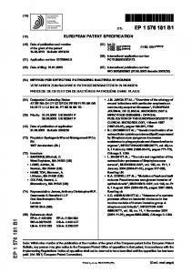
Figure 1. Methylhydrazine reaction with acetone.
300
Results and Discussion Methylhydrazine is a powerful reducing agent. It reacts quickly with acetone. The reaction proceeds as a SN2 nucleophilic substitution reaction and is shown in Figure 1. A literature report indicates that the reaction between hydrazine and acetone proceeds quantitatively in less than 2 min in aqueous media (9). The resulting acetone methylhydrazone (the imine in Figure 1) is more stable than methylhydrazine and is suitable for GC–MS analysis. In addition to the acetone methylhydrazone derivative, a second compound, the enamine (Figure 1), is possible. The enamine forms when the nitrogen attached to the methyl group in methylhydrazine becomes the nucleophile. The more thermodynamically favored product is the imine due to the acidic nature of the nitrogen-bound hydrogen atom on the unmethylated end of the methylhydrazine molecule, which promotes water as the leaving group. The use of acetone as the solvent and the derivatizing agent offers several advantages over other derivatizing agents. Acetone is a common solvent and easy to handle. Other derivatizing agents often need hydrophobic solvents in which the experimental drug substance does not have the needed solubility for an acceptable limit of detection. Because acetone is a good solvent for the experimental drug substance, derivatization happens immediately as soon as methylhydrazine contacts with acetone and finishes as soon as the drug substance sample is fully dissolved into the acetone. This minimizes the possibility of evaporation of methylhydrazine in dilution steps and, therefore, ensures quick and quantitative derivatization. The derivative, acetone methylhydrazone, is soluble and stable in acetone, and acetone is compatible with GC analysis. There is no need for post-derivatization extraction, as is often needed for other derivatization methods. Acetone is very volatile and minimally retained on the gas chromatographic column, so it does not interfere with the detection of acetone methylhydrazone. The GC–MS conditions minimized interference from the acetone and the experimental drug substance. Mass spectrometry in SIM mode (m/z 86, the molecular mass of acetone methylhydrazone) was used for detection due to its high sensitivity and specificity. In this case, a solution of 7.6 ng/g methylhydrazine spiked in 7.6 mg/g drug substance sample has 1 part per million methylhydrazine relative to the drug substance. An injection of 2 µL of this solution has an injection mass of 12 picograms of methylhydrazine equivalent (~ 22 picograms as acetone methylhydrazone).
Journal of Chromatographic Science, Vol. 48, April 2010
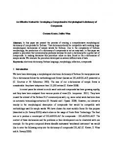
The stock standard solution (7.6 µg/g methylhydrazine equivalent) was injected. The separation was monitored by TIC mode. The identity of the acetone methylhydrazone peak at 12.8 min was verified by comparison of its mass spectrum to the reference mass spectrum of acetone methylhydrazone shown in Figure 2. The fragmentation patterns in the two spectra matched. The peak at m/z 30 was not measured due to the m/z cutoff setting of the mass spectrometer. The chromatogram of the standard solution of 7.6 ng/g methylhydrazine equivalent is shown in Figure 3. The acetone blank is shown in Figure 4. It can be seen that, even when the SIM mode was used, there were three peaks in the chromatogram of the acetone blank at high sensitivity. They are toluene at 12.5 min, 4-hydroxy-4-methyl-2-pentanone at 13.4
Figure 2. Comparison of the mass spectrum for the peak at 12.8 min in the chromatogram for the standard solution of 6 ng/mL methylhydrazine equivalent to the reference mass spectrum of acetone methylhydrazone.
Figure 3. The chromatogram of the standard solution of 6 ng/mL methylhydrazine equivalent in SIM mode (m/z 86). The peak at 12.8 min is from acetone methylhydrazone.
min, and 2-heptanone at 13.85 min. Their identities were established by analyzing possible impurities in acetone and confirmed by comparing with respective authentic samples. These peaks do not interfere with the detection of acetone methylhydrazone, which elutes out at 12.8 min. However, acetone can form 4hydroxy-4-methyl-2-pentanone, commonly known as diacetone alcohol, which is a dimerization product of acetone over time in storage. 4-Hydroxy-4-methyl-2-pentanone could potentially further convert to mesityl oxide, which would elutes out at 13.0 min from the column at the chromatographic condition. If mesityl oxide is formed in acetone solution, it potentially could interfere with the detection of acetone methylhydrazone. It is, therefore, very important to check the freshness of the acetone by running the acetone blank. Absence of mesityl oxide peak would confirm the suitability of the acetone solvent for the analysis. Furthermore, the spiked sample is used not only for recovery calculation but also for checking the resolution of the separation between acetone methylhydrazone and 4-hydroxy-4methyl-2-pentanone. The chromatogram of the spiked sample of 7.6 mg/g drug substance with 7.6 ng/g methylhydrazine equivalent is shown in Figure 5. The signal-to-noise ratio for the peak of acetone methylhydrazone at 12.8 min in Figure 3 is about 27to-1. The signal-to-noise ratio for the peak of acetone methylhydrazone at 12.8 min in Figure 5 is about 25-to-1. In both cases, the signal-to-noise ratios are larger than 3-to-1; therefore, the experiments establish LOD for methylhydrazine in the drug substance sample at 1 part per million methylhydrazine relative to the drug substance. Finally, a sample solution of 7.6 mg/g drug substance “as is” was injected. The chromatogram is shown in Figure 6. It has a similar baseline profile to that of the acetone blank. It is, therefore, concluded that the methylhydrazine was not detected in the drug substance with an LOD of 1 part per million methylhydrazine relative to the drug substance. Additional validation was performed, which included a sonication study, recovery, and stability in solution. The sonication
Figure 4. The chromatogram of the acetone blank in SIM mode (m/z 86).
301
Journal of Chromatographic Science, Vol. 48, April 2010
Conclusion A GC–MS method has been developed as a limit test of trace level methylhydrazine in an experimental drug substance. It utilizes acetone as both a dissolving solvent and a derivatizing agent, eliminating the need for post-derivatization extraction and allowing direct injection. The derivatization reaction between methylhydrazine and acetone is quick and quantitative. GC–MS detection in SIM mode offers good separation for acetone methylhydrazone from matrix interference and sufficient sensitivity for detecting 1 part per million methylhydrazine relative to the drug substance in the sample matrix.
Acknowledgment Figure 5. The chromatogram of the spiked sample of 6 mg/mL drug substance with 6 ng/mL methylhydrazine equivalent in SIM mode (m/z 86). The peak at 12.8 min is from acetone methylhydrazone.
The authors thank Yong Tao and Chris Foti for helpful discussion on derivatization chemistry. We would also like to thank Steve Richoll for help in using the GC–MS instrumentation, and Wendy Wang for help in identifying the impurities in acetone using GC–MS.
References 1. 2. 3. 4. 5. 6. 7.
Figure 6. The chromatogram of the sample solution of 6 mg/mL drug substance “as is” in SIM mode (m/z 86).
8.
experiment was performed to determine if sonication of the drug substance sample for 15 min after constitution with acetone had any detrimental effect on the recovery of acetone methylhydrazone. One of the two identically prepared samples of 7.6 mg/g drug substance with 76 ng/g methylhydrazine spike was subject to the sonication of 15 min, while the other was not. Both samples were analyzed. Based on the 96% recovery from area response comparison and 99% recovery from peak-height comparison, sonication for 15 min does not have any noticeable detrimental effect on the assay result. Supplemental recovery experiments for 7.6 ng/g spiked drug substance samples were between 90–120% of the theory, which is adequate for the trace level analysis (13,14). Solution stability was demonstrated to be acceptable for 24 h under refrigeration conditions. Repeated injections of the 76 ng/g intermediate standard solution yielded 8% relative standard deviation in area counts consistently.
9.
302
10. 11. 12. 13. 14.
S. Budavari (Editor). Merck Index, 12th ed., Merck & Co., Inc. Whitehouse Station, NJ, 1996, pp. 6167. NIOSH Pocket Guide to Chemical Hazards (NIOSH Publication 2005-149, September 2005, accessed in http://www.cdc.gov/niosh/npg/) Documentation for Immediately Dangerous to Life or Health Concentrations (IDLH): Methyl hydrazine, accessed in http://www.cdc.gov/niosh/idlh/ 60344.html. L. Muller, et. al. A rationale for determining, testing, and controlling specific impurities in pharmaceuticals that possess potential for genotoxicity. Regul. Toxicol. Pharmacol. 44: 198–211 (2006). EMEA Guideline on the Limits of Genotoxic Impurities, accessed in http://www.emea.europa.eu/pdfs/human/swp/519902en.pdf S.L. Larson and A.B. Strong. Ion Chromatography with Electrochemical Detection for Hydrazine Quantitation in Environmental Samples Technical Report IRRP-963 (March 1996), WES-TR-IRRP-96-3; Order No. AD-A306785. Methods for Determination of Hazardous Substance, Health and Safety Executive, Suffolk, UK, August 1997 MDHS Method No. 86. J.R. Holtzclaw, S.L. Rose, J.R. Wyatt, D.P. Rounbehler, and D.H. Fine. Simultaneous Determination of Hydrazine, Methylhydrazine, and 1,1Dimethylhydrazine in Air by Derivatization/Gas Chromatography. Anal. Chem. 56: 2952–2956 (1984). S. Selim and C.R. Warner. Residue Determination of Hydrazine in Water by Derivatization and Gas Chromatography. J. Chromatogr. 166: 507–511 (1978). M.A. Rutschmann and H-R. Buser. Determination of Daminozide and Dimethylhydrazine Residues in Swiss Apple Juice Concentrates Using Gas Chromatography-Mass Spectrometry. J. Agric. Food Chem. 39: 176–181 (1991). A. Mozayani, R.T. Coutts, and T.J. Danielson. Gas Chromatographic Analysis of Monoalkylhydrazines. J. Chromatogr. 423: 131–137 (1987). L.A. Dee. Gas Chromatographic Determination of Aqueous Trace Hydrazine and Methylhydrazine as Corresponding Pyrazoles. Anal. Chem. 43: 1416–1419 (1971). E. Rozet, A. Ceccato, C. Hubert, et. al. Analysis of recent pharmaceutical regulatory documents on analytical method validation. J. Chromatogr. A 1158: 111–125 (2007). Guidance for Industry: Bioanalytical Method Validation, US Department of Health and Human Services, Food and Drug Administration, Center for Drug Evaluation and Research (CDER), Center for Biologics Evaluation and Research (CBER), Rockville, May 2001. Accessed at http://www.fda.gov/downloads/Drugs/ GuidanceComplianceRegulatoryInformation/Guidances/UCM070107.pdf
Manuscript received April 6, 2009; revision received June 19, 2009.