May 30, 1995 - of the Porcine Stomach. Andrew J. Mackin, Robert M. Friendship, Brian P. Wilcock, Ronald 0. ... acute cases may present with pale skin colour ...
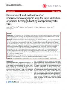
Development and Evaluation of an Endoscopic Technique Permitting Rapid Visualization of the Cardiac Region of the Porcine Stomach Andrew J. Mackin, Robert M. Friendship, Brian P. Wilcock, Ronald 0. Ball, and Heather L. Ayles
ABSTRACT
endoscopic and postmortem findings occurred because endoscopy Our study was designed to ascer- was possibly more sensitive at tain whether a flexible videoscope detecting small and superficial could be used to efficiently monitor ulcerations. However, further studulcers of the pars esophagea in a ies are needed to verify the accularge group of grower-finisher swine. racy of endoscopic diagnosis of gasGastroscopy was performed on tric ulcers in the live pig. 2 separate occasions in 32 pigs following anesthesia with intravenous RESUME pentobarbital, and ulcers of the pars esophagea were subjectively Dans le but de determiner si un graded. The pigs were then necropsied. Grades from the second endo- videoscope flexible pouvait etre scopic examination were compared utilise pour evaluer les ulceres for agreement with grades derived gastro-oesophagiens chez le porc a from gross inspection of the pars I'engraissement, on proceda en esophagea at necropsy, and with deux occasions distinctes a une grades derived from histopathologic gastroscopie chez 32 porcs anesexamination of sections of the same thesies au pentobarbital. L'ulceration observee au niveau gastroregion. The pars esophagea was ade- oesophagien fut cotee de faqon quately visualized in all endoscopic subjective. Les cotes obtenues lors examinations. The average duration de la seconde gastroscopie furent of each examination, from anes- comparees aux cotes octroyees lors thetic induction, was approximately de l'examen macroscopique de 8 min. Gastroscopy permitted appre- l'estomac effectue au moment de la ciation of a wide range of focal and necropsie des animaux a la suite de diffuse superficial and deep ulcera- leur euthanasie, de meme qu'aux tive lesions of the pars esophagea, cotes obtenues lors de l'examen but failed to unequivocally identify histopathologique de sections de la parakeratosis of the pars esophagea. region examinee. La duree moyenne pour chaque Agreement between endoscopic and subsequent necroscopic and histo- examen etait d'environ 8 minutes a pathologic gradings of ulcerations partir du moment de l'induction, et l'examen endoscopique permettait was poor. We concluded that the use of a de visualiser de faqon adequate la flexible videoscope permitted rapid region investiguee. Lors de la gastroinspection of the pars esophagea, scopie, on observa une vaste gamme and was therefore a practical method de lesions superficielles et proof experimentally monitoring the fondes focales et diffuses, mais on progression of spontaneous gastric ne put identifier avec certitude une ulcers in pigs. We also postulated parake'ratose de la portion oesophathat the poor agreement between gienne. La correlation entre les
cotes obtenues lors de l'examen endoscopique et celles des examens macroscopique et microscopique e'tait faible. L'utilisation d'un videoscope flexible a permis une inspection rapide et s'est avere' une methode experimentale pratique pour suivre l'evolution des ulceres gastrooesophagiens chez le porc. La faible correspondance entre l'examen endoscopique et les trouvailles postmortem pourraient resulte du fait que l'endoscopie est une methode plus sensible pour detecter les ulcerations superficielles et de petite taille. Toutefois, des etudes supplementaires sont necessaires pour verifier la veracite d'un diagnostic d'ulceration gastrique chez le porc vivant pose par examen endoscopique. (Traduit par docteur Serge Messier)
INTRODUCTION Naturally occurring gastric ulceration can be a significant cause of economic loss for producers of growerfinisher swine (1). Although ulcer formation has been associated with the feeding of finely-ground, pelleted feeds to rapidly-growing pigs, the precise pathogenesis of gastric ulceration in swine has not been determined (1-3). Sudden death due to acute blood loss is often the first clinical indication of ulceration (1-3). Less acute cases may present with pale skin colour, melena, and increased respiratory rate and effort. Severe hemorrhage from a deep ulcer is presumably a late sequel to a chronic and progressive pathological process
Department of Clinical Studies (Mackin), Population Medicine (Friendship), Pathobiology (Wilcock), Department of Animal and Poultry Science (Ball, Ayles), Ontario Veterinary College, University of Guelph, Guelph, Ontario NIG 2W1. Address correspondence and reprint requests to Dr. Robert M. Friendship. Supported by a grant from the Ontario Ministry of Agriculture, Food and Rural Affairs, the Ontario Pork Producers Marketing Board, and Ralston Purina Canada. Received May 30, 1995.
Can J Vet Res 1997; 61: 121-127
121
TABLE I. System for scoring and grading endoscopic findings Score 0 I 2 3 4 5 6
Endoscopic appearance No visible lesions Shallow erosions < 10% of pars esophagea Shallow erosions 10-20% of pars esophagea Shallow erosions > 20% of pars esophagea Deep elcerations < 10% of pars esophagea Deep ulcerations 10-20% of pars esophagea Deep elcerations > 20% of pars esophagea
TABLE II. System for scoring and grading findings at gross necroscopic inspection
Score 0 1 2 3
Necroscopic appearance No visible lesions Parakeratosis Focal, shallow erosions Diffuse or deep ulcerations
Grade A B C
involving the gastric mucosa (3). Experimental and clinical investigations of the etiology and prevention of gastric ulceration in swine have been hampered by the occult nature of early lesions (2). With the increasing availability of technologically advanced flexible fiberscopes and videoscopes (4-6), physicians are routinely using gastroscopy in human patients as a sensitive and practical means of monitoring the incidence and progression of gastric ulcers (4,7). Although the same technology is available to veterinarians, the use of gastroscopy for the clinical investigation of naturallyoccurring gastric ulceration in animals has been predominantly limited to companion animals (8,9). Gastric ulcerations in swine typically affect the pars esophagea, a small glandless area of stratified squamous epithelium encircling the junction of the esophagus and the stomach (1-3). Given that ulcerations in the pig predictably occur near the entrance of the esophagus into the stomach, we speculated that typical lesions could be readily visualized using a modern flexible endoscope. This study was designed to determine whether a flexible videoscope could be used experimentally to efficiently monitor the incidence and progression of ulcers of the pars esophagea in a large group of pigs. MATERIALS AND METHODS EXPERIMENTAL ANIMALS
Endoscopic examinations were performed on 2 separate occasions in 32 healthy Yorkshire pigs enrolled in 122
Grade A
B
C
a therapeutic trial evaluating the effects of a feed additive on the incidence of gastric ulceration in growerfinisher swine (10). Endoscopy was initially performed prior to the commencement of the feed trial, at which time the pigs were 10 wk old, and had a mean weight of 31 kg (range 24.5 kg to 37.5 kg). Endoscopy was repeated 3 wk later at the completion of the feed trial, by which time the pigs had attained a mean weight of 50 kg (range 41 kg to 64 kg). Pigs were fed a moderately finely-ground pelleted ration (mean particle size of 763 microns) in an attempt to induce stomach lesions. The experimental protocol was approved by the University of Guelph Animal Care Committee and was carried out in accordance with the principles published in the Canadian Council on Animal Care "Guide to the Care and Use of Experimental Animals." ENDOSCOPIC EXAMINATIONS
The pigs were housed in groups of 8 in 4 large pens. Feed was removed from the pens 12 h prior to the scheduled time of endoscopy. Water was freely available until the pigs were removed from the pens immediately prior to anesthesia. Anesthetic induction was performed in a working area adjacent to the pens. General anesthesia was induced with intravenous pentobarbital (Somnotol, MTC Pharmaceuticals, Cambridge, Ontario) administered via a butterfly catheter into a marginal ear vein at a dose of 30 mg/kg. Small batches of 2 or 3 pigs were induced and then transported on a large trolley to a nearby room for endoscopic examination. Further intravenous pentobarbital was given as required to maintain a moderate depth of anesthesia throughout the endoscopic procedure. For endoscopic examination, anesthetized pigs were placed in right lateral recumbency on a stainless steel surgical table, and a metal speculum
(1 1) was used to keep the mouth open. Endoscopy was performed with a forward viewing flexible videoscope with a working length of 100 cm and an outer diameter of 10 mm (GIF type 100 EVIS Gastrointestinal Videoscope, Olympus Corporation, Lake Success, New York, USA). Each examination was recorded on video tape to enable reassessment of endoscopic findings at a later date. The identity of each pig examined was permanently registered on video tape by recording ear tag numbers at the commencement of each examination. The endoscope was then rapidly passed per os into the cardiac region of the stomach. Accurate passage of the endoscope through the oropharynx and esophagus was facilitated by continuous visual assessment of the television monitor. Following entry into the cardiac region, the stomach was partially inflated with room air passed via the air/water nozzle of the endoscope. The distal end of the endoscope was then maximally retroflexed and pulled gently in an oral direction to enable visualization of the pars esophagea (Figures 1 and 2). After thorough endoscopic examination of the pars esophagea and adjacent cardiac mucosa, the stomach was completely deflated by depressing the suction valve of the endoscope, and the endoscope was removed. The pigs were then returned to their pens for anesthetic recovery. All endoscopic examinations were performed by one endoscopist (AJM). Several weeks after the completion of the study, the endoscopist carefully reviewed the video recordings of the second series of endoscopic examinations, and numerically scored lesions of the pars esophagea on a scale of 0 to 6 (Table I). POSTMORTEM
Two days after the 2nd endoscopic examination, the pigs were electrically stunned and humanely slaughtered, and the complete gastrointestinal tract was removed and submitted for postmortem inspection. All necropsy inspections were performed by one pathologist (BPW). Gross lesions of the pars esophagea were numerically scored on a scale of 0 to 3 (Table II). No magnification aids were used in determining the lesion score.
Figure 2
Figure 1. Ante mortem radiograph illustrating the position of the retroflexed endoscope within the inflated stomach during examination of the pars esophagea. Figure 2. Postmortem photograph illustrating the approximate position of the retroflexed endoscope within the stomach during examination of the pars esophagea. The exact anatomic orientation of the endoscope is slightly different than ante mortem positioning because of deflation of the stomach during postmortem.
Figure 3. Typical appearance of the normal porcine pars esophagea as visualized through the retroflexed endoscope. The shaft of the endoscope can be seen entering the stomach from the esophagus through the center of the rectangular pars esophagea. Figure 4. Pars esophagea obscured by slurry of gastric juices and wood shavings.
a
A linear strip of epithelium (approxcm to 6 cm in length) was collected across the junction of the pars esophagea to include the junction with granular stomach. If an erosion or ulcer was grossly detected, the strip was oriented to include the lesion. The strip was fixed in 10% neutral buffered formalin and processed routinely for paraffin-embedding and light microscopy. One longitudinal section from each strip was examined. Microscopic lesions of the pars esophagea were numerically scored on a scale of 0 to 3 (Table III). Parakeratosis was defined as hyperplasia of the sqpuamous epithelium with retention of a thickened hypereosinophilic superficial layer with ghost-like
Figure 3
imately 4
Figure 4
123
TABLE III. System for scoring and grading histopathologic findings Score 0 1 2 3
Histopathologic findings No visible lesions Parakeratosis Erosions Ulcerations
Grade A B C
nuclear outlines. Erosions were defined as partial-thickness epithelial loss, usually accompanied by increased basophilia and hyperplasia of the stratum basale. Ulcerations were defined as loss of epithelium to and through the basement membrane, typically accompanied by neutrophilic inflammation and by immature granulation tissue within the exposed submucosa. STUDY DESIGN
The initial endoscopic examination of each pig was used to develop and refine endoscopy techniques. The second endoscopic examination was utilized to compare endoscopic observations with ensuing postmortem findings. Numerical endoscopic, necroscopic and histopathologic scores were converted into a 3-tiered alphabetical grading system (Tables I, II, and III) to enable comparison of results. Endoscopic grades were then assessed for agreement with necroscopic and histopathologic grades by calculating a weighted kappa (Ciccetti method) and the variance of kappa (12). Neither the endoscopist nor the pathologist were aware of each other's results at the time of scoring lesions of the pars esophagea. The measures were blinded to the treatments.
RESULTS ENDOSCOPIC EXAMINATIONS
Over the period of the study, 64 endoscopic examinations were performed. Partial gastric inflation permitted adequate visualization of the pars esophagea and adjacent glandular cardiac mucosa in all examinations performed (Figure 3). The normal pars esophagea was visualized as a small rectangular area of roughened and slightly raised epithelium surrounding the opening of the esophagus into the stomach. Epithelial roughenings typically formed ridges radiating outwards from the esophageal opening, and were often stained yellow by gastric juices. Complete 124
TABLE IV. Summary of endoscopic, necroscopic and histopathologic findings
Pig number 17 18 19 20 21 22 23 24 41 42 43 44 45 46 47 48 49 50 51 52 53 54 55 56 57 58 59 60 61 62 63 64
Endoscopic score 2 2 0 0 5 2 0 1 6 2 6 2 6 1 1 1 1 3 1 6 3 6
3 3 2 6 3 0 1 0 1
3
Endoscopic grade B B
Necropsy score
A A C B A B C B C B C B B B B C B C C C C C B C C A B A B C
gastric inflation did not improve visualization of the pars esophagea, and was often associated with a discernible decrease in the depth of the animal's respiration. In most examinations, thorough inspection of the par esophagea could be achieved by clockwise and anticlockwise rotation of the distal end of the retroflexed endoscope. Prior to the initial endoscopic examination, several pens of pigs had access to bedding material composed of wood shavings. In these pigs, the pars esophagea was frequently obscured by an adherent slurry of gastric juices and shavings (Figure 4). Attempted removal of this slurry by suction rapidly and permanently occluded the endoscope's suction apparatus. In all affected pigs, however, satisfactory dislodgement of the adherent slurry was achieved by a combination of vigorous flushing with 50 mL to 100 mL of sterile water through the instrument channel of the endoscope, elevating the cardiac region of the stomach by transiently tilting the trunk of the pig into a partial sternal position, and firmly slap-
2 2 1 1 1 2 1 1 2 1
2 I 2 1 2 1 1 2 2 3 1 3 2 2 2 2 3 2 2 2 1 2
Necropsy grade B B
Histologic grade 3 3
A A A B A A B A B A B A B A A B B C A C B B B B C B B B A B
0 0 I 3 1 1 3 1 3 1 2 1 1 1 2 3 2 3 3 3 3 1 1 2 3 3 1 3 2 2
Histologic score C C A A A C A A C A C A B A A A B C B C C C C A A B C C A C B B
ping the left flank. Complete removal of bedding material 12 h prior to anesthesia prevented this problem during the 2nd endoscopic examination. Porcine gastric mucosa was observed to be exquisitely sensitive to endoscopic manipulation. Linear mucosal bruising was frequently observed at sites of contact with the retroflexed endoscope (Figure 5). No other lesions were observed during examination of the cardiac mucosa. Thorough endoscopic examination of the remaining gastric mucosa was not attempted in this study. However, even after 12 h of fasting, the gastric body and pyloric antrum were frequently observed to be obscured by considerable amounts of gastric juice and slurried feed. The complete endoscopic examination of each pig was usually completed within 20 min of anesthetic induction. The average time for the procedure was 8 min per pig. ENDOSCOPIC VS. POSTMORTEM FINDINGS
Compared with visual inspection at necropsy, endoscopy permitted
appreciation of a wider range of focal and diffuse superficial and deep ulcerative lesions of the pars esophagea. Typical superficial erosions of the pars esophagea were visualized endoscopically as focal or linear areas of surface granularity and reddish discoloration radiating outwards from the junction between the esophagus and the stomach (Figure 6). Deeper ulcerations of the pars esophagea were visualized endoscopically as focal dark red to black depressions, and occurred most commonly along the junction between the pars and the adjacent cardiac mucosa (Figure 7). Deep ulcerations were occasionally observed to be actively bleeding during endoscopy (Figure 8). Parakeratotic thickening of the pars esophagea, although easily recognized during necropsy, was not appreciated with certainty during the preceding endoscopic examination. Since endoscopy, gross necroscopic inspection and histopathology emphasized dissimilar aspects of lesions affecting the pars esophagea, different numerical scoring systems were developed for each method of inspection. Endoscopic, necroscopic and histopathologic numerical scores and alphabetical grades are summarized in Table 4. Comparing the endoscopic and gross necroscopic grades, it was shown that the agreement was only fair (k = 0.217, V(k) = 0.011). Similarly, there was only fair agreement between endoscopic and histologic examination results (k = 0.251, V(k) = 0.016). The results of histologic examination and gross necroscopic grades were in moderate agreement (k = 0.410, V(k) = 0.010).
DISCUSSION Experimental studies reporting endoscopy of the porcine stomach have previously been published in both the human and veterinary literature. Most studies were published in human medical journals, and utilized the pig as a model for the development of advanced endoscopic techniques for the investigation, diagnosis, and treatment of human gastropathies (13-18). In these studies, the endoscopist typically concentrated on the mucosa of the gastric body. Studies concentrating on the structures of the
cardiac region of the porcine stomach are much less common. Within the veterinary literature, several authors have reported the use of endoscopy to examine the porcine pars esophagea and adjacent cardiac mucosa. Over 25 y ago, veterinarians at the University of Wisconsin described evaluation of the porcine gastric mucosa using either a flexible fiberscope or a specialized gastrocamera mounted on a flexible connecting tube passed per os and guided to the various regions of the stomach using fluoroscopy (19). Detailed examination of the pars esophagea was hampered by the poor manoeuvrability of these early instruments. More recently, veterinarians in Japan described a technique for serially inspecting and photographing the porcine gastroesophageal region utilizing a rigid metal fiberscope passed through a cylindrical plastic cannula inserted into the proximal greater curvature of stomach via the left abdominal wall (20). Although this technique permitted detailed inspection of the pars esophagea, preceding mid-line laparotomy was necessary to insert the plastic cannula. In both of these previous studies by veterinary researchers, the limited manoeuvrability of the endoscopes that were employed necessitated the use of cumbersome or invasive procedures such as fluoroscopy and gastric cannulation. Our study documents that the use of more advanced endoscopic technology permits rapid and detailed non-invasive visualization of the pars esophagea, thereby providing a practical means of following the progression of spontaneous gastric ulceration in pigs. Endoscopy is widely recognized as a sensitive means of detecting and monitoring early gastric ulcers in both humans and companion animals (4,7-9). Given the lack of histopathologic confirmation of suspected ulcers at the time of endoscopy, and the poor agreement between the endoscopic and the subsequent postmortem grading of lesions, it is valid to question the accuracy of our original endoscopic diagnoses. However, we postulate that the poor agreement between endoscopic and necropsy findings may be primarily due to difficulty in identifying small or superficial ulcerations of the pars esophagea at gross postmortem. Modern videoscopes can
magnify small ulcers to fill the screen of a television monitor without appreciable loss of detail, thereby permitting close scrutiny of the contrast in color and surface texture between denuded, vascular submucosa and adjacent intact epithelial surfaces. Given the poor agreement between findings at endoscopy and subsequent gross inspection at necropsy, it is therefore hardly surprising that our study also documented poor agreement between endoscopic and histopathologic findings. Methodical histopathologic examination of multiple serial sections of each pars esophagea, a time-consuming procedure not attempted in our study, would be necessary to definitively establish the accuracy of endoscopic findings. In addition, the lack of agreement between endoscopic examination and gross postmortem findings may have been caused to some extent by the delay between the final endoscopic examination and the time of slaughter. Studies have shown that ulcers may be produced within 1 or 2 d (21), particularly following a period of fasting such as occured in this experiment. We suspect that endoscopic examination of the live pig may be a more accurate assessment of gastric ulceration than gross pathological examination, however, further studies are required to verify this hypothesis. Because endoscopic examination requires a period of fasting prior to performing the procedure, and fasting may lead to the formation of ulcers, researchers must be cautious in interpreting results of experiments using serial endoscopic examinations, in that the procedure may influence the severity of the lesions being studied. The endoscopic technique described in this study was designed to meet the requirements of a specific research project: namely, to visualize the pars esophagea of a large group of pigs as rapidly and cost-effectively as possible. Our technique, however, could easily be modified to suit the particular needs of other projects. Controlled anesthesia utilizing endotracheal intubation, and subsequent maintenance with inhalant anesthetic agents would facilitate time-consuming procedures such as the collection of multiple endoscopically-guided biopsies. Since we were only interested in lesions of the pars esophagea, the pigs in our 125
Figure 5. Traumatic linear bruising of the cardiac mucosa due to contact with the shaft of the endoscope.
Figure 6. Typical endoscopic appearance of multifocal superficial erosions of the pars esophagea. The most obvious erosion is located within the right upper corner of the pars esophagea, although close inspection reveals several other subtle reddish discolorations within the central portion of the pars.
study were placed in right lateral recumbency in order to drain gastric contents into the pyloric antrum. Positioning the animal in left lateral recumbency would presumably facilitate endoscopic examination of the gastric body and pylorus. Since the stomachs of many of the pigs in our study still retained gastric juices and feed despite prior fasting for 12 h, thorough examination of the entire gastric lumen would probably require fasting for a minimum of 24 h (13).
REFERENCES 1. CONSTABLE PD. Gastric ulcers of swine. In: Howard JL, ed. Current Veterinary Therapy 3: Food Animal Practice. Philadelphia: WB Saunders. 1993: 735-736.
126
Figure 7. Typical endoscopic appearance of multifocal deep ulcerations of the pars esophagea. The margins of the pars esophagea appear depressed relative to the adjacent cardiac mucosa, and most of the pars epithelium is disrupted by dark red/black discolorations.
Figure 8. Active hemorrhage is visualized as a swirl of dark red blood oozing from a focal deep ulcer adjacent to the shaft of the endoscope.
2. O'BRIEN JJ. Gastric ulcers. In: Leman AD, Straw BE, Glock RD, Mengeling WL, D'Allaire S, Taylor DJ, eds. Diseases of Swine. 7th ed. Ames: Iowa State Univer-
sity Press, 1992: 680-691. 3. BARKER IK, VAN DREUMEL AA, PALMER N. The alimentary system. In: Jubb KVF, Kennedy PC, Palmer N, eds. Pathology of Domestic Animals: Volume 2. 4th ed. San Diego: Academic Press, 1993: 1-318. 4. SCHUMAN BM. Upper gastrointestinal endoscopy. In: Berk JE, ed. Bockus Gastroenterology. 4th ed. Philadelphia: WB Saunders, 1985: 564-580. 5. AXON AT. Endoscopy. In: Shearman DJC, Finlayson NDC, eds. Diseases of the Gastrointestinal Tract and Liver. 2nd ed. Edinburgh: Churchill Livingstone, 1989: 89-112. 6. BARLOW DE. Fiberoptic instrument technology. In: Tamms TR, ed. Small Animal Endoscopy. St. Louis: CV Mosby, 1990: 1-24. 7. BLACKSTONE MO. Benign gastric ulcers. In: Blackstone MO, ed. Endoscopic Interpretation: Normal and Pathologic
8. 9.
10.
11.
12. 13.
14.
Appearances of the Gastrointestinal Tract. New York: Raven Press, 1984: 12 1-136. TAMMS TR. Gastroscopy. In: Tamms TR, ed. Small Animal Endoscopy. St. Louis: CV Mosby, 1990: 89-166. TWEDT DC. Vomiting. In: Anderson NV, ed. Veterinary Gastroenterology. 2nd ed. Philadelphia: Lea & Febiger, 1992: 336-367. AYLES HL, BALL RO, FRIENDSHIP RM, BUBENIK GA. Effect of fineness of feed and dietary melatonin on gastric ulcers in pigs (Abstract). Can Soc Anim Sci, Regina, 1994. LEAHY JR, BARROW P. Restraint of Animals, 2nd ed. Ithaca: Cornell Campus Store, Inc, 1953: 147. FLEISS J. Statistical Methods for Rates and Proportions, 2nd ed. New York: J Wiley Publishing, 1981: 212-236. HEY H, WAMBERG T, RASMUSSEN M, J0RGENSEN F. Endoscopic evaluations of potentially ulcerogenic drugs: A new in vivo porcine test model. Eur J Surg Res 1987; 19: 312-317. ENGSTRAND L, GUSTAVSSON S, JORGENSEN A, SCHWAN A, SCHEYNIUS
A. Inoculation of barrier-born pigs with Helicobacter pylori: A useful animal model for gastritis type B. Infect Immun 1990; 58: 1763-1768. 15. FRIMBERGER E, CLASSEN M. A new pull-through trocar technique for percutaneous operative endoscopy. Endoscopy 1991; 23: 338-341. 16. ENGSTRAND L, ROSBERG K, HUBINETTE R, BERGLINDH T, ROLFSEN W, GUSTAVSSON S. Topographic mapping of Helicobacter pylori colonization in long-term-infected pigs. Infect Immun 1992; 60: 653-656.
17. SCHNEIDER TA, WITTGEN CM, ANDRUS CH, KAMINSKI DL. Comparison of minimally invasive methods of parietal cell vagotomy in a porcine model. Surgery 1992; 112: 649-655. 18. MEYER-ROSBERG K, GUSTAVSSON S. '3C-urea breath test for diagnosis of experimental Helicobacter pylori infection in barrier born pigs. Gut 1993; 34: 594-598. 19. KOWALCZYK T, TANAKA Y, MUGGENBURG BA, OLSON WG, MORRISSEY JF. Endoscopic examination of
stomach of swine. Am J Vet Res 1968; 29: 729-736. 20. KOKUE E, KUREBAYASHI Y, SHIMODA M, HAYAMA T. Serial endoscopic observation of swine gastroesophageal ulceration induced by injection of a histamine-oil-beeswax mixture. Am J Vet Res 1981; 42: 1807-18 10. 21. STRAW B, HENRY S, NELSSEN J, DOSTER A, MOXLEY R, ROGERS D, WEBB D, HOGG A. Prevalence of lesions in the pars esophagea of normal and sick pigs. Proc Int Pig Vet Soc Congress, 1992: 286.
127