Nov 22, 1988 - and College of Veterinary Medicine, Cornell University, Ithaca, New York 148532. Received .... from the University of California were obtained from ani- mals with ..... D1 10 htipt*q. t t p47 gp40. D36 .. fa du m. _ @ o22. FIG. 5.
Vol. 27, No. 3
JOURNAL OF CLINICAL MICROBIOLOGY, Mar. 1989, p. 474-479 0095-1137/89/030474-06$02.00/0 Copyright © 1989, American Society for Microbiology
Development and Evaluation of Immunoassay for Detection of Antibodies to the Feline T-Lymphotropic Lentivirus (Feline Immunodeficiency Virus) THOMAS P. O'CONNOR, JR.,l* SHERYL TANGUAY,' ROBIN STEINMAN,l ROBERTA SMITH,' MARGARET C. BARR,2 JANET K. YAMAMOTO,3 NIELS C. PEDERSEN,3 PHILIP R. ANDERSEN,' AND QUENTIN J. TONELLI1 IDEXX Corp., Portland, Maine 041011; School of Veterinary Medicine, University of California, Davis, California 956163; and College of Veterinary Medicine, Cornell University, Ithaca, New York 148532 Received 19 September 1988/Accepted 22 November 1988
The feline T-cell lymphotropic lentivirus (feline immunodeficiency virus) is a recently described felinespecific retrovirus that can produce chronic immunodeficiency-like disorders in cats. A microdilution plate format enzyme-linked immunosorbent assay has been developed to detect the presence of antibody to the virus in feline serum or plasma. Temporal studies performed with experimentally infected animals show that seroconversion can be demonstrated 3 to 4 weeks after exposure to the virus. Results of a serosurvey (n = 1,556 samples) indicate that infection is fairly common in both clinic (5.2%) and sick cat (15.2%) populations. Western blot (immunoblot) and sodium dodecyl sulfate radioimmunoprecipitation assays were developed to confirm microdilution plate test results and to identify peptides specific for the feline immunodeficiency virus. All microdilution plate test positive results and selected negative results were confirmed by one or both of these procedures. These data demonstrate that this microassay plate enzyme-linked immunosorbent assay is a very sensitive and specific test for detection of antibody to the feline immunodeficiency virus.
animals but
A recently identified retrovirus has been shown to be the causitive agent of a chronic immunodeficiency-like syndrome in cats (21). In initial reports the virus was named feline T-lymphotropic lentivirus to reflect the tropism of the virus for feline T cells and the classification of the agent as a member of the lentivirus subfamily of retroviruses (12, 22). The name of the virus has been recently changed to feline immunodeficiency virus (FIV) to conform with international nomenclature for immunodeficiency-linked viruses (24). The FIV agent has a strong but not absolute tropism for the feline T-lymphocyte cell line, which may be responsible for the immunosuppressive nature of the virus. Viral particle morphology and the Mg2+ metal requirement of the viral reverse transcriptase are supportive of the classification of the virus as a lentivirus. The virus is distinct from previously described feline retroviruses and represents the first report of a feline-specific lentivirus (21). Members of the lentivirus subfamily infecting other species include the human immunodeficiency virus (HIV) (8, 15), visna virus in sheep (19), caprine arthritisencephalitis virus (7), equine infectious anemia virus (20), bovine immunodeficiency virus (9), and simian immunodeficiency virus (3). The clinical course of FIV infection appears to be characterized by a lengthy asymptomatic phase persisting several months or perhaps years, during which viral infection can be demonstrated but clinical symptoms are not apparent (24). This period of apparent viral latency is typical of lentiviral infections and frequently precedes the development of clinical abnormalities (10). Clinical symptoms most commonly associated with FIV infection include rhinitis and gingivitis, anemia, diarrhea, pustular dermatitis, and generalized lymphadenopathy (12, 22). Symptoms vary among infected
*
are
characterized by
a
chronic and persistent
nature.
The FIV is infectious within feline populations and can be transmitted after intimate and prolonged contact. Initial reports suggest that biting may be an important mode of viral transmission (24). Isolates of FIV have been identified in the United States (21), the United Kingdom (11), and Japan (13). A limited serosurvey in the United States reported that 1 in 18 healthy cats and 10 of 25 unhealthy cats were seropositive for FIV infection (21). The continuous propagation of FIV in an established Crandell feline cell line has permitted the isolation and purification of large quantities of virus. The highly purified virus was used to develop a sensitive microdilution platebased enzyme-linked immunosorbent assay (ELISA) for detection of feline antibody specific for the FIV agent. In this report we describe the development of the FIV microdilution plate ELISA, partially characterize immunoreactive viral peptides, and present data on the prevalence of viral infection in several populations (n = 1,556). The results of this survey demonstrate that FIV infection is relatively common and widespread in the United States.
MATERIALS AND METHODS Virus and cell culture. The FIV was propagated in chronically infected Crandell feline kidney (Crfk) cells (6). The virus was concentrated from tissue culture fluids by precipitation with polyethylene glycol (4) and purified by density gradient centrifugation on glycerol gradients as previously described (18). The FIV microdilution plates were prepared by coating microdilution wells with 100 ,ul of an inactivated detergentdisrupted preparation of FIV antigen (5.0 ,ug/ml). Samples of feline serum or plasma were diluted (1:100) in 20 mM sodium phosphate-150 mM NaCl (pH 7.4) containing calf serum and bovine serum albumin. Diluted samples (100 pt) were ap-
Corresponding author. 474
VOL. 27, 1989
plied to individual microdilution wells and incubated for 30 min at room temperature. The contents of the microdilution plates were aspirated, and the plates were washed (five times) and incubated (30 min, room temperature) with goat anti-feline horseradish peroxidase conjugate (Kirkegaard and Perry). The contents of the microdilution plates were aspirated, the plates were washed (five times), and substrate solution (100 ,ul) was added. The substrate solution was prepared immediately before the assay by mixing equivolume portions of 0.1% 3,3',5,5'-tetramethylbenzidine in 60% methanol-40% glycerol with 0.04% hydrogen peroxide in 0.1 M dibasic potassium phosphate-citrate (pH 4.8) containing 0.01% Thimerosal. The enzymatic reaction was carried out for 15 min at room temperature and then stopped by addition of 100 ,ul of dilute hydrofluoric acid solution (1:400). Individual microdilution well optical density values were determined spectrophotometrically at 650 nm. To minimize interassay variability, ELISA optical density values were normalized by using positive and negative control reagents assayed on each microdilution plate. The presence or absence of antibody to FIV was determined by relating the A650 of the sample to the A6,( of the positive control reagent. The positive control contains a standardized level of antibody to FIV in feline serum. The relative level of FIV-specific antibody in the sample was determined by calculating the sample-to-positive (S/P) ratio in the following manner: (sample A650 - negative control A650)!(positive control A65O - negative control A650). Samples with S/P ratios less than 0.5 are classified negative. S/P ratios equal to or greater than 0.5 are positive for antibody to FIV. Clinical samples. The FIV ELISA was used to screen a total of 1,556 samples obtained from five sources: 223 from the Boston Refuge League (Boston, Mass.), 130 from the Chicago Cat Clinic (Chicago, 111.), 459 from Clinipath Laboratories (Valparaiso, Ind.), 145 from the Veterinary Reference Laboratory (Dallas, Tex.), and 599 from the University of California (San Diego). Samples from the Boston Refuge League, Chicago Cat Clinic, Clinipath, and the Veterinary Reference Laboratory were not selected on the basis of previous disease status and represent a collection of samples received by these clinics and laboratories. Samples obtained from the University of California were obtained from animals with a previous unspecified disease or illness. This set of samples should be regarded as comprising a sick cat population. Samples for the seroconversion studies were obtained from the Cornell Feline Health Center (Ithaca, N.Y.) and the University of California (Davis). These were obtained from specific-pathogen-free animals after inoculation with either infectious cell free tissue culture fluid (1 ml), FIV-infected whole blood (1 ml), or 0.44-,um-filtered plasma (2 ml) from infected animals. Western blot assay. The Western blot (immunoblot) protocol used was a modification of the procedure initially described by Towbin et al. (23). Purified FIV was disrupted with sodium dodecyl sulfate (SDS) and mercaptoethanol and separated with a 10% SDS-polyacrylamide gel. Viral proteins were transferred to nitrocellulose sheets, which were then blocked with detergent and calf serum. Individual strips were cut from each sheet and incubated for 2 h with a 1:100 dilution of sample prepared in a phosphate-buffered saline solution containing 0.05% Tween 20, 1% bovine serum albumin, and 30% calf serum. Strips were washed exhaustively with phosphate-buffered saline-0.05% Tween 20 and incubated for 1 h with goat anti-feline horseradish peroxidase conjugate. The wash cycle was repeated, and the
IMMUNOASSAY FOR ANTIBODIES TO FIV
475
precipitating substrate 4-chloronaphthol was added. Reactions were stopped by washing with deionized H20, and results were immediately interpreted. The presence of two or more FIV viral bands was necessary to confirm a sample as positive for antibody to FIV. The molecular weights of FIV-reactive peptides were determined by comparison of immunoperoxidase-stained Western blot strips with Coomassie blue-stained polyacrylamide gels and amido blackstained nitrocellulose strips (R. Steinman, T. O'Connor, Q. Tonelli, K. Lawrence, C. Seymour, J. Goodness, N. Pedersen, and P. R. Andersen, submitted for publication). Uninfected Crfk host cell extract was used to prepare Western blot strips to identify reactive bands unrelated to FIV infection. These were used in the identification of nonspecific bands appearing on FIV blot strips. RIPA-SDS-PAGE. For the radioimmunoprecipitation assay (RIPA) with SDS-polyacrylamide gel electrophoresis (PAGE), the FIV productively growing in cell culture was metabolically labeled with [35S]methionine and [35S]cysteine at 37°C for 4 h. Cells were lysed in 10 mM phosphate buffer (pH 7.5) containing 100 mM NaCI, 1% Triton X-100, 0.5% sodium deoxycholate, 0.1% SDS, 0.1 mM phenylmethylsulfonyl fluoride, and 100 Kallikren inactivator units of aprotinin per ml (lysis buffer). Cell lysates were clarified by centrifugation at 100,000 x g for 30 min before use. Generally, 100 ,ul of viral extract was mixed with 5 ,ul of serum sample and incubated for 18 h at 3°C. Samples were concentrated by adding 0.2 ml of a 5% suspension of protein A-Sepharose CL-4B (Pharmacia Fine Chemicals) in 10 mM phosphate (pH 7.5)-100 mM NaCl-1% Triton X-100-0.1% SDS and centrifuging. The pellet was washed three times in lysis buffer, heated in sample buffer, and applied to a 12% SDS-polyacrylamide gel (14). Gels were processed for fluorography and exposed at -70°C to Kodak XAR-5 film. RESULTS FIV ELISA detection of seroconversion in response to experimental infection. Serum samples from a total of 14 experimentally infected cats were obtained to assess the sensitivity of the microdilution plate test with respect to seroconversion. Temporal samples were obtained for each animal and assayed by the FIV ELISA. Western blots and/or RIPA-SDS-PAGE were used to confirm both positive and negative results. Seroconversion was demonstrated in all animals by 4 weeks after the initial exposure. Microdilution plate data for cats 2429 and 2838 are shown in Fig. 1 and are typical of the entire series of experimentally infected animals. Western blots and RIPA-SDS-PAGE confirmatory test data are shown in Fig. 2 through 5 and demonstrate seroconversion paralleling the microdilution plate test results. With the exception of one cat, which died during the study (week 14), all cats remained seropositive during the testing period. Most of the experimentally infected animals showed similar clinical manifestations, including an initial neutropenia lasting 4 to 5 weeks, fever lasting several days postinfection, and a generalized enlargement of peripheral lymph nodes occurring 4 weeks postinfection and persisting for 2 to 9 months. However, with the exception of the single cat cited above, none of the experimentally infected animals developed symptoms associated with late-stage FIV infections
(24).
The Western blot assay of the two temporal series showed initial reaction to the FIV gag proteins piS and p26 (Fig. 2 and 3). Reaction to additional FIV proteins (plO, p32, gp4O,
J. CLIN. MICROBIOL.
O'CONNOR ET AL.
476
B
A
FTLV TEMPORAL SAMPLE
C
E
D
F
G
H
K
L
2429 -r
I
p65 _lu
gp4O p32 p26
_
-
:
pl5
-
_
-
L li.i.LL 0r 0 0.5
I
1
2-
1.5
2 2.5 3 S 7 9 il MIONTHS AFTER INOCULATION
13
15
FTLV TEMPORAL SAMPLE
_n
_.
_ |-_
ÈLGLLL LL
FIG. 2. Western blot strips after assay of serum samples obtained sequentially during the course of experimentally induced FIV infection in cat 2429. Results are shown for samples obtained at the following times (months) postinfection (lanes): A, preimmunization bleed; B, 0.75; C, 1.0; D, 1.25; E, 2.0; F, 3.0; G, 4.0; H, 5.0; t, 7.0; J, 9.0; K, 11.0; L, 14.0.
2838
3.5ri 15
0-3
o o.s 1
_M
_
1.5
2
2.5
3
5
7
9
il
13
15
MONTHS AFTER INOCULATION
FIG. 1. Microdilution plate assay results of serum samples obtained sequentially during the course of experimentally induced FIV infection in cats 2429 and 2838. S/P ratios were calculated as described in Materials and Methods.
Western blot and/or RIPA-SDS-PAGE confirmatory assays were conducted on all samples that tested positive by the microdilution plate test. Each of these samples was positive by the confirmatory procedures. Western blot assays of FIV antibody-positive samples typically showed a strong reaction to the FIV gag proteins p26 and pi5 and variable reactivity to additional FIV proteins (10,000, 32,000, 40,000, 47,000, and 65,000 daltons) (Steinman et al., submitted). Staining of two or more FIV peptides was required for positive result confirmation by Western blotting, whereas reaction to gpl3O was required for confirmation by RIPA-SDS-PAGE. The RIPA-SDS-PAGE assays of positive samples demonstrated variable reactions to proteins of 130,000, 110,000, 47,000, 40,000, 36,000, 22,000, and 15,000 daltons; however, all plate test-positive samples immunoprecipitated gpl3O. A small percentage (3%) of microdilution plate test-positive samples were reactive to only a single FIV peptide (p26 or piS) in the Western blot assay. These samples were positive when tested using the RIPASDS-PAGE assay. A set of 150 plate test-negative samples were negative when tested by the Western blot assay. A
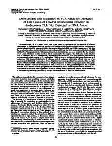
p47, and p65) occurred during the course of infection (Steinman et ai., submitted). Serum samples from specificpathogen-free cats with experimentally induced FIV infection were flot reactive on immunoblots of HIV, HIV type 2, simian immunodeficiency virus, caprine arthritis-encephatitis virus, or feline leukemia virus (24). The RIPA-SDSPAGE assay showed positive results after approximately the same time period as the microdilution plate test and Western blot assay (Fig. 4 and 5). FIV proteins immunoprecipitated by the temporal samples were identified as gpl3O, pilO, p47, gp4O, p36, and p22 (Steinman et ai., submitted). FIV ELISA: clinical evaluation. The frequency distributions for the clinic and sick cat populations are shown in Fig. 6 and 7, respectively. Results are plotted as frequency versus the S/P ratio. As described in Materials and Methods, an S/P ratio greater than 0.5 indicates that the sample contains significant levels of antibody to FIV. The seropositive rate was 5.2% for the clinic population (n = 957) and 15.2% for the sick cat population (n = 599).
B
C
D
E
F
G
J
H
L
K
p65 gp4O
_çw
4
p26 p15
plo
-
i
-r
I âw '[
'
à"
_
I
FIG. 3. Western blot strips after assay of serum samples obtained sequentially during the course of experimentally induced FIV infection in cat 2838. Results are shown for samples obtained at the following times (months) postinfection (lanes): A, preimmunization bleed; B. 0.5; C, 1.0; D, 1.5; E, 2.0; F, 2.5; G, 3.0; H, 4.0; I, 5.0; J, 6.0; K. 8.0; L, 13.0.
VOL. 27, 1989 A
B C D E
t
gpl30
p110 p47
gp4O
p36
p22
.
F G
H
w i
`
K
J
M
L
P
N O
Q
CLINIC POPULATION
R
FTLV NEGATIVE N _ 907
to -
-
i
1
450 -
4so400
350z w
gpO30
D1 10
p47
gp40 D36
o22
D
E
F 5
250-
w U.
00
150 100 50 -
DISCUSSION The seroconversion studies demonstrate that a significant antibody level to FIV can be measured 3 to 4 weeks after initial exposure to the virus. The long time course (13 to 14 months) of continued antibody response shown for cats 2429 and 2838 demonstrate the persistent nature of infection. The extended disease-free period observed for 13 of 14 experimentally infected cats may reflect the long incubation period of the virus. A similar disease-free state has been well documented after seroconversion to HIV in humans (16). Western blot assays show the initial reaction to the FIV gag proteins (primarily piS and p26), whereas the RIPASDS-PAGE technique showed an initial reaction to the FIV env proteins (gp130, pllO, gp40). Reaction to additional viral proteins was observed during the course of infection with both techniques. This pattern of antibody response, and the differing capacity of the Western blot and the RIPA-SDSPAGE techniques to detect sets of virus-specific antibodies, B
C) 300 -
o
FIG. 4. RIPA-SDS-PAGE results for serum samples obtained sequentially during the course of experimentally induced FIV infection in cat 2429. Results are shown for samples obtained at the following times (months) postinfection (lanes): A, preimmunization bleed; B, 0.25; C, 0.75; D, 0.80; E, 1.0; F, 1.25; G, 2.0; H, 2.25, I, 3.0; J, 4.0; K, 5.0; L, 7.0; M, 9.0; N, 10.0; O, 11.0; P, 13.0; Q, 14.0; R, 16.0.
A
477
IMMUNOASSAY FOR ANTIBODIES TO FIV
"
K
M
N
0
P
a
R
t t
htip t*q .. fa
L
du
m
_ @
FIG. 5. RIPA-SDS-PAGE results for serum samples obtained sequentially during the course of experimentally induced FIV infection in cat 2838. Results are shown for samples obtained at the following times (months) postinfection (lanes): A, preimmunization bleed; B, 0.25; C, 0.50; D, 1.0; E, 1.25; F, 1.50; G, 1.75; H, 2.0; 1, 2.25; J, 2.50; K, 3.0; L, 4.0; M, 5.0; N, 6.0; 0, 8.0; P, 9.75; Q, 13.0; R, 14.0.
o
L 0.5
1
1.5
2
2.5
3
3.5
4
3.5
4
S/P RATIO
CLINIC POPULATION FTLV POSIVE N = 50 6-
4z w I
3
w
CE
2-
o O
0.5
1
1.5
2
2.5
3
S/P RATIO
FIG. 6. FIV microdilution plate assay results showing frequency plotted versus S/P ratio for the FIV-negative (n = 907) and FIVpositive (n = 50) clinic population. S/P ratios were calculated as described in Materials and Methods. Samples with S/P ratios greater than 0.5 are classified as positive.
has been observed in HIV antibody studies in humans (1, 2, 5). The naturally occurring antibody response to FIV in cats may serve as a useful model for the similar response to HIV in humans. The FIV microdilution plate test can be evaluated by examining the distribution of assay results for a collection of positive and negative samples relative to the assay cutoff. ELISA plate test data have been reported for a total of 1,556 samples. The mean S/P ratio was 0.019 (standard deviation, 0.048) for the combined negative population and 2.46 (standard deviation, 1.18) for the overall positive population. The assay cutoff value (S/P ratio, 0.5) is positioned 0.481 units or 10.02 standard deviations above the mean of the S/P ratio for the negative samples. Similarly, the assay cutoff is below assay results for all 141 positive samples tested and substantially below the mean S/P ratio for the positive population. None of the samples included in this study was incorrectly identified by the microdilution plate test when assayed by
J. CLIN. MICROBIOL.
O'CONNOR ET AL.
478
disease is as high as reported, the benefit of FIV screening for veterinary clinicians would be substantial.
SICK CAT POPULATION FTLV NEGATIVE N = 508
26
K
LITERATURE CITED 1. Allain, J. P., D. A. Paul, Y. Laurian, and D. Senn. 1986. Serological markers in early stages of human immunodeficiency virus infection in hemopheliacs. Lancet ii:1233-1236. 2. Barin, F., M. F. McLane, J. S. Allan, T. H. Lee, J. E.
l
24w
221O
-
20O
M
-
l O le l ;o w 16Mo 14 a a Io C)
-
12 si 10 a1o eo 4o-
L
2Io-
oO
|Ul§|l§||||| 2.5 3 2.5 S/P RATIO
lTIl,.,,,,,,,1,,,,,,,|,,,,,2 I 2 1.5 0.5
Groopman, and M. Essex. 1985. Virus envelope protein of HTLV-III represents major target antigen for antibodies in AIDS patients. Science 228:1094-1096. 3. Benveniste, R. E., L. O. Arthur, C.-C. Tsai, R. Sowder, T. D. Copeland, L. E. Henderson, and S. Oroszlan. 1986. Isolation of a lentivirus from a macaque with lymphoma: comparison with HTLV-III/LAV and other lentiviruses. J. Virol. 60:483-490. 4. Bishop, D. H. L., R. Ruprecht, R. W. Simpson, and S. Spiegelman. 1971. Deoxyribonucleic acid polymerase of Rous sarcoma virus: reaction conditions and analysis of the reaction product J. Virol. 8:730-741. acids. nucleic P. Putman, and S. S. Alexander. R. Redfield, S., R. Burke, D. llllllglll5. 3.5 4 3.5 1987. Variations in Western blot banding patterns of human T-cell lymphotropic virus type III/lymphadenopathy-associated
Microbiol. virus. J. Clin. A., of 6. Crandell, 1959. Cytopathology and E. 25:81-84. Q. Despeau. of feline renal in tissue cultures virus feline viralR.rhinotracheitis cells. Proc. Soc. Exp. Biol. Med. 154:1403-1418. 7. Dahlberg, J. E., J. M. Gaskin, and K. J. Perk. 1981. Morphological and immunological comparison of caprine arthritis encephalitis and ovine progressive pneumonia viruses. J. Virol.
SICK CAT POPULATION FTLV POSmVE N = 91
s5-
39:914-919. 8. Gallo, R. C., S. Z. Salahuddin, M. Popovic, M. Kaplan, B. F. Haynes, R. Redfield, J. Oleske, B. Sofai, G. M. Shearer, J. T. C.>z Palker, G. White, P. Foster, and P. D. Markham. 1984. Frequent w detection and isolation of cytopathic retroviruses (HTLV-III) 3a from patients with AIDS and at risk for AIDS. Science 224: wL cc M. 500-502. 2 -9. Gonda, M. A., M. J. Braun, S. G. Carter, T. A. Kost, J. W. Bess, Jr., L. G. Arthur, and M. J. Van der Maaten. 1987. Characterization and molecular cloning of a bovine lentivirus related to 1"i human | | | immunodeficiency virus. Nature (London) 330:388-392. 10. Haase, A. T. 1986. Pathogenesis of lentivirus infections. Nature (London) 322:130-136. r r r ntmlT 011. Harbour, D. A., P. D. Williams, T. J. Gruffyd-Jones, J. BurG bridge, and G. R. Pearson. 1988. Isolation of a T-lymphotropic 3.5 3 4 2.5 2 1.5 0.5 1 lentivirus from a persistently leucopenic domestic cat. Vet. Rec. S/P RATIO FIGY. 7. FIV microdilution plate assay results showing frequency 122:8Hd86. 12. Hardy, W. D. 1988. Feline T-lymphotropic lentivirus: retroviplotted d versus S/P ratio for the FIV-negative (n = 508) and FIVas rus-induced immunosuppression in cats. J. Am. Anim. Hosp. ratios were calculated = S/P positif ve (n 91) sick cat population. Assoc. 24:241-243. descri ibed in Materials and Methods. Samples with S/P ratios greater 13. Ishida, T., T. Washizu, K. Toriyabe, S. Motoyoshi, and I. than O ).5 are classified as positive. Tomoda. 1988. Detection of feline T-lymphotropic lentivirus (FTLV) infection in Japanese domestic cats. Jpn. J. Vet. Sci. 50:39-44. 14. Laemmli, U. K. 1970. Cleavage of structural proteins during the the Western blot or RIPA-SDS-PAGE procedures. The assembly of the head of bacteriophage T4. Nature (London) relati'vely large standard deviation in S/P ratio observed for 227:680-685. the p)ositive population reflects variation in antibody re4-
-
-
riTlr
sponsse in individual animals. Tht results of this serosurvey indicate that FIV infection is wigdespread in both the clinic (5.2%) and the sick cat %'o) populations. The substantially higher incidence of (15.2' viral infection in the sick cat population apparently reflects the diisease-causing potential of the virus. The overall seropositiive rate is similar to the feline leukemia virus seropositive irate reported for similar populations (17). The identificatiorn of FIV as a transmissable disease-causing agent and the dlemonstration of the extent of infection in cat populations has serious implications in veterinary medicine. The the clinic .oth the .ositive rate the sick and the clinic and bot ound for rate found sick cat serop positive nforinfection a provide will FIV by that suggests nations popul mean ingful characterization for previously undiagnosed disof the virus to cause ease syndromes in cats. If the potential jrcat
,
1
--
---
- -- - - ---
-- - -
- - - - - - - - -- -
--- -
-- --
--
- --- -
15. Levy, J. A., A. D. Hoffman, S. M. Kramer, J. A. Lendis, J. M. and L. S. Oshiro. 1984. Isolation of lymphocytoShimabukuro, pathic 225:840-842.from San Francisco patients with AIDS. Scienceretroviruses 16. Lui, K., D. N. Lawrence, W. M. Morgan, T. A. Peterman,
H. W. Haverkos, and D. J. Bregman. 1986. A model-based approach for estimating the mean incubation period of transfusion-associated acquired immunodeficiency syndrome. Proc. Natl. Acad. Sci. USA 83:3051-3055. 17. McMichael, J. C., S. Stiers, and S. Coffin. 1986. Prevalence of feline leukemia virus infection among adult cats at an animal center: association of viremia with phenotype and control Am. Vet.N.Res. 47:765-768. B. Parekh, E. W. Blakeney, and Lohrey, Montelaro, R.J. C., 18. season. C. J. Issel. 1982. Isolation and comparative biochemical properties of the major internal polypeptides of equine infectious anemia virus. J. Virol. 42:1029-1038.
VOL. 27, 1989 19. Narayan, O., D. E. Griffin, and J. Chase. 1977. Antigenic shift in visna virus in persistently infected sheep. Science 197:376-378. 20. Parekh, B., C. J. lssel, and R. C. Montelaro. 1980. Equine infectious anemia virus, a putative lentivirus, contains polypeptides analogous to prototype-C oncornaviruses. Virology 107: 520-525. 21. Pedersen, N. C., E. W. Ho, M. L. Brown, and J. K. Yamamoto. 1987. Isolation of a T-lymphotropic virus from domestic cats with an immunodeficiency-like syndrome. Science 235:790-793. 22. Sparger, E. E. 1988. Feline T-lymphotropic lentivirus infection.
IMMUNOASSAY FOR ANTIBODIES TO FIV
479
Feline Med. (Vet. Learning Systems) 4:9-14. 23. Towbin, H., T. Staehelin, and J. Gordon. 1979. Electrophoretic transfer of proteins from polyacrylamide gels to nitrocellulose sheets: procedure and some applications. Proc. Natl. Acad. Sci. USA 76:4350-4354. 24. Yamamoto, J. K., E. Sparger, E. W. Ho, P. R. Andersen, T. P. O'Connor, C. P. Mandell, L. Lowenstine, R. Munn, and N. C. Pedersen. 1988. The pathogenesis of experimentally induced feline immunodeficiency virus (FIV) infection in cats. Am. J. Vet. Res. 49:1246-1258.