J Cachexia Sarcopenia Muscle DOI 10.1007/s13539-013-0108-8
ORIGINAL ARTICLE
Development and relative validity of a new field instrument for detection of geriatric cachexia: preliminary analysis in hip fracture patients Anthony M. Villani & Michelle D. Miller & Ian D. Cameron & Susan Kurrle & Craig Whitehead & Maria Crotty
Received: 10 December 2012 / Accepted: 14 April 2013 # Springer-Verlag Berlin Heidelberg 2013
Abstract Background Geriatric cachexia is distinct from other agerelated muscle wasting syndromes; however, detection and therefore treatment is challenging without the availability of valid instruments suitable for application in the clinical setting. This study assessed the sensitivity and specificity of a newly developed screening instrument utilising portable assessments against previously defined and commonly accepted diagnostic criteria for detection of geriatric cachexia. Methods Cross-sectional analyses from 71 older adults’ post-surgical fixation for hip fracture were performed. The diagnostic criteria required measures of appendicular skeletal muscle index derived from dual-energy X-ray absorptiometry and anorexia assessed by ≤70 % of estimated energy requirements. These assessments were replaced with midupper arm muscle circumference and the Simplified Nutritional Appetite Questionnaire, respectively, to create a field instrument suitable for screening geriatric cachexia. Sensitivity, specificity and positive and negative predictive values were calculated. Results The current diagnostic algorithm identified few patients as cachectic (4/71; 5.6 %). The sensitivity and A. M. Villani : M. D. Miller (*) Department of Nutrition and Dietetics, School of Medicine, Flinders University, GPO Box 2100, Adelaide, SA 5001, Australia e-mail:
[email protected] I. D. Cameron Rehabilitation Studies Unit, University of Sydney, Sydney, NSW, Australia S. Kurrle Hornsby Ku-Ring-Gai Hospital, Sydney, NSW, Australia C. Whitehead : M. Crotty Department of Rehabilitation and Aged Care, Flinders University, GPO Box 2100, Adelaide, SA 5001, Australia
specificity of the geriatric cachexia screening tool was 75 and 97 %, respectively. The screening tool had a positive predictive value of 60 % and a negative predictive value of 99 %. Conclusions Given the unexpected prevalence of cachexia in such a vulnerable group, these results may suggest problems in operationalising of the consensus definition and diagnostic criteria. Although the application of a newly developed screening tool using portable field measures looks promising, the authors recommend additional research to identify the prevalence of geriatric cachexia, which captures all diagnostic criteria from the consensus definition. Future investigation may then be positioned to explore the predictive validity of screening tools using portable field measures, which potentially achieve higher sensitivity. Keywords Cachexia . Older adults . Hip fracture . Validity . Reliability
1 Introduction Age-related diseases associated with skeletal muscle atrophy have become one of the most extensively developed topics of clinical investigation. Despite much research, the aetiology of geriatric syndromes associated with skeletal muscle wastage remains unclear [1, 2]. Despite progressive losses in skeletal muscle mass (SMM) and function, i.e. sarcopenia being a common characteristic of ageing [1, 3], muscle wasting is not specific to age-related sarcopenia. Physical inactivity [4], starvation [5, 6] and frailty [7, 8] are all associated with skeletal muscle atrophy and therefore should be distinguished from other forms of muscle wasting syndromes such as cachexia.
J Cachexia Sarcopenia Muscle
Older adults with recent hip fracture are an important clinical group at increased risk of muscular disuse, immobilisation, progressive disability, institutionalisation and subsequent mortality post-surgery [9, 10]. Low SMM is often present upon hospital admission among hip fracture patients [11, 12], which often worsens throughout the admission [13]. Appropriate therapy is therefore paramount to facilitate improved health outcomes and increased participation in rehabilitation in this cohort [14]. The effectiveness of a more traditional nutritional approach such as energy and protein supplementation in this patient group, however, is not convincing [11, 13]. Current evidence suggests that nutritional supplementation alone may not prevent skeletal muscle wasting in this group secondary to the pathophysiology of cachexia [15, 16]. Therefore, the provision of appropriate therapy depends on the accurate identification of cachexia. In an older adult population, cachexia is infrequently diagnosed and therefore rarely treated. A recent secondary analysis from a sample of older adults aged ≥65 years participating in ambulatory rehabilitation showed that one in five participants (37/187) were defined as cachectic [17]. At the present time, cachexia lacks a universally accepted definition, which represents a key issue for identification and the provision of treatment. Independent of this, however, it is generally accepted that cachexia is a multi-factorial syndrome associated with underlying illness characterised by ongoing loss of body weight and skeletal muscle (with or without fat mass), anorexia and systemic inflammation [2, 15, 16, 18]. The most commonly accepted consensus definition and set of diagnostic criteria published by the Society of Sarcopenia, Cachexia and Wasting Disorders (SCWD) allows clinicians and researchers to make a definitive diagnosis of cachexia [18]. For a clear diagnosis of cachexia, however, one of the challenges for clinicians is the application of easy, non-invasive portable field assessments. While there are multiple clinical tools used for the assessment of cancer cachexia [19–22], this is yet to be explored nor validated in post-surgical older adults with limited mobility. The purpose of this cross-sectional analysis was to present preliminary evidence for the assessment of construct validity of a newly developed screening tool utilising portable field methods against the previously defined diagnostic criteria for detection of geriatric cachexia in communitydwelling older adults’ post-surgical fixation for hip fracture.
2 Methods
programme in community-dwelling older adults’ postsurgical fixation for hip fracture [23]. Participants were recruited from three acute care settings including Flinders Medical Centre, Adelaide, SA, Flinders Private Hospital, Adelaide, SA and Hornsby Ku-ring-gai Hospital, Sydney, NSW [23]. Data contributing to the final analyses were from baseline measures, routinely performed within 14 days post-surgery. Participants aged ≥65years were eligible for the study if they were admitted with a diagnosis of hip fracture confirmed by radiology report, had a Mini-Mental State Examination score of ≥18, had a body mass index (BMI) between 18.5 and 35 kg/m2, and were community dwelling within existing local service boundaries. Exclusion criteria included a pathological fracture or malignancy, those residing in residential care, non-English speaking, limited to stand transfers only post-surgery or non-ambulatory pre-fracture, unable to provide informed consent or deemed medically unstable within 14 days post-surgery. This study was conducted according to the guidelines described in the Declaration of Helsinki, and all procedures involving human subjects were approved by the Flinders Clinical Research Ethics Committee (Protocol 110/067). Written informed consent was obtained from all subjects. 2.2 Measurements and procedures 2.2.1 Cachexia diagnostic algorithm In 2008, the SCWD published a commonly accepted consensus definition in addition to a set of diagnostic criteria for a diagnosis of cachexia [18]. Weight loss (e.g. BMI ≤20kg m2, or loss of body weight ≥5 % within the previous 12 months or less) is the cornerstone of this consensus definition [18]. Furthermore, the presence of at least three of the following criteria, i.e. decreased muscle strength, fatigue, anorexia, low appendicular skeletal muscle (ASM) index and abnormal biochemical parameters (inflammation, anaemia or low albumin), is required for a diagnosis of cachexia [18]. The diagnostic algorithm and diagnostic cut-offs established by the SCWD are shown in Fig. 1. Measures of ASM index (male, ≤7.25kg; female, ≤5.45kg), recorded dietary intake ≤70 % of estimated energy requirements and isometric hand-grip strength (lowest tertile for grip strength; male, ≤27.3 kg; female, ≤16.3 kg) coupled with a BMI ≤22 kg m2 were used as the diagnostic algorithm for the detection of cachexia in the present study (Fig. 1). A BMI ≤ 22kg m 2 , rather than ≤20 kg m2 was used as the critical level of BMI for older adults in the present study [24–26].
2.1 Patients and recruitment 2.3 Weight and height This was a cross-sectional analysis performed as part of the INTERACTIVE trial (ACTRN 12607000017426), a prospective randomised controlled trial of a nutrition and exercise
Body weight was recorded to the nearest 0.1 kg using calibrated digital scales (Tanita, BF-681 Scale and Body
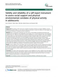
J Cachexia Sarcopenia Muscle Fig. 1 Diagnostic algorithm for cachexia established by the Society of Sarcopenia, Cachexia and Wasting Disorders (SCWD) and the proposed new screening tool for detection of geriatric cachexia in hip fracture patients. In the present study, the newly developed screening tool was assessed against three diagnostic measures from the diagnostic algorithm for cachexia. BMI body mass index, FFM fat-free mass, ASM appendicular skeletal muscle, DEXA dual energy X-ray absorptiometry, SNAQ Simplified Nutritional Appetite Questionnaire, MUAMC midupper arm muscle circumference. a Diagnostic cutoffs from the proposed diagnostic algorithm for cachexia were derived from Evans et al. [18]. b Screening tool diagnostic criteria cut-offs for isometric hand-grip strength and MUAMC were obtained from wave 1 of the Australian Longitudinal Study of Ageing (ALSA) [35]. c A score ≤14 for SNAQ identifies persons with anorexia at risk of significant weight loss of at least 5 % within 6 months [38]
Fat Monitor, Tokyo, Japan) with participants wearing light clothing and without footwear. Participants unable to mobilise were weighed using a calibrated weigh chair. Knee height was measured on the non-injured leg with participants wearing no footwear with the participant in a supine or seated position. Height was estimated from knee height using validated age and gender specific equations [27, 28]. BMI was calculated as weight (kilogram) divided by the square of height (metre). 2.4 Dual-energy X-ray absorptiometry Whole-body and regional body composition was estimated using dual-energy X-ray absorptiometry (DEXA) (Lunar Prodigy, GE Healthcare, UK) with the automated reporting GE EnCORE bone densitometry software (version 10.51.006).
The system software provides estimates of FFM, lean soft tissue, fat mass and bone mineral density for total body and body segments including both arms, both legs and the trunk. ASM was calculated as the sum of lean soft tissue mass in both arms and legs [29] with the ASM index calculated using the formula, ASM/height2 (kg/m2) [30]. Prior to all DEXA scans, all participants underwent a DEXA screening checklist to ensure safety and validity of the technique. Participants were excluded from the DEXA scan if they reported a history of nuclear scans or other X-ray examinations in the previous 0– 14 days or had a recorded body weight ≥130 kg. Prior to the scan, all participants were asked to remove all metal accessories, were asked to identify any medications taken in the previous 24 h (including calcium or iron supplements) and were asked to identify any history of previous fracture and/or metal implants. The software recognises metal in the body,
J Cachexia Sarcopenia Muscle
such as artificial joints, allowing exclusion from calculations prior to analysis. Participants were dressed in hospital gowns and positioned in a supine position on the table top with their feet in a neutral position with hands flat by their sides. All DEXA scans were performed by a licenced technician. 2.5 Dietary intake and analyses Dietary intake was assessed using a single multiple-pass 24h dietary recall. Hospital ready-reckoners were used to estimate energy and protein intake. Individual estimated energy requirements were calculated using gender-specific Harris–Benedict equations [31] to estimate resting metabolic rate, with adjustments made for physical activity (1.2), trauma (1.35) and weight gain (0.25 kg/week) [32]. All dietary analyses were performed by an accredited practising dietitian. 2.6 Isometric hand-grip strength Isometric hand-grip strength was used as a reliable and valid surrogate measure of muscle strength [33, 34] for the diagnostic algorithm and in the development of the screening tool. Grip-strength was measured in the dominant hand with a calibrated Smedley Hand Dynamometer (Tokyo, Japan). All measures were performed on three separate occasions with ~60-s rest intervals between each measure with the mean of the three measures used for analyses. All measures were recorded to the nearest 0.1 kg. 2.7 Development of cachexia screening tool In keeping with the cachexia diagnostic algorithm, BMI ≤22 kg m 2 and isometric hand-grip strength were maintained in the newly developed screening tool. Measures of ASM index derived from DEXA and anorexia assessed by 24-h dietary recall in the diagnostic algorithm were replaced with portable assessments of body composition and anorexia including mid-upper arm muscle circumference (MUAMC) and the Simplified Nutritional Appetite Questionnaire (SNAQ), respectively. Diagnostic cut-off criteria for isometric hand-grip strength (lowest tertile for grip strength; male, ≤27.3 kg; female, ≤16.3 kg) and MUAMC (≤25th percentile; male, ≤23.49 cm; female, ≤20.84 cm) were derived from age and gender specific data obtained from wave 1 of the Australian Longitudinal Study on Ageing [35]. The newly developed screening tool and specific diagnostic cut-offs are shown in Fig. 1. 2.8 Upper-arm anthropometry MUAMC is derived from two upper-arm anthropometric techniques, MUAC and triceps skinfold thickness (TSF).
MUAC was measured to the nearest 0.1 cm using a flexible steel measuring tape (KDS Corporation, Kyoto, Japan), and TSF was measured to the nearest 0.2 mm using a calibrated Harpenden skinfold calliper (Baty, International Sussex, UK). All anthropometric measures were performed by trained dietitians and/or physiotherapists, with each measure performed on three separate occasions with the mean of the three measures used for analyses. Unless affected by injury, all anthropometric measures were taken on the right-hand side of the body. MUAMC was estimated from TSF (mm) thickness and MUAC (cm) using the formula: MUAMC= MUAC(cm) −0.3142×TSF(mm) [36, 37]. 2.9 Appetite Appetite was assessed using the SNAQ; a quick and feasible four-item derivative of the eight-item Council on Nutrition Appetite Questionnaire developed by the Council for Nutritional Strategies in Long-Term Care in institutionalised and community-dwelling older adults [38]. The SNAQ takes approximately ~1 min to complete and was administered either by trained dietitians and/or physiotherapists. Participants were asked to respond to the four-item questionnaire, which included a self-rating of appetite, satiation, gustation and the frequency of meals eaten throughout the day. The total SNAQ score is the sum of scores from the four items, with lower scores indicating deterioration in appetite. Possible scores range from 4 (worst) to 20 (best). A score of ≤14 for the four-item questionnaire identifies persons with anorexia at risk of significant weight loss of at least 5 % within 6 months [38]. 2.10 Statistical analyses Analyses were performed using SPSS for Windows 17.0 software (SPSS Inc, Chicago, IL, USA). Contingency tables were used to determine the specificity, sensitivity and predictive values for the screening tool as a function of its ability to predict geriatric cachexia against the commonly accepted diagnostic algorithm. For continuous data, mean (SD) were reported with frequencies and percentages reported for categorical data. Independent samples t tests were used to assess gender differences for each diagnostic parameter of cachexia. Significance was set at P