ANTIMICROBIAL AGENTS AND CHEMOTHERAPY, Feb. 2011, p. 854–859 0066-4804/11/$12.00 doi:10.1128/AAC.01126-10 Copyright © 2011, American Society for Microbiology. All Rights Reserved.
Vol. 55, No. 2
Development and Validation of a High-Performance Liquid Chromatography Method for Determination of Cefquinome Concentrations in Sheep Plasma and Its Application to Pharmacokinetic Studies䌤 Kamil Uney,1* Feray Altan,2 and Muammer Elmas1 Department of Pharmacology and Toxicology, Faculty of Veterinary Medicine, University of Selcuk, 42031 Konya, Turkey,1 and Department of Pharmacology and Toxicology, Faculty of Veterinary Medicine, University of Dicle, 21280 Diyarbakir, Turkey2 Received 13 August 2010/Returned for modification 2 November 2010/Accepted 14 November 2010
Cefquinome has a broad spectrum of antibacterial activity and was developed especially for use in animals. A simple and sensitive high-performance liquid chromatography (HPLC) method with UV-visible detection for quantification of cefquinome concentrations in sheep plasma was developed and validated. Separation of cefquinome from plasma components was achieved on a Phenomenex Gemini C18 column (250 mm by 4.6 mm; internal diameter [i.d.], 5 m). The mobile phase consisted of acetonitrile and 0.1% trifluoroacetic acid in water and was delivered at a rate of 0.9 ml/min. A simple and rapid sample preparation involved the addition of methanol to 200 l of plasma to precipitate plasma proteins followed by direct injection of 50 l of supernatant into the high-performance liquid chromatography system. The linearity range of the proposed method was 0.02 to 12 g/ml. The intraday and interday coefficients of variation obtained from cefquinome were less than 5%, and biases ranged from ⴚ3.76% to 1.24%. Mean recovery based on low-, medium-, and high-quality control standards ranged between 92.0 and 93.9%. Plasma samples were found to be stable in various storage conditions (freeze-thaw, postpreparative, short-term, and long-term stability). The method described was found to be readily available, practicable, cheap, rapid, sensitive, precise, and accurate. It was successfully applied to the study of the pharmacokinetics of cefquinome in sheep. This method can be very useful and an alternate to performing pharmacokinetic studies in the determination of cefquinome for clinical use.
The parameter best correlated with the treatment efficiency of cefquinome, like other -lactam antibiotics, is the time that the plasma concentration remains above the MIC, which is the pharmacodynamic parameter for efficacy (19). It has been suggested that this time should be equal to 60% to 70% of the interval time between doses for cephalosporins (12). Since the period during which the plasma concentration remains above the MIC is one of the most important parameters for the success of the treatment, monitoring the concentration of cefquinome in plasma is useful to make appropriate drug dosage regimens, to maximize its clinical efficacy, and to reduce the selection of resistant pathogens. In addition, simple analytical conditions and sample preparation methods are useful in routine clinical practice. Widespread use of cefquinome in clinical practice accentuates the need for a rapid and reliable method of its determination in biofluids. However, a number of methods for the determination of cefquinome were reported for milk and animal tissues. These methods include high-performance liquid chromatography with UV detector (HPLC-UV) (17, 18, 21), HPLC with diode array detector (5), liquid chromatography combined with tandem mass spectrometry (1), and screening methods (7, 8, 13, 14, 18). To the authors’ knowledge, only two published papers involving analytical methods for the determination of cefquinome in plasma, one employing liquid chromatography combined with electrospray tandem mass spectrometry (LC-ESI-MS-MS) (11) and one using HPLC-UV (9), reported complete validation procedures. Although LC-ESI-
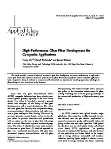
Cefquinome, 1-[[(6R,7R)-7-[[(2Z)-(2-amino-4-thiazolyl)(methoxyimino)acetyl]amino]-2-carboxy-8-oxo-5-thia-1azabicyclo[4.2.0]oct-2-en-3-yl]methyl]-5,6,7,8-tetrahydroquinolinium inner salt (Fig. 1), is an aminothiazolyl cephalosporin that was developed especially for use in animals (4, 6, 19). The chemical modifications of the basic cephalosporin structure provide cefquinome’s zwitterionic property. This property of cefquinome can facilitate rapid penetration across biological membranes, including the porins of the bacterial cell wall, enhance bioavailability, and improve the spectrum of antimicrobial activity compared with the second- and third-generation cephalosporins (6, 15, 16, 19). Cefquinome is stable against chromosomally and plasmid-encoded -lactamases that are produced by a majority of clinically important bacteria (10). It has been approved for treatment of respiratory tract diseases, acute mastitis and foot rot in cattle, calf septicemia, respiratory diseases in pigs, metritis-mastitis-agalactia syndrome in sows, foal septicemia, and respiratory tract diseases in horses (2–4). Potentially it will also be of therapeutic value in many sheep diseases. However, there are no published data on the clinical efficacy and the pharmacokinetics of cefquinome in sheep.
* Corresponding author. Mailing address: Department of Pharmacology and Toxicology, Faculty of Veterinary Medicine, University of Selcuk, 42031 Konya, Turkey. Phone: (90332) 2232733. Fax: (90332) 2410063. E-mail:
[email protected]. 䌤 Published ahead of print on 22 November 2010. 854
VOL. 55, 2011
HPLC ANALYSIS OF CEFQUINOME IN SHEEP PLASMA
FIG. 1. Chemical structure of cefquinome.
MS-MS has a higher detection sensitivity, it is expensive and not readily available. HPLC-UV is a readily available and more economical instrument for measuring the drug concentration in plasma. However, the technique described for HPLC-UV methods developed by Li et al. (9) uses complex procedures for sample preparation, including solid-phase extraction. In this study, a newly developed and validated method that includes a simple and single-step extraction procedure using inexpensive chemicals, a small sample volume, and less organic solvent consumption and that is readily available, practicable, rapid, and sensitive is described for the determination of cefquinome in sheep plasma using HPLC and a UV detector. The applicability of the method for pharmacokinetic study of sheep is also demonstrated. MATERIALS AND METHODS HPLC analysis. (i) Chemicals and reagents. Cefquinome was kindly supplied from Provet (Turkey). All reagents were of analytical grade. Acetonitrile (ACN) and methanol (MeOH) were purchased from Merck (Darmstadt, Germany). Trifluoroacetic acid (TFA) was of spectrophotometric grade (⬎99%; Aldrich, WI). Ultrapure analytical grade type I water for HPLC was produced by a water purification system (aquaMax-Ultra system; Younglin Instrument Co. Ltd., South Korea). (ii) Instrumentation and chromatographic conditions. The HPLC system (Shimadzu, Tokyo, Japan) consists of a pump (LC-10AD with CBM-20A system controller) with a low-pressure-gradient flow control valve (FCV-10AL) and a degasser (DGU-14A) to pump the mobile phase, an autosampler (SIL 10AD), and a column oven (CTO 10A). The detection was performed using an SPD10Avp UV-VIS detector set at 268 nm. The column and autosampler temperatures were kept at 40°C and room temperature, respectively. The reverse-phase chromatography was performed with an analytical Gemini C18 column (250 mm by 4.6 mm; internal diameter [i.d.], 5 m; Phenomenex, Torrance, CA). The optimized method used a binary-gradient mobile phase with water containing 0.1% TFA as mobile phase A and ACN as mobile phase B; the time program of the gradient is listed in Table 1. Mobile phase was filtered, degassed by passage through a 0.45-m nylon filter (Millipore, Bedford, MA) under a vacuum, and sonicated (T840DH; Elma, Germany) for 30 min. The flow rate was 0.9 ml/min, and the injection volume was 50 l. The LC solution software program (Shimadzu, Japan) running on an Asus PC was used for instrument control and data analysis. Plasma samples were stored at ⫺70°C in an Operon ultralow temperature freezer (Operon Co. Ltd., South Korea). (iii) Stock solutions and standards. A standard solution of cefquinome was prepared by the direct weighing of the standard substance with subsequent dissolution in water. The concentration of the standard stock solution was 1 mg/ml, and the solution was stored at ⫺70°C. The primary stock solution of cefquinome was diluted quantitatively with water for the preparation of calibration standards and quality control (QC) samples. Cefquinome calibration standards were prepared fresh daily at concentrations of 0, 0.02, 0.04, 0.10, 0.40, 1, 2, 4, 10, and 12 g/ml by spiking 190 l blank sheep plasma with 10 l of water (for the “zero” standard sample) or cefquinome working solutions. In the same manner, QC samples with cefquinome at low (0.4 g/ml), medium (2.0 g/ml), and high (10 g/ml) concentrations were freshly prepared to evaluate the accu-
855
racy and precision of this HPLC method. Prior to HPLC analysis, these calibration standards and QC samples were processed according to the method that is outlined as follows. (iv) Sample preparation procedure. Aliquots of calibrators, QC samples, and other plasma samples (200 l) were added to 1.5-ml microcentrifuge tubes. Subsequently, to all samples, 400 l of MeOH was added and mixed for 10 s, and the samples were centrifuged at 4,000 ⫻ g for 10 min. After centrifugation, 300 l of clear supernatant was pipetted into a fresh vial, 150 l of water was added and mixed for 10 s, and the mixed clear supernatant (200 l) was pipetted into an autosampler vial. The extracted cefquinome sample (50 l) was injected into the HPLC system. HPLC method validation. (i) Selectivity. Selectivity of the method was evaluated by analyzing blank sheep plasma, plasma spiked with cefquinome, and plasma samples obtained from cefquinome pharmacokinetic studies of the sheep. For all plasma samples, chromatograms were visually examined for chromatographic interference from endogenous compounds. (ii) Linearity of calibration curve. The linearity of the method was evaluated by a calibration curve in the range of 0.02 to 12 g/ml cefquinome. Calibration standards were run before and after the samples; both sets of standard peak areas were used to calculate the linear regression equation as well as the coefficient of determination. Blank samples were included with each set. Six calibration curves constructed on six separate days were analyzed to evaluate the linearity of each calibration curve. The peak area of cefquinome was plotted against the corresponding concentrations. The calibration curve was constructed by weighted (1/y) least-squares linear regression analysis. The calibration curves were described by the following linear equation: y ⫽ ax ⫾ b, where y is the analyte area and x is the concentration (g/ml). The slope, intercept, and correlation coefficient were calculated for each standard curve. Unknown concentrations were computed from the equation of the calibration curve. (iii) Sensitivity. The limits of detection (LOD) and quantification (LOQ) were determined by signal-to-noise ratio evaluations of samples spiked from 0.01 to 0.1 g/ml. The LOD is a parameter that provides the lowest concentration in a sample that can be detected from background noise but not quantitated. The LOD was defined as the lowest concentration with a signal-to-noise ratio of at least 3. LOQ is defined as the lowest concentration of analyte with a signal-tonoise ratio of at least 10 and acceptable accuracy and precision (⬍15% for each criterion). (iv) Precision and accuracy. Cefquinome QC samples at low, medium, and high concentrations were spiked for the determination of precision and accuracy and prepared as described above. Six replicates of each level of QC samples were assayed in one run for the intraday experiment. Six replicates of each level of QC samples were assayed within six different days for the interday experiment. The intra- and interday precision and accuracy of the assay were determined by percent coefficient of variation (CV) and percent bias values, respectively. The coefficient of variation was calculated as follows: CV (%) ⫽ (standard deviation/mean) ⫻ 100. The percent bias values from the theoretical concentration were calculated by the following equation: Bias (%) ⫽ [(calculated concentration ⫺ theoretical concentration)/theoretical concentration] ⫻ 100. Acceptance criteria for accuracy and precision were as follows: the CV was lower than 15%, and the bias was within ⫾15%. Inter- and intraday precision, in terms of the CV, was obtained by subjecting the data to one-way analysis of variance (ANOVA). (v) Recovery. For calculation of absolute recoveries of cefquinome, spiked QC samples were prepared at low (0.4-g/ml), medium (2-g/ml), and high (10-g/ ml) concentrations. Six replicates of each QC sample were extracted by the above-mentioned sample preparation and injected into the HPLC system. The concentration of cefquinome was determined from the linear regression of
TABLE 1. HPLC mobile-phase gradient conditions for analysis of cefquinomea Time (min)
Flow rate (ml/min)
%A
%B
0 7 10 11 15
0.9 0.9 0.9 0.9 0.9
90 50 50 90 90
10 50 50 10 10
a A, mobile phase A (0.1% trifluoroacetic acid in water); B, mobile phase B (acetonitrile).
856
UNEY ET AL.
ANTIMICROB. AGENTS CHEMOTHER.
FIG. 2. Representative chromatograms of blank sheep plasma (A); sheep plasma spiked with cefquinome (2 g/ml) (B); and sheep plasma sample at 20 min after intravenous administration of cefquinome at dose of 2 mg/kg (C). CFQ, cefquinome.
the analytical standard curve. The recovery was calculated by comparing the observed concentration with the spiked concentrations. (vi) Stability. The stability of stock standard solution was investigated under two conditions: after storage at room temperature for 24 h and at ⫺70°C for 90 days. The stability was measured by comparison between the peak area of the stock standard solution and that of freshly prepared solution. For each storage condition, five replicates were analyzed in one analytical batch. Three freeze-thaw cycles and short-term, long-term, and postpreparative stabilities of cefquinome in sheep plasma were investigated using low (0.4-g/ml) and high (10-g/ml) QC samples and evaluated by the calculated bias between observed and theoretical concentrations. The freeze-thaw stability of the cefquinome was determined over three freeze-thaw cycles within 3 days. In each freeze-thaw cycle, the spiked plasma samples were frozen at ⫺70°C for 24 h and
thawed at room temperature. The short-term stability was examined by analyzing triplicates of the frozen low and high QC samples (⫺70°C) kept at room temperature for 24 h before sample preparation. The long-term stability was evaluated by keeping in triplicate the two concentration levels of routine QC samples, with storage at ⫺70°C for 90 days. The postpreparative stability was evaluated by keeping in triplicate the two extracted QC samples in the autosampler under normal analytical conditions for 24 h. The samples were analyzed, and the results were compared with those obtained for the freshly prepared samples. For all stability studies, the acceptance criterion was as follows: ⫾15% bias from the theoretical value. Pharmacokinetic application. Five healthy sheep (female, 28 to 32 kg body weight, 13 months) were used. Animals were not pregnant or lactating. They were housed in individual pens and were fed on barley grains, stalks, and dry
VOL. 55, 2011
HPLC ANALYSIS OF CEFQUINOME IN SHEEP PLASMA
TABLE 2. Calibration curve parameters and statistics for cefquinome in sheep plasma Curve no. or parametera
Slope
Curve no. 1 2 3 4 5 6
25,211 24,857 24,863 25,469 25,383 26,257
Parameters Mean (n ⫽ 6) SD CV (%)
25,340 517 2.04
a
857
TABLE 3. Calibration curve precision and accuracy for cefquinome in sheep plasmaa
Intercept
Correlation coefficient
Theoretical concn (g/ml)
Found concn (g/ml)
CV (%)
Calculated concn (g/ml)
Bias (%)
1,111 417 233 956 500 359
0.9991 0.9999 0.9998 0.9998 0.9999 0.9999
596
0.9997 0.0004 0.0348
0.02 0.04 0.1 0.4 1 2 4 10 12
0.0192 0.0374 0.0928 0.3769 0.9273 1.8651 3.7170 9.2266 10.8817
2.08 2.14 1.62 2.15 2.66 2.87 2.65 1.71 2.53
0.0206 0.0402 0.0998 0.4053 0.9970 2.0054 3.9965 9.9213 11.7006
2.79 0.57 ⫺0.16 1.33 ⫺0.30 0.27 ⫺0.09 ⫺0.79 ⫺2.50
a
n ⫽ 6. SD, standard deviation; CV, coefficient of variation.
SD, standard deviation; CV, coefficient of variation.
grass. Drinking water was available ad libitum. The Ethics Committee of the Faculty of Veterinary Medicine (University of Selcuk, Konya, Turkey; report no. 2010/049) approved the study protocol. In the pharmacokinetic study, the crossover design was performed. The withdrawal interval between the phases of the study was 15 days. Cefquinome (Cobactan injection, 25 mg/ml; Intervet) was given as a bolus intravenous (i.v.) injection (into the vena jugularis) at a dose of 2 mg/kg body weight. After the withdrawal period, cefquinome was administered as an intramuscular (i.m.) injection at the same dose. Intramuscular injections were administered into the hind leg between the semitendinosus and the semimembranosus muscles. Blood samples (2 ml) were collected via a jugular catheter at 5, 10, 15, 20, and 30 min and 1, 1.5, 2, 3, 4, 6, and 8 hours following cefquinome administration. Plasma was separated immediately by centrifugation and stored at ⫺70°C until analysis. The obtained plasma concentration data of cefquinome were analyzed using WinNonlin 4.1 software (Pharsight, CA) to obtain the appropriate pharmacokinetic parameters. Pharmacokinetic parameters measured included absorption half-life (t1/2a), distribution half-life (t1/2␣), elimination half life (t1/2), total body clearance (CLtot), apparent volume of distribution at steady-state (Vss), and area under the curve (AUC). Data obtained following analysis of plasma concentrations were plotted on a log-linear graph to determine the best-fit model for compartmental analysis. Based on this and on Aikaike’s information criterion (AIC) (20), the two-compartmental model was chosen for analysis of the data. The maximum plasma concentration (Cmax) and time to reach Cmax (Tmax) after i.m. administration were determined by direct observation of the plasma concentration-time curve of cefquinome in each animal. Bioavailability (F) was calculated by the method of corresponding areas: F (%) ⫽ (AUCi.m./AUCi.v.) ⫻ 100. All data were expressed as the mean ⫾ standard deviation (SD). Harmonic means were calculated for t1/2a, t1/2␣, and t1/2. The Wilcoxon rank sum test was used to test for significant differences in t1/2␣ and t1/2. The other pharmacokinetic data were analyzed using the paired t test. Statistical significance was assigned at a P value of ⬍0.05. The SPSS (version 10.0) statistical software was used.
RESULTS AND DISCUSSION Chromatographic conditions. The chromatographic conditions and sample preparation for the proposed method were optimized to suit the preclinical pharmacokinetic studies. Before we began the proposed method, chromatograms of cefquinome standards and QC samples analyzed by HPLC analytical columns, including a Gemini C18 and a PLRP-S polymeric column, were compared. The Gemini C18 column was selected for the assay based on its given cefquinome retention time, peak shape/symmetry, and ability to separate cefquinome from endogenous substances (selectivity). The composition of the mobile phase is also a critical factor in separating cefquinome from endogenous substances. Initial tests were performed in the isocratic mode using several combinations of 0.1% TFA
and ACN. Under these conditions, cefquinome was not efficiently separated from endogenous substances. Optimal chromatographic conditions were obtained in the gradient elution mode (0 to 50% ACN from 0 to 15 min), allowing better separation of cefquinome without interference from endogenous substances. Under these conditions, the retention time was about 7.1 min, with a total run time of 15 min. Representative chromatograms of blank plasma, a plasma sample spiked with 2 g/ml of cefquinome, and plasma at 20 min after i.v. administration of cefquinome at a dose of 2 mg/kg are shown in Fig. 2. System suitability parameters for the method were as follows: the theoretical plate value was 13,844, and the tailing factor was less than 1.22. A proper selection of solvent was essential for yielding maximum recoveries. A protein precipitation method was utilized to extract cefquinome from plasma samples. Protein precipitation was selected because it had very clear advantages, such as fewer steps, shorter processing time, and good sample cleanup. Because most -lactam antibiotics are unstable in media of high or low pH, the deproteinization of plasma by the use of strong acids, like perchloric acid and trichloroacetic acid, is excluded. We tested the change of sample preparation by adding organic solvents (MeOH and ACN). The best result was obtained with MeOH, without compromising recoveries. Linearity of calibration curve. The linearity of the method was observed in the concentration range of 0.02 to12 g/ml, demonstrating its suitability. Each experiment at all concentrations was repeated two times on six separate days to obtain the calibration data. Regression results and statistics from calibration standard curves are shown in Table 2. The correlation coefficients (r), indicating the functional linear relationship between the concentration of analyte and the area under the peak, were above 0.999 across the concentration range used. The calibration curve precision and accuracy are presented in Table 3, demonstrating that the accuracy and the precision are ⬍15% for all concentration points. Sensitivity. The LOQ was established by determining the concentrations of four spiked calibration standards from 0.01 to 0.1 g/ml. The LOQ of the method was found to be 0.02 g/ml for cefquinome in sheep plasma, with acceptable accuracy and precision (⬍15% for each criterion). The LOQ for the next concentration assayed, 0.01 g/ml, could not be determined with acceptable accuracy and precision (22.8 and
858
UNEY ET AL.
ANTIMICROB. AGENTS CHEMOTHER.
TABLE 4. Intra- and interday precision and accuracy for cefquinome in sheep plasma Parametera
TABLE 5. Stability of cefquinome in sheep plasma under various storage conditionsa
Value for theoretical concn (g/ml) of: 0.4
2
10
Intraday Overall mean (n ⫽ 6) SD CV (%) Bias (%)
0.407 0.016 4.02 ⫺3.76
1.935 0.066 3.31 ⫺0.74
10.124 0.232 2.29 1.24
Interday Overall mean (n ⫽ 36) SD CV (%) Bias (%)
0.400 0.020 4.90 ⫺0.04
1.999 0.098 4.88 ⫺0.06
9.960 0.410 4.12 ⫺0.41
a
SD, standard deviation; CV, coefficient of variation.
27.2%, respectively). The LOD was determined to be 0.01 g/ml, based on a signal-to-noise ratio of 3:1. Precision and accuracy. The accuracy and precision of the method were evaluated with QC samples at concentrations of 0.4, 2, and 10 g/ml. Intra- and interday precision and accuracy are reported in Table 4. At all levels, intra- and interassay precision levels were lower than 4 and 5%, respectively. The intra- and interassay accuracy levels ranged from ⫺3.76 to 1.24% and from ⫺0.41 to ⫺0.04%, respectively. These results indicated that the proposed method was precise and accurate. Recovery. The absolute recovery was calculated by comparing the peak areas of the prepared QC samples with those of the standard solutions. The extraction recoveries of cefquinome from plasma were 92.0 ⫾ 4.56, 93.9 ⫾ 4.59, and 93.6 ⫾ 3.85% for the low, medium, and high QC samples, respectively. The recovery of cefquinome using the described procedure was consistent and efficient. Stability. Stock solutions of cefquinome, stored at room temperature for 24 h and at ⫺70°C for 90 days, were stable under working conditions. The peak area ratios of stock standard solutions stored at room temperature for 24 h and at ⫺70°C for 90 days to freshly prepared solution (means [CV values]) were 102.9% (2.1%) and 99.8% (3.7%), respectively. No significant changes were present in the peak areas of analytes. Plasma QC samples of cefquinome at two concentrations (0.4 and 10 g/ml) were used for the stability experiments. The results of the stability test that included freeze-thaw, shortterm, long-term, and postpreparative stabilities are summarized in Table 5. Three freeze-thaw cycles and 24-h, room temperature storage of the QC samples did not appear to affect the quantification of cefquinome. The QC samples stored in a freezer at ⫺70°C remained stable for 90 days. The testing of the postpreparative stability of QC samples indicated that cefquinome was stable when kept in the autosampler for up to 24 h. This suggests that sheep plasma samples containing cefquinome can be handled under normal laboratory conditions without any significant loss of compound. Pharmacokinetic application. The method described here was successfully applied to the study of the pharmacokinetics of cefquinome in sheep. General adverse reactions were not observed with any sheep. Following i.v. and i.m. administra-
Storage condition
Theoretical concn (g/ml)
Bias (%)
CV (%)b
Freeze-thaw cycle 1
0.4 10
3.61 2.17
3.53 4.81
Freeze-thaw cycle 2
0.4 10
0.83 ⫺1.32
2.65 2.38
Freeze-thaw cycle 3
0.4 10
⫺5.92 1.95
5.24 4.67
Postpreparative stability (24 h at room temp)
0.4 10
⫺4.64 0.77
6.91 1.98
Short-term stability (24 h at room temp)
0.4 10
6.08 2.46
5.54 7.42
Long-term stability (90 days at ⫺70oC)
0.4 10
⫺6.73 ⫺3.48
4.25 3.86
a b
n ⫽ 3. CV, coefficient of variation.
tions, plasma concentration-time curves of cefquinome best fit a two-compartment open model in all animals. Plasma concentration-time curves are shown in Fig. 3. The pharmacokinetic parameters estimated are shown in Table 6. When both routes of administration were compared, significant differences were found for the t1/2␣, t1/2, and AUC parameters (P ⬍ 0.05) (Table 6). Cefquinome was rapidly absorbed after i.m. administration, with a t1/2a of 0.31 ⫾ 0.05 h. The absolute bioavailability (F) of cefquinome after i.m. administration was 89.31 ⫾ 6.06%. This value indicates good absorption of the drug from the site of injection. In conclusion, an effective, rapid, and sensitive quantification of plasma levels of a therapeutic agent is necessary for effective monitoring of drug levels. The HPLC-UV method described in this paper is readily available, practicable, cheap, rapid, and sensitive and is a precise and accurate method. All results obtained were within the acceptable ranges for bioanalytical purposes. The straightforward sample preparation pro-
FIG. 3. Mean ⫾ SD semilogarithmic plots of the plasma concentrations in relation to time data of cefquinome in sheep (n ⫽ 5) after single intravenous and intramuscular administrations of cefquinome at a dosage of 2 mg/kg of body weight.
VOL. 55, 2011
HPLC ANALYSIS OF CEFQUINOME IN SHEEP PLASMA
TABLE 6. Mean ⫾ SD for pharmacokinetic parameters for cefquinome in sheep (n ⫽ 5) after single intravenous and intramuscular administrations of cefquinome at a dosage of 2 mg/kg of body weight Parameterb
t1/2a (h) (HO) t1/2␣ (h) (HO) t1/2 (h) (HO) AUC (g h ml⫺1) CLtot (liter h kg⫺1) Vss (liter kg⫺1) Cmax (g ml⫺1) Tmax (h) F (%)
7.
Values for: i.v.a
0.06 ⫾ 0.04* 0.78 ⫾ 0.19* 5.83 ⫾ 0.45* 0.34 ⫾ 0.03 0.36 ⫾ 0.06
6.
i.m.
0.31 ⫾ 0.05 0.31 ⫾ 0.05 1.88 ⫾ 0.40 5.19 ⫾ 0.25 2.60 ⫾ 0.14 0.50 89.31 ⫾ 6.06
ⴱ, significantly different at P ⬍ 0.05. t1/2a, absorption half-life; t1/2␣, distribution half-life; t1/2, elimination halflife; AUC, area under the plasma concentration-time curve; CLtot, total body clearance; Vss, apparent volume of distribution at steady-state; Cmax, maximum plasma concentration; Tmax, time to reach peak concentration; F, bioavailability; HO, harmonic mean. a b
8.
9. 10.
11.
12.
13.
14.
cedure, a simple deproteinization step for plasma, prevents analyte degradation and allows the extraction of many samples a day. This method can be very useful and an alternate to performing pharmacokinetic studies in the determination of cefquinome for clinical use. REFERENCES 1. Becker, M., E. Zittlau, and M. Petz. 2004. Residue analysis of 15 penicillins and cephalosporins in bovine muscle, kidney and milk by liquid chromatography-tandem mass spectrometry. Anal. Chim. Acta 520:19–32. 2. CVMP. 1995. Cefquinome. Summary report. EMEA/MRL/005/95. European Agency for the Evaluation of Medicinal Products, London, United Kingdom. 3. CVMP. 1999. Cefquinome (extension to pigs). Summary report (2). EMEA/ MRL/545/99-final. European Agency for the Evaluation of Medicinal Products, London, United Kingdom. 4. CVMP. 2003. Cefquinome (extension to horses). Summary report (3). EMEA/MRL/ 883/03-final. European Agency for the Evaluation of Medicinal Products, London, United Kingdom. 5. Ehinger, A. M., H. Schmidt, and M. Kietzmann. 2006. Tissue distribution of
15.
16.
17.
18.
19.
20.
21.
859
cefquinome after intramammary and ‘⬘systemic’’ administration in the isolated perfused bovine udder. Vet. J. 172:147–153. Gue´rin-Fauble´e, V., G. Carret, and P. Houffschmitt. 2003. In vitro activity of 10 antimicrobial agents against bacteria isolated from cows with clinical mastitis. Vet. Rec. 152:466–471. Lamar, J., and M. Petz. 2007. Development of a receptor-based microplate assay for the detection of beta-lactam antibiotics in different food matrices. Anal. Chim. Acta 586:296–303. Le Breton, M. H., M. C. Savoy-Perroud, and J. M. Diserens. 2007. Validation and comparison of the Copan milk test and Delvotest SP-NT for the detection of antimicrobials in milk. Anal. Chim. Acta 586:280–283. Li, X. B., et al. 2008. Pharmacokinetics and bioavailability of cefquinome in healthy piglets. J. Vet. Pharmacol. Ther. 31:523–527. Limbert, M., et al. 1991. Antibacterial activities in vitro and in vivo and pharmacokinetics of cefquinome (HR IIIV), a new broad-spectrum cephalosporin. Antimicrob. Agents Chemother. 35:14–19. Maes, A., et al. 2007. Determination of cefquinome in pig plasma and bronchoalveolar lavage fluid by high-performance liquid chromatography combined with electrospray ionization mass spectrometry. J. Mass Spectrom. 42:657–663. Pea, F., and P. Viale. 2009. Bench-to-bedside review: appropriate antibiotic therapy in severe sepsis and septic shock—does the dose matter? Crit. Care 13:214. doi:10.1186/cc7774. Pikkemaat, M. G., et al. 2009. Nouws antibiotic test: validation of a postscreening method for antibiotic residues in kidney. Food Control 20:771– 777. Pikkemaat, M. G., S. O. V. Dijk, J. Schouten, M. Rapallini, and H. J. van Egmond. 2008. A new microbial screening method for the detection of antimicrobial residues in slaughter animals: the Nouws antibiotic test (NATscreening). Food Control 19:781–789. Sader, H. S., and R. N. Jones. 1993. The fourth-generation cephalosporins: antimicrobial activity and spectrum definitions using cefpirome as an example. Antimicrob. Newsl. 9:9–16. Shpigel, N. Y., et al. 1997. Efficacy of cefquinome for treatment of cows with mastitis experimentally induced using Escherichia coli. J. Dairy Sci. 80:318– 323. Sørensen, L. K., and L. K. Snor. 2000. Determination of cephalosporins in raw bovine milk by high-performance liquid chromatography. J. Chromatogr. A 882:145–151. Suhren, G., and K. Knappstein. 2003. Detection of cefquinome in milk by liquid chromatography and screening methods. Anal. Chim. Acta 483:363– 372. Thomas, E., V. Thomas, and C. Wilhelm. 2006. Antibacterial activity of cefquinome against equine bacterial pathogens. Vet. Microbiol. 115:140– 147. Yamaoka, K., T. Nakagawa, and T. Uno. 1978. Application of Akaike’s information criterion (AIC) in the evaluation of linear pharmacokinetic equations. J. Pharmacokinet. Biopharm. 6:165–175. Zhang, X., J. Li, H. Jiang, and J. Shen. 2007. Residue depletion of cefquinome in swine tissues after intramuscular administration. J. Agric. Food Chem. 55:10493–10498.