Received: 2 March 2017
Revised: 26 June 2017
Accepted: 30 June 2017
DOI: 10.1002/bmc.4046
RESEARCH ARTICLE
Development and validation of a high‐performance liquid chromatography method for the quantification of talazoparib in rat plasma: Application to plasma protein binding studies Mahendra Kumar Hidau1
|
Srikanth Kolluru2
|
Srinath Palakurthi1
1
Department of Pharmaceutical Sciences, Irma Lerma Rangel College of Pharmacy, Texas A&M Health Science Center, Kingsville, Texas, USA
2
Department of Pharmaceutical Sciences, KGI School of Pharmacy, Claremont, California, Texas, USA Correspondence Srinath Palakurthi, Department of Pharmaceutical Sciences, Irma Lerma Rangel College of Pharmacy, Texas A&M Health Science Center, Kingsville, TX 78363, USA. Email:
[email protected]
Abstract A sensitive and selective RP‐HPLC method has been developed and validated for the quantification of a highly potent poly ADP ribose polymerase inhibitor talazoparib (TZP) in rat plasma. Chromatographic separation was performed with isocratic elution method. Absorbance for TZP was measured with a UV detector (SPD‐20A UV–vis) at a λmax of 227 nm. Protein precipitation was used to extract the drug from plasma samples using methanol–acetonitrile (65:35) as the precipitating solvent. The method proved to be sensitive and reproducible over a 100–2000 ng/mL linearity range with a lower limit of quantification (LLQC) of 100 ng/mL. TZP recovery was found to be >85%. Following analytical method development and validation, it was successfully employed to determine the plasma protein binding of TZP. TZP has a high level of protein binding in rat plasma (95.76 ± 0.38%) as determined by dialysis method. KEY W ORDS
chromatography, PARP inhibitor, protein binding
1
|
I N T RO D U CT I O N
inhibitor, was shown to be effective against a variety of cancer cells with the EC50 in nanomolar concentrations (Wang et al., 2016). A clin-
Poly(ADP‐ribose)polymerase (PARP) enzymes are critical for recogni-
ical study with TZP monotherapy showed efficacy in a cohort of 39
tion and repair of DNA breaks (Murai et al., 2012). PARP inhibitors
patients with an objective response rate of 65% in ovarian and perito-
are particularly effective in homologous recombination (HR)‐deficient
neal tumors, and 33% in breast cancer patients. It was well tolerated
cancers, but are also expected to have wide applications beyond BRCA
(Engert, Kovac, Baumhoer, Nathrath, & Fulda, 2016; Livraghi & Garber,
mutations, such as in sporadic tumors with ‘BRCAness’ (Lord &
2015; Pulliam, Taverna, Lyons, & Nephew, 2015; Roche et al., 2015;
Ashworth, 2008). More than 200 clinical trials on PARP inhibitors are
Smith et al., 2015). Like olaparib, TZP traps PARPs to DNA (Murai
either completed or currently active for various cancers, such as ovar-
et al., 2012). The minimum toxic dose of TZP was 1 mg/day, with
ian and breast cancers, glioblastoma and others (https://clinicaltrials.
common adverse events of neutropenia, thrombocytopenia and ane-
gov). At present, many PARP inhibitors like olaparib, talazoparib
mia. Currently three Phase III clinical trials on TZP are in progress
(TZP), veliparib, niraparib and rucaparib are in clinical trials for various
(Murai et al., 2012; Smith et al., 2015).
cancerous conditions (Bryant et al., 2005; Lord & Ashworth, 2008;
Quantitation of a new compound in various analytical or
O0 Connor, 2015; Rouleau, Patel, Hendzel, Kaufmann, & Poirier,
bioanalytical matrices is a crucial step for drug discovery and develop-
2010). The US Food and Drug Administration (FDA) has approved
ment in a timely and cost‐effective way (Shah, 2007; Shah et al., 2000).
the PARP inhibitor olaparib for the treatment of ovarian cancer in
Chromatography is always a preferred option for quantitative analysis
2014 and also classified olaparib as having advanced remedy status
of the drug in a robust way. Our research laboratory has been develop-
for BRCA or ATM mutated castration‐resistant prostate cancer
ing novel nanoparticle formulation for targeted delivery of TZP to
(Helleday, 2016; Liu & Matulonis, 2016). TZP, the most potent PARP
triple negative breast cancer. To the best of our knowledge, no
Abbreviations: ACN, acetonitrile; DFBA, difluprednate; FDA, US Food and Drug Administration; PARP, poly(ADP‐ribose)polymerase; PBS, phosphate‐buffered saline; TZP, talazoparib.
Biomedical Chromatography. 2017;e4046. https://doi.org/10.1002/bmc.4046
bioanalytical method has been published yet for TZP. We anticipate that development of a sensitive analytical method would accelerate the development of TZP and its formulations. The present study aimed
wileyonlinelibrary.com/journal/bmc
Copyright © 2017 John Wiley & Sons, Ltd.
1 of 5
2 of 5
HIDAU
to develop and validate a sensitive and selective RP‐HPLC method and apply this method to determine the plasma protein binding of TZP per FDA guidelines for industry on bioanalytical method validation.
2.4
|
ET AL.
HPLC setup and bioanalysis
RP‐HPLC pump arrangement (LC‐20A, Shimadzu) accompanied by a degassing unit (DGU‐20A, Shimadzu, Kyoto, Japan) and an autosampler (SIL‐20A, with a 100 μL loop) was applied to inject the
2
EXPERIMENTAL
|
bioanalytical samples onto a Luna C18 column (5 μm, 250 × 3 mm internal diameter, Phenomenex, USA) connected to a C18 pre‐column
2.1
|
Material and methods
for the protection of the main C18 column. An isocratic elution method was used at a flow rate of 1 mL/min with 60:40 (v/v) ratios of metha-
TZP and difluprednate (DFBA) were purchased from Selleckchem
nol–ACN (65:35) to deionized water. Absorbance for TZP was
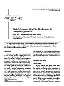
(Houston, TX, USA; Figure 1). Regenerated cellulose dialysis
measured with a UV detector (SPD‐20A UV–vis) at a λmax of 227 nm.
membrane with molecular weight cut‐off of 50 kDa was purchased
Total run time was 10 min and retention times for TZP and DFBA were
from Spectrum Laboratories (Rancho Dominguez, CA, USA). Metha-
7.65 and 6.5 min, respectively (Figure 2). The calibration curve was
nol and acetonitrile (HPLC grade), ammonium acetate and glacial
found to be linear over the concentration range of 100–2000 ng/mL
acetic acid were purchased from VWR International, Sugar Land,
(y = 55.021× − 1317.9, r2 = 0.9995).
TX, USA. Phosphate‐buffered saline (PBS, pH 7.4) was purchased from Mediatech Inc. (Manassas, VA, USA). Buffer was filtered through a 0.45 μm cellulose membrane (Whatman International Ltd, Mailstone, England).
2.5
|
Method validation
The RP‐HPLC method validation was performed with regard to recovery from biosamples, accuracy, precision, intra and inter‐day var-
2.2 | Stock solutions, calibration and quality control standards preparation
iability, sensitivity, specificity and reproducibility (Singh et al., 2015). The RP‐HPLC method was validated as per FDA guidelines for industry on bioanalytical method validation (Matuszewski, Constanzer, &
TZP and DFBA (used as internal standard, IS) were weighed accurately
Chavez‐Eng, 2003; US Department of Health and Human Services,
and stock solutions (1 mg/mL) of both drugs were made: 1 mg of TZP
2001). Calibration standard linearity was determined between 100
and DFBA dissolved in 1 mL of methanol and acetonitrile (ACN),
and 2000 ng/mL concentration for 3 days. Inter‐ and intra‐day
respectively. Mother stock solutions were used to make working
precisions were estimated using analysis of variance (one way) in terms
stocks of TZP and DFBA (100 μg/mL). A parallel dilution method was
of relative standard deviation (RSD) (Singh et al., 2015). The recoveries
used to make analytical standards of TZP over the concentration range
of TZP from the extracted samples were estimated by correlating the
of 15.6, 31.25, 62.5, 125, 250, 500, 1000 and 2000 ng/mL in methanol
known concentrations of plasma samples. This whole process was
for the estimation of TZP recovery in the rat plasma. Three replicates
recapitulated for concentration levels of 100, 800 and 1600 ng/mL,
each of quality control (QC) samples (100, 800 and 1600 ng/mL as
which represent the low, medium and high quality control (LQC,
low, medium and high quality control, respectively) were made sepa-
MQC and HQC) standards respectively (Singh et al., 2015).
rately. The calibration curve for TZP was made by adding 10 μL of working solution to 90 μL blank rat plasma over a concentration range of 100–2000 ng/mL.
2.6
|
Stability studies
The chemical stability of any compound in stock solution and biometrix
2.3
|
Drug extraction
is a critical parameter for its further development. Various stability studies like freeze–thaw stability, bench‐top stability, dry residue
A simple protein precipitation method was used for the extraction of
stability and long‐term stability were conducted to determine the
TZP from the plasma sample, using methanol–ACN (65:35 ratios). To
stability of TZP in stock solution and in rat plasma. For determination
extract the control, QC plasma standard (90 μL rat plasma added with
of TZP stability, spiked controls in rat plasma were made at 100, 800
TZP and IS) and matrix blank samples (100 μL rat plasma without TZP
and 1600 ng/mL concentrations, representing LQC, MQC, HQC
and IS), 200 μL methanol–ACN (65:35) was added as a precipitating
respectively, each with three sets of replicates (Singh et al., 2015).
solvent. The resulting mixture was vortexed for about 2 min and centrifuged at 8500 rpm for 10 min. A 100 μL aliquot of supernatant used for HPLC analysis.
2.7
|
Protein binding determination
Plasma protein binding of TZP was determined by equilibrium dialysis bag method using spectrum dialysis membranes of cellulose ester nature and molecular weight cut‐off (50 kDa) with PBS buffer (pH 7.4) as the dissolution medium. The study was carried out using 1 mg/mL of TZP dissolved in Sprague–Dawley rat plasma placed in the dialysis bag (Barre, Chamouard, Houin, & Tillement, 1985; Giacomini, Abang, & Blaschke, 1982). The dialysis bag was dipped in a glass bottle containing 75 mL of the dissolution medium. The glass FIGURE 1
Chemical structure of talazoparib (a) and IS difluprednate (b)
bottle was placed in an incubating orbital shaker (VWR, Houston TX,
HIDAU
3 of 5
ET AL.
FIGURE 2
HPLC chromatograms: Blank extracted plasma (a); 50 ng/mL (b); calibration standard talazoparib at 125 ng/mL (c); and spiked IS and talazoparib at 1000 ng/mL (d)
USA) maintained at 37 ± 0.5°C and 150 rpm. Samples of volume 500
values determined by uniform weighting and weighting factor 1/x
μLwere withdrawn at 0.25, 0.5, 0.75, 1, 2, 3, 4, 5, 18 and 24 h and
(Kumar Hidau, Singh, Shahi, Mounika, & Kumar Singh, 2015; Singh
analyzed using HPLC.
et al., 2015). The r2 values were always >0.999. Intra‐ and inter‐batch variation for TZP accuracy and precision were evaluated from quality
3
control samples at 100, 800 and 1600 ng/mL concentrations for 5 days
RESULTS A ND DIS CUS SION
|
(Singh et al., 2015). The method accuracy and precision were determined by percentage nominal values and percentage co‐efficient of variance
3.1
|
Extraction and recovery of the sample
(CV), respectively. Accuracy and precision values for 5 days are shown in
Initially, various drug extraction procedures including protein precipita-
Table 2. The back‐calculated concentration for the lower limit of quanti-
tion with different organic solvents, liquid–liquid extraction and solid‐
fication must be within ±20% of nominal concentrations and all other
phase extraction methods were tested and used to identify an optimal
quality standard (LQC, MQC and HQC) concentration values must be
method (Singh et al., 2015). Finally, a single‐step protein precipitation
within ±15% of their nominal concentrations, according to the present
method was considered, using methanol and ACN as precipitating
FDA guidelines (Singh et al., 2015; US Food and Drug Administration,
solvent at 65:35 ratio (Singh et al., 2015). TZP recoveries from the
2013). The data values were within the acceptance limits.
quality control samples were consistent and >85%. Values are shown in Table 1. Recovery of TZP was found higher in 2 times dilution with
3.3
|
Specificity of the method
precipitating solvent in comparison with 3 times dilution. Method specificity was determined by comparison between the
3.2
|
chromatograms obtained for the plasma spiked with TZP at LOQ and
Precision and accuracy
those acquired from plasma samples without drug. Specificity of this
A concentration range of 100–2000 ng/mL was set for the calibration
HPLC method was determined by preparing and analyzing individual
range to quantitate TZP. A 1/x2 weighting factor was used over this
as well as pooled rat plasma samples without the drug. Chromatogram
range of calibration curve and variance was comparable with different concentration values related to the r2 (least‐squares linear regression) TABLE 1
Recovery of spiked plasma samples at low, medium and high concentrations respectively
TABLE 2
Intra‐ and inter‐assay accuracy and precision for the talazoparib (TZP) QC samples in the rat plasma Precision (CV, %) (n = 5)
QC standard
Concentration (ng/mL)
Day1
Day2
Day3
Mean ± SD
Theoretical nominal concentration QC standard (ng/mL)
LQC
100
98.56
85.35
91.02
91.65 ± 6.6
LQC
100
−12.13
−11.18
6.7
−9.1
MQC
800
98.63
90.50
105.73
98.29 ± 7.6
MQC
800
12.39
12.24
9.4
6.8
HQC
1600
91.80
88.44
97.94
92.73 ± 4.8
HQC
1600
13.85
14.44
8.0
5.9
Accuracy (%) (n = 5)
Intra‐day Inter‐day Intra‐day Inter‐day
4 of 5
HIDAU
ET AL.
interference was also detected for all blank plasma samples visually.
plasma proteins is a dynamic process with excess of PBS buffer around
No interference was found in the area of TZP and DFBA retention
the dialysis bag acting as a sink for free drug removal. The percentage
times after analyzing plasma samples (Figure 2). These data reveal that
binding was estimated from the percentage of drug remaining in the
the method was specific for the quantification of TZP in different QC
plasma after 24 h and the free amount of drug was evaluated from
samples.
the dissolution medium. Protein binding for TZP was estimated to be 95.76 ± 0.38% (Figure 3).
3.4
|
Stability studies
Different stability studies were performed for the TZP. In a
4
|
CO NC LUSIO N
freeze–thaw stability study, three freeze–thaw cycles between −80°C and melting ice temperatures were performed for QC samples.
A sensitive, selective and precise RP‐HPLC bioanalytical method has
For bench‐top stability studies, QC samples were placed at room
been successfully developed and validated for quantification of TZP
temperature for up to 8 h. In dry residue stability studies, QC standards
in rat plasma. The lower limit of quantification was 100 ng/mL and
were extracted by a single‐step protein precipitation method and the
linearity covered a concentration range of 100–2000 ng/mL. A time
dry residues were stored at −80°C for 3 days. The dry residues were
and cost‐effective protein precipitation method for sample extraction
reconstituted with methanol–ACN (65: 35) and analyzed by HPLC.
was developed with absolute recovery of >85%. Stability studies
Long‐term stability was determined by storing the QC samples for
revealed that TZP has good stability throughout the storage period
30 days at −80°C (Singh et al., 2015). Various chemical stability data
and sample processing under various conditions. The analytical
are presented in Table 3. All stability data points were found within
method was successfully applied for the plasma protein binding study.
the acceptance limit.
The results indicate that TPZ is highly protein‐bound compound. The validated HPLC method may be applied to various in‐vitro in‐vivo, pre-
3.5
|
clinical, clinical, regulatory and exploratory studies like toxicokinetic,
Protein binding determination
pharmacokinetics and drug–drug interaction studies (Gautam, Singh,
The validated method was successfully applied to estimate plasma
Pratap, & Singh, 2010; Kumar Hidau et al., 2015; Singh et al., 2015).
protein binding of TZP in Sprague–Dawley rats at 1 mg/mL concentration (n = 3). Association and dissociation of drug compounds with
CONFLIC T OF IN TE RE ST
TABLE 3
There is no conflict of interest to disclose.
Stability of TZP in rat plasma after freeze–thaw cycles, bench‐top stability, dry residue stability and long‐term stability
ORCID
Nominal concentration (ng/mL)
Concentration recovered (ng/mL)
Recovery (%)
After 3 freeze–thaw stability
100 800 1600
86.11 ± 3.25 732.74 ± 48.3 1900 ± 109.2
85.57 91.59 114.34
Bench‐top stability for 8 h at ambient temperature
100 800 1600
86.24 ± 9.12 701.92 ± 22.16 1810.49 ± 44.05
85.69 87.74 113.15
Dry residue stability for 3 days
100 800 1600
89.04 ± 7.44 721.24 ± 37.67 1820.14 ± 48.97
89.04 90.15 113.75
Long‐term stability for 30 days
100 800 1600
88.57 ± 8.12 723.29 ± 20.85 1745.15 ± 61.53
88.57 90.40 109.19
Storage conditions
Mahendra Kumar Hidau Srinath Palakurthi
http://orcid.org/0000-0003-1455-5392
http://orcid.org/0000-0002-9509-9511
RE FE RE NC ES Barre, J., Chamouard, J., Houin, G., & Tillement, J. (1985). Equilibrium dialysis, ultrafiltration, and ultracentrifugation compared for determining the plasma‐protein‐binding characteristics of valproic acid. Clinical Chemistry, 31, 60–64. Bryant, H. E., Schultz, N., Thomas, H. D., Parker, K. M., Flower, D., Lopez, E., … Helleday, T. (2005). Specific killing of BRCA2‐deficient tumours with inhibitors of poly (ADP‐ribose) polymerase. Nature, 434, 913–917. Engert, F., Kovac, M., Baumhoer, D., Nathrath, M., & Fulda, S. (2016). Osteosarcoma cells with genetic signatures of BRCAness are susceptible to the PARP inhibitor talazoparib alone or in combination with chemotherapeutics. Oncotarget, 5. Gautam, N., Singh, R., Pratap, R., & Singh, S. (2010). Liquid chromatographic–tandem mass spectrometry assay for quantitation of a novel antidiabetic S002‐853 in rat plasma and its application to pharmacokinetic study. Biomedical Chromatography, 24, 692–698. Giacomini, K., Abang, A., & Blaschke, T. (1982). Calculation of drug concentration in plasma after equilibrium dialysis. British Journal of Clinical Pharmacology, 14, 752–754. Helleday, T. (2016). PARP inhibitor receives FDA breakthrough therapy designation in castration resistant prostate cancer: Beyond germline BRCA mutations. Annals of Oncology, 27, 755–757.
FIGURE 3
after 24 h
Release profile of percent drug remaining in the plasma
Kumar Hidau, M., Singh, Y., Shahi, S., Mounika, P., & Kumar Singh, S. (2015). LC‐MS/MS assay for quantification of a novel antitubercular molecule S006‐830: Pharmacokinetic and plasma protein binding studies in rats. Current Pharmaceutical Analysis, 11, 35–42.
HIDAU
5 of 5
ET AL.
Liu, J. F., & Matulonis, U. A. (2016). What is the place of parp inhibitors in ovarian cancer treatment? Curr Oncol Rep, 18, 29. Livraghi, L., & Garber, J. E. (2015). PARP inhibitors in the management of breast cancer: Current data and future prospects. BMC Medicine, 13, 188. Lord, C. J., & Ashworth, A. (2008). Targeted therapy for cancer using PARP inhibitors. Current Opinion in Pharmacology, 8, 363–369. Matuszewski, B., Constanzer, M., & Chavez‐Eng, C. (2003). Strategies for the assessment of matrix effect in quantitative bioanalytical methods based on HPLC‐MS/MS. Analytical Chemistry, 75, 3019–3030. Murai, J., Huang, S. Y., Das, B. B., Renaud, A., Zhang, Y., Doroshow, J. H., … Pommier, Y. (2012). Trapping of PARP1 and PARP2 by clinical PARP inhibitors. Cancer Research, 72, 5588–5599. 0
O Connor, M. J. (2015). Targeting the DNA damage response in cancer. Molecular Cell, 60, 547–560. Pulliam, N., Taverna, P., Lyons, J., & Nephew, K. P. (2015). Novel combination therapy of DNMT inhibitor SGI‐110 and PARP inhibitor BMN‐673 (talazoparib) for BRCA‐proficient ovarian cancer. Cancer Research, 75, 2943–2943. Roche, H., Blum, J., Eiermann, W., Im, Y.‐H., Martin, M., Mina, L., … Lokker, N. (2015). P1: 01 a phase 3 study of the oral PARP inhibitor talazoparib (BMN 673) in brca mutation subjects with advanced breast cancer (Embraca). Annals of Oncology, 26(Supplement 2), ii16. Rouleau, M., Patel, A., Hendzel, M. J., Kaufmann, S. H., & Poirier, G. G. (2010). PARP inhibition: PARP1 and beyond. Nature Reviews Cancer, 10, 293–301.
novel triethylamine containing thiophene S006‐830 – An antitubercular molecule and its application to pharmacokinetic and bioavailability studies in SD rats. Drug Testing and Analysis, 7, 721–726. Smith, M. A., Reynolds, C. P., Kang, M. H., Kolb, E. A., Gorlick, R., Carol, H., … Billups, C. A. (2015). Synergistic activity of PARP inhibition by talazoparib (BMN 673) with temozolomide in pediatric cancer models in the pediatric preclinical testing program. Clinical Cancer Research, 21, 819–832. US Department of Health and Human Services (2001). Guidance for Industry, Bioanalytical Method Validation. Retrieved from: http://www.fda. gov/cvm. US Food and Drug Administration (2013). Guidance for Industry – Bioanalytical Method Validation. Silver Spring, MD: Center for Drug Evaluation and Research, Department of Health and Human Services, US Food and Drug Administration. Retrieved from: http://www. fda. gov/downloads/Drugs/GuidanceComplianceRegulatoryInformation/ Guidances/UCM070107. pdf. Wang, B., Chu, D., Feng, Y., Shen, Y., Aoyagi‐Scharber, M., & Post, L. E. (2016). Discovery and characterization of (8S,9R)‐5‐fluoro‐8‐(4‐ fluorophenyl)‐9‐(1‐methyl‐1H‐1,2,4‐triazol‐5‐yl)‐2,7,8,9‐te trahydro‐ 3H‐pyrido[4,3,2‐de]phthalazin‐3‐one (BMN 673, talazoparib), a novel, highly potent, and orally efficacious poly(ADP‐ribose) polymerase‐1/2 inhibitor, as an anticancer agent. Journal of Medicinal Chemistry, 59, 335–357.
Shah, V. P. (2007). The history of bioanalytical method validation and regulation: Evolution of a guidance document on bioanalytical methods validation. The AAPS Journal, 9, E43–E47.
How to cite this article: Hidau MK, Kolluru S, Palakurthi S.
Shah, V. P., Midha, K. K., Findlay, J. W., Hill, H. M., Hulse, J. D., McGilveray, I. J., … Powell, M. L. (2000). Bioanalytical method validation – A revisit with a decade of progress. Pharmaceutical Research, 17, 1551–1557.
in rat plasma: Application to plasma protein binding studies.
Singh, Y., Hidau, M. K., Misra, A., Kushwaha, H. N., Tiwari, A., Sharma, A. K., & Singh, S. K. (2015). UFLC method development and validation of a
Development and validation of a high‐performance liquid chromatography method for the quantification of talazoparib Biomedical
Chromatography.
10.1002/bmc.4046
2017;e4046.
https://doi.org/