Introduction: Chest radiography is one of the most frequently performed diagnostic radiographic examinations in the United. Kingdom1. Despite its dominance ...
Development and validation of a psychometric scale for assessing PA chest image quality: a pilot study Mraity H1,2, England A1, Akhtar I1, Aslam A3, De Lange R4, Momoniat H1, Nicoulaz S5, Ribeiro A6, Mazhir S2, Hogg P1. University of Salford1, United Kingdom; University of Kufa2, Iraq; Oslo and Akershus University College of Applied Sciences3, Norway; Hanzehogeschool Groningen4, The Netherlands; Haute École de Santé Vaud5, Switzerland; Escola Superior de tecnologias da Saúde de Lisboa6, Portugal
Introduction:
A series of seven PA chest X-ray images were acquired using an anthropomorphic phantom with a range of image qualities. Image quality perception was initially confirmed for the seven images using signal-to-noise ratio (SNR) and group consensus. Participants were invited to independently score each of the images using the initial image quality perception scale. Cronbach’s alpha was used to test interval reliability.
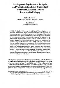
Chest radiography is one of the most frequently performed diagnostic radiographic examinations in the United Kingdom1. Despite its dominance the interpretation of a chest radiograph can be difficult2. There is the possibility of improving diagnostic yield with improvements in image quality. However for this to be possible there needs to be robust mechanisms for image quality assessment. Results: A robust observer performance Fifty three participants (student and qualified measurement is needed for measuring the radiographers and trainee radiologists) used actual clinical quality of an image. Image the scale to grade image quality perception quality perception assessment involves on each of the seven images (SNR range, 17.2 observers considering how much image to 36.5). Aggregated mean image quality detail they can perceive. These perceptual scale score increased with increasing SNR measurements can be undertaken by from 42.1 to 87.7 (r=0.98, P0.7 was obtained across each of the seven often been based on the Commission of images (Table 1). European Communities (CEC) 1996 image criteria4 or a modified version. However, Figure 1. Mean image quality scores for individual items there are problems when using the CEC criteria, as they are outdated and researchers tend to adjust the image criteria according to their own needs. The approach used to measure perception of image quality often involves observers rating criteria using a Likert scale. This often considers two issues: physical information within an image (stimulus or signal) and perceptual effect (psychological) that is related to human analysis of the perceived image5,6. For the latter, the approach we have taken for scale creation and validation utilises Bandura’s theory for self-efficacy7. Currently, there is no published and Table 1. Demonstrates internal reliability coefficients for scale validated perceptual scale for assessing items across all images. image quality of a posterior-anterior (PA) X- Image set No. Signal-to-Noise Ratio (SNR) Alpha Coefficients 17.2 1 0.792 ray image.
Aim: With specific reference to exposure factors the aim of this study was to create a psychometric scale to assess image quality perception of PA chest Xray images.
Method: Bandura’s theory7 was used to guide scale development. A review of the literature was undertaken to identify items/factors which could be used to evaluate image quality using a perceptual approach. A draft scale was then created (22 items) and presented to a focus group (student and qualified radiographers). Within the focus group the draft scale was discussed and modified accordingly.
2
20.9
0.833
3
24.7
0.874
4
27.6
0.896
5
30.6
0.837
6
21.3
0.854
7
36.5
0.869
Discussion: The aim of this project was to use self-efficacy to develop and validate an image quality perception scale for PA chest radiography. For our 22 item scale, we found a strong relationship between SNR and the scale score. This suggests that the perceptual measure of image quality is relatively valid. In all cases Cronhbach’s alpha coefficients were greater than 0.7 for all images. This suggests the scale has good reliability. The scale items are consistent in measuring perception of image quality.
Due to a lack of publications which discuss the creation and validation of a scale for assessing image quality perception of PA chest X-ray images it is impossible to compare our work with similar studies. However, because our scale has validation data associated with it we believe it represents a more robust way of assessing visual image quality for chest X-ray images than anything currently published. To the author’s knowledge this is the first CXR scale that has validation data associated with it. Other reports have only stated criteria for assessing image quality without any actually testing whether they work (e.g CEC criteria4). Images were generated using a phantom; therefore, it was not possible to assess the effects of pathology. Other factors include problems with malpositioning, the use of high kVp, AEC and an anti-scatter grid. Further work should consider taking these factors into account.
Limitations: The sample size of the participants could be considered a limitation. Psychological studies have used in excess of 150 participants for scale development8,9. Adequate sample size would ensure reducing the random error that might be associated with human based studies. However, for our work 53 was considered acceptable as a pilot to primarily testing the scale and obtaining a considerable feedback regarding the suitability of items for measuring what they are designed to measure and their reliability10.
Conclusion: This study represents the first development of a chest image quality perception scale based on Bandura’s theory. There was excellent correlation between the image quality perception scores derived using the scale and the SNR and group consensus. Further research will involve a more detailed item and factor analysis.
Acknowledgments: The authors wish to thank Erasmus Programme for funding this project. References 1. D. Hart, B. F. Wall, M. C. Hillier, and P. C. Shrimpton, Frequency and Collective Dose for Medical and Dental X-ray Examinations in the UK , 2008, no. December 2010. 2008. 2. Van Ginneken, B., Ter Haar Romeny, B. M., Viergever, M. A. Computer-aided diagnosis in chest radiography: a survey. Medical Imaging, 2001;20(12):1228-1241. 3. Thompson J, Hogg P, Thompson S, Manning D, Szczepura K. ROCView: prototype software for data collection in jackknife alternative free-response receiver operating characteristic analysis. The British journal of radiology. 2012 Sep;85(1017):1320-6. 4. Commission of European Communities. European guidelines on quality criteria for diagnostic radiographic images. EUR 16260EN. Brussels, Belgium, The European Commission, 1996. 5. Mansson LG. Methods for the evaluation of image quality . Radiation protection dosimetry. 2000;90(1–2):89–99.
6.
Sharp FP. Quantifying image quality. Clinical Physics and Physiological Measurement. 1990;11:21-6. 7. Bandura A. Social foundations of thought and action: Asocial cognitive theory. New Jersey: Prentice Hall. ; 1986. 8. Tingberg, A. M. Quantifying the quality of medical x-ray images: An evaluation based on normal anatomy for lumbar spine and chest radiography. Thesis(PhD). Lunds Universitet (Sweden), 2000. 9. Kitching J, Cassidy S, Eachus P, and Hogg P, “Creating and validating selfefficacy scales for students.,” Radiologic technology, vol. 83, no. 1, pp. 10–9, 2011. 10. Fliess-Douer O, van der Woude LH, Vanlandewijck YC. Development of a new scale for perceived self-efficacy in manual wheeled mobility: a pilot study. Journal of rehabilitation medicine : official journal of the UEMS European Board of Physical and Rehabilitation Medicine. 2011 Jun;43(7):602-8.