Clinical Chemistry 46, No. 11, 2000
References 1. Domachowske JB, Malech HL. Phagocytes. In: Rich RR, Fleisher TA, Shearer WT, Schwartz BD, Strober W, eds. Clinical immunology principles and practice, 1st ed. St. Louis, MO: Mosby, 1996:392– 407. 2. Abramson SL. Phagocyte deficiencies. In: Rich RR, Fleisher TA, Shearer WT, Schwartz BD, Strober W, eds. Clinical immunology principles and practice, 1st ed. St. Louis, MO: Mosby, 1996:677–93. 3. Carulli G. Effects of recombinant human granulocyte colony-stimulating factor administration on neutrophil phenotype and functions. Haematologica 1997;82:606 –16. 4. Hoglund M, Hakansson L, Venge P. Effects of in vivo administration of G-CSF on neutrophil functions in healthy volunteers. Eur J Haematol 1997;58:195– 202. 5. Vanholder R, DeSmet R, Waterloos MA, Landschoot N, Vogeleere P, Hoste E, et al. Mechanisms of uremic inhibition of phagocyte reactive species production: characterization of the role of p-cresol. Kidney Int 1995;47: 510 –7. 6. Canturk NZ, Vural B, Canturk Z, Oktay G, Kirkali G, Solakoglu S. Effects of granulocyte-macrophage colony-stimulating factor on incisional wound healing in an experimental diabetic rat model. Endocr Res 1999;25:105–16. 7. Cross CE, Collins HL, Bancroft GJ. CR3-dependent phagocytosis by murine macrophages: different cytokines regulate ingestion of a defined CR3 ligand and complement-opsonized Cryptococcus neoformans. Immunology 1997; 91:289 –96. 8. Woodman RC, Erickson RW, Rae J, Jaffe HS, Curnutte JT. Prolonged recombinant interferon-g therapy in chronic granulomatous disease: evidence against enhanced neutrophil oxidase activity. Blood 1992;79:1558 – 62. 9. Buchwald UK, Geerdes Fenge HF, Vockler J, Ziege S, Lode H. Interleukin-10: effects on phagocytosis and adhesion molecule expression of granulocytes and monocytes in a comparison with prednisolone. Eur J Med Res 1999;4: 85–94. 10. Girard D, Paquet ME, Paquin R, Beaulieu AD. Differential effects of interleukin-15 (IL-15) and IL-2 on human neutrophils: modulation of phagocytosis, cytoskeleton rearrangement, gene expression, and apoptosis by IL-15. Blood 1996;88:3176 – 84. 11. Yuan L, Inoue S, Saito Y, Nakajima O. An evaluation of the effects of cytokines on intracellular oxidative production in normal neutrophils by flow cytometry. Exp Cell Res 1993;209:375– 81. 12. Novella A, Bergamaschi G, Canale C, Fossati G, Gritti D, Lucotti C, et al. Expression of adhesion molecules and functional stimulation in human neutrophils: modulation by GM-CSF and role of the Bcr gene. Br J Haematol 1997;98:621– 6. 13. Wilson CB. Developmental immunology and role of host defenses in neonatal infant. In: Remington JS, Klein JO, eds. Infectious diseases of the fetus and newborn infant, Philadelphia: WB Saunders, 1990:17– 67. 14. Canturk Z, Canturk NZ, Onen F. Effects of rhG-CSF on neutrophil functions and survival in sepsis induced diabetic rats. Endocr Res 1998;24:141–57. 15. Esparza B, Sanchez H, Ruiz M, Barranquero M, Sabino E, Merino F. Neutrophil function in elderly persons assessed by flow cytometry. Immunol Invest 1996;25:185–90. 16. Gessler P, Nebe T, Birle A, Mueller W, Kachel W. A new side effect of inhaled nitric oxide in neonates and infants with pulmonary hypertension: functional impairment of the neutrophil respiratory burst. Intensive Care Med 1996; 22:252– 8. 17. Stahl D, Haensch GM. Granulozyten-Funktionspru¨fung. In: Thomas L, ed. Labor und Diagnose, 5th ed. Frankfurt, Main: TH Books Verlagsgesellschaft, 1998:804 –11. 18. O’Gorman MRG, Gilman-Sachs A, Baum LL, Fletcher MA. Cellular immune function tests. In: Nakamura RM, Burek CL, Cook L, Folds JD, Server JL, eds. Clinical diagnostic immunology: protocols in quality assurance and standardization. Berlin: Blackwell Wissenschaftsverlag, 1998:127– 46. 19. Ochs HD, Igo RP. The NBT slide test: a simple method for detecting chronic granulomatous disease and female carriers. J Pediatr 1973;83:77– 82. 20. Hasui M, Hirabayashi Y, Hattori K, Kobayashi Y. Increased phagocytic activity of polymorphonuclear leukocytes of chronic granulomatous disease as determined with flow cytometric assay. J Lab Clin Med 1991;117:291– 8. 21. Roesler J, Hecht M, Freihorst J, Lohmann Matthes ML, Emmendorffer A. Diagnosis of chronic granulomatous disease and of its mode of inheritance by dihydrorhodamine 123 and flow microcytofluorometry. Eur J Pediatr 1991;150:161–5. 22. Emmendorffer A, Hecht M, Lohmann Matthes ML, Roesler J. A fast and easy method to determine the production of reactive oxygen intermediates by human and murine phagocytes using dihydrorhodamine 123. J Immunol Methods 1990;131:269 –75. 23. Vowells SJ, Sekhsaria S, Malech HL, Shalit M, Fleisher TA. Flow cytometric analysis of the granulocyte respiratory burst: a comparison study of fluorescent probes. J Immunol Methods 1995;178:89 –97. 24. Franke L, Pizzulli A. Normal values for the function of monocytes and granulocytes in children. Clin Lab 1997;43:995–7. 25. Prince HE, Lape Nixon M. Influence of specimen age and anticoagulant on
26. 27.
28.
29.
1839
flow cytometric evaluation of granulocyte oxidative burst generation. J Immunol Methods 1995;188:129 –38. Hirt W, Nebe T, Birr C. Phagotest and Bursttest (Phagoburst), test kits for study of phagocyte functions. Wien Klin Wochenschr 1994;106:250 –2. Rothe G, Oser A, Valet G. Dihydrorhodamine 123: a new flow cytometric indicator for respiratory burst activity in neutrophil granulocytes. Naturwissenschaften 1988;75:354 –5. Miyagawa B, Klingemann HG. Phagocytosis and burst activity of granulocytes and monocytes after stem cell transplantation. J Lab Clin Med 1997;129:634 –7. Serke S, van Lessen A, Huhn D. Quantitative fluorescence flow cytometry: a comparison of the three techniques for direct and indirect immunofluorescence. Cytometry 1998;33:179 – 87.
Development and Validation of an Automated and Ultrasensitive Immunoturbidimetric Assay for C-Reactive Protein, Luis Borque,1* Laura Bellod,1 Antonio Rus,1 M. Luisa Seco,1 and Francisco Galisteo-Gonza´lez2 (1 Laboratorio Central, Hospital “San Milla´n-San Pedro”, Avenida de la Autonomia de la Rioja no. 3, Logron˜o 26004, Spain; 2 Departamento Fı´sica Aplicada, Universidad de Granada 18071, Spain; * author for correspondence: e-mail
[email protected]) C-Reactive protein (CRP) is a marker of acute phase response to inflammation that increases rapidly within hours of disease onset. CRP measurements have been used for many years in the management of a variety of clinical situations, such as bacterial infections, ischemic necrosis of tissue, and active inflammatory conditions (1 ). Recent studies of CRP concentrations within the conventional reference interval have suggested new clinical applications. CRP is a prognostic risk factor for coronary heart disease in both patients with angina (stable or not) and in apparently healthy subjects (2 ), and distinguishes rapidly destructive vs slowly progressive osteoarthritis (3 ). In neonates, CRP ⬎1–2 mg/L could be associated with serious disease, usually bacterial infection (4 ). In most of these studies, the authors have proposed a cutoff point for CRP of 2–3 mg/L. Recent reports show median values in healthy controls of 0.58 –1.72 mg/L, and 95 percentile ranges of ⬃0.1– 6.0 mg/L (5, 6 ). Nevertheless, these results can be influenced by the sensitivity of the methodology used for CRP quantification. Among the many commercially available assays for CRP, the most common use fluid-phase or particle-enhanced turbidimetric or nephelometric procedures (7 ). Such assays are suitable for the measurement of proteins at concentrations ⬎5–10 mg/L. New methods of measuring CRP have lower limits of detection one-tenth those of earlier methods (8, 9 ). Some use expensive monoclonal antibodies, and others use higher sample volumes and antibody concentrations in the reaction mixtures, which can increase nonspecific reactions (10 ). We describe a microparticle reagent that we use in an optimized turbidimetric procedure with immunopurified polyclonal antibodies against CRP. It allows lower antibody coverage on the microparticle reagent and provides highly sensitive and robust particle-enhanced turbidimet-
1840
Technical Briefs
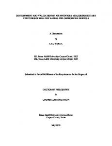
ric immunoassay for serum CRP. In addition, we present a new procedure of sequential covalent coupling of IgG and bovine serum albumin (BSA) that improves the reagent colloidal stability and could eliminate most of the drawbacks of the light-scattering immunoassays, such as nonspecific agglutination. On the basis of clinical data, the aim of the present study has been the development of a highly colloidally stable turbidimetric reagent with a detection limit ⬍0.3 mg/L for CRP and an analytical range up to 50 –100 mg/L, which may be applicable to most of the turbidimetric clinical chemistry analyzers. An IgG fraction of a goat polyclonal antiserum (International Enzymes, Inc.) and an affinity-purified IgG (SCIPAC) against human CRP were used. The microparticle reagents were prepared according to a previously published method by the carbodiimide [1-ethyl-3-(3-dimethylamino-propyl)carbodiimide chloride] procedure (11 ). The anti-human CRP immunoglobulin was covalently coupled to carboxyl-modified polystyrene microparticles, with diameters between 100 and 330 nm, purchased from Polymer Laboratories Ltd and Bangs Laboratories as a 100 g/L suspension. For the immunopurified anti-CRP antibodies, we used a modified procedure consisting of two sequential covalent coupling steps. Antibody solutions (0.08 – 0.9 mg/m2 of latex) were incubated with 1-ethyl-3(3-dimethylamino-propyl)carbodiimide chloride microparticles at 4 °C. After 60 min, free surfaces were saturated with BSA added at a final concentration of 15 mg/m2, and the particles were then incubated at 4 °C for 6 h. After centrifugation, the coated microparticles were resuspended (5 g/L) in glycine-buffered saline-BSA containing 0.17 mol/L NaCl, 0.1 mol/L glycine, 3 g/L BSA, 1 g/L Tween 20, pH 8.2. Coverage was calculated by measuring the uncoupled IgG present in the supernatants of the first centrifugation, after filtering with a Nucleopore polycarbonate filter (pore diameter, 0.1 m), using the copper reduction/ bicinchoninic acid reaction (BCA method; Pierce) and/or the measurement of the absorbance at 280 nm (E1280cm ⫽ 1.4 kg/L). The extent of covalent coupling was determined by displacement of passively adsorbed protein with sodium dodecyl sulfate (10 g/L in 0.1 mol/L Tris, pH 10). After elution, the released protein was measured using the BCA procedure. The immunoagglutination reaction was measured on a Cobas Mira (Hoffman-La Roche) by a turbidimetric assay and on an IMMAGETM immunochemistry system (Beckman Coulter) by a near-infrared particle immunoassay (NIPIA) method (12 ). The experimental details are summarized in Table 1. In both procedures, sample CRP values were automatically calculated from a multipoint calibration curve using a log/logit calculation procedure. As calibrator, we used a CRP reference standard containing 180 mg/L CRP (CRP T Standard, ref. 0737224; Hoffmann-La Roche), according to IFCC reference material CRM 470. The covalently coupled isotherms of IgG anti-CRP on carboxyl-modified polystyrene particles (100-nm diameter) are shown in Fig. 1A. The maximum adsorption
plateau is achieved at a polyclonal antibody concentrations of 3.6 mg/m2. The immunopurified antibody showed a similar trend, having a covalently coupled adsorption isotherm according to that of the nonimmunopurified antibody in the range studied (0.08 – 0.9 mg/ m2). The covalent coupling efficiency rate was always ⬎92% (IgG chemically bound/IgG chemically plus passively adsorbed). The microparticle reagent response of the different antibodies to increasing CRP concentrations can be seen in Fig. 1B, where immunopurified antibodies show the best results. The most striking feature in Fig. 1B is that the curve for the immunopurified antibody at 0.3 mg/m2 was equivalent to that obtained for nonimmunopurified antibody at 3.6 mg/m2. No further improvement was obtained when IgG loadings for immunopurified antibody concentrations ⬎0.3 mg/m2 were tested. We found that at any given working dilution of microparticles studied, the initial absorbance decreases with increasing wavelength and increases with microparticle diameter, according to the light-scattering theory. The effect of varying the particle size on a reagent with a constant charge of 0.3 mg/m2 anti-CRP immunopurified antibody indicates that the analytical sensitivity increases with size and decreases with wavelength, whereas the upper measuring limit shows an opposite behavior (Fig. 1C). Thus, a compromise between sensitivity and measurement range is demanded. A microparticle size of 150 nm and a wavelength of 600 nm were selected as the best choice because of the high slope and wide dynamic range showed. We chose this microparticle reagent, with a coverage of 0.3 mg/m2 anti-CRP immunopurified antibody, for method validation. Polyethyleneglycol (PEG) is commonly used to increase the immunoagglutination kinetics. However, a negative influence on reagent stability was clearly visible at PEG concentrations ⱖ30 g/L in the reaction buffer because of nonspecific aggregation of the reagent. Because PEG can also produce nonspecific precipitation of endogenous and exogenous serum components in the reaction medium, we fixed the final PEG concentration in the reaction buffer at 25 g/L. We monitored the reaction kinetics at pH values of 6.5–9.5, observing that immunoagglutination decreased with increasing pH (data not shown); no significant differences were observed when several nonionic detergents were used at concentrations up to 1 mL/L, although the addition of nonionic detergent increases the Table 1. Conditions for CRP determination. Assay mode Sample, L Diluent (water), L Reaction buffer, L Latex reagent, L Preincubation time, min Time interval, min Wavelength, nm Temperature, °C
Cobas Mira
IMMAGE
Fixed-time 3 30 250 60 1 5 600 37
Kinetic rate 4 250 60 5 2.5 940 37
Clinical Chemistry 46, No. 11, 2000
microparticle stability by decreasing the nonspecific binding. We then selected 0.1 mol/L Tris (pH 6.5) containing 0.15 mol/L NaCl, 3 g/L BSA, 25 g/L PEG, and 1 g/L Tween 20 as the reagent buffer for method validation. The intra- and interassay CVs (n ⫽ 20) for three pooled samples studied were 2.5– 6% and 1.8 –5.2% for the turbidimetric and NIPIA measurements, respectively. The turbidimetric method affords a measuring range of 0.3– 45 mg/L for CRP in human serum on the Cobas Mira analyzer. In the Beckman IMMAGE instrument, the upper assay range was extended to 100 mg/L, with a substantial increase in the dynamic range, by means of the nearinfrared wavelength capability of the system (940 nm reading). Samples with CRP values above the upper limit of the calibration curve can be reanalyzed after automated or manual sample dilution. The prozone effect did not occur for CRP concentrations up to 260 mg/L. The detection limits for CRP, defined as the mean plus 3 SD of the zero signal (saline solution), were 0.25 and 0.14 mg/L on the Cobas Mira and Beckman IMMAGE analyzers, respectively. Rheumatoid factor at concentrations up to 650 kIU/L did not interfere with the assay (CRP recovery within 100% ⫾ 5%). The comparison study between the turbidimetric and
1841
NIPIA assays (y) and a monoclonal latex-enhanced nephelometric method (x; Dade-Behring N-Latex mono CRP) gave correlation coefficients ⬎0.99, with no significant differences between regression parameters (P ⬎0.05), indicating a close relationship between these procedures even at low CRP values (⬍5 mg/L). Parameter values (a, b), correlation coefficients (r), and confidence regions were as follows: Cobas Mira, b ⫽ 0.99 (range, 0.96 –1.01); a ⫽ 0.25 (⫺0.11 to 0.32); r ⫽ 0.997; n ⫽ 68; and IMMAGE, b ⫽ 0.95 (0.92–1.05); a ⫽ 0.10 (⫺0.17 to 0.15); r ⫽ 0.99; n ⫽ 68. The most important features of the method presented here are high analytical sensitivity (detection limit ⬍0.3 mg/L CRP) and good precision (CVs ⬍7%) in the measuring range up to 45 mg/L. This range is wide enough to determine the CRP values found in 72% of our hospitalized patients and ⬎90% of our outpatient population. Nevertheless, a further increase of the dynamic range up to 100 mg/L may be gained on the IMMAGE analyzer by use of a 940 nm wavelength. Both procedures display a prozone phenomenon at CRP concentrations ⬎260 mg/L, with 99.4% of our patients included. The results obtained by these methods were well correlated (r ⬎0.99) with those determined by a nephelometric ultrasensitive
Fig. 1. Covalent isotherm (A) and dose–response curves as a function of antibody type and coverage (B) and particle size (C). (A), covalent adsorption of whole (f) and immunopurified (E) anti-CRP IgG antibodies on 100-nm microparticles. The dashed line indicates the theoretical maximum coverage efficiency (100% of IgG chemically bound). Jads, amount of antibody adsorbed; Jin, amount of antibody added. (B), absorbance at 405 nm with 100-nm microparticles coated with different coverages of anti-CRP whole antibody (䡺, 3.6 mg/m2; ƒ, 2.9 mg/m2; E, 0.9 mg/m2) and with immunopurified antibody (0.3 mg/m2; F). (C), effect of particle size on absorbance at 600 nm. Particle diameter: 䡺, 100 nm; F, 150 nm; f, 336 nm.
1842
Technical Briefs
monoclonal CRP immunoassay, opening the door to a less expensive automated method with similar sensitivity and a wide measuring range. For practical use, it is essential to obtain microparticle reagents colloidally stable under reaction and storage conditions, with the stability often depending on protein coverage. Colloidal particles coated with polyclonal IgG usually are not very stable, mainly because of the high antibody coverage needed to obtain good sensitivities (3.6 mg/m2 in our case). Posttreatment with additives (BSA, surfactants) has been proposed (13, 14 ), although it reduces IgG charge and reactivity. We suggest an alternative approach that uses a small amount of immunopurified antibody and stabilizes the reagent by saturation of free surface with BSA in a two-step sequential covalent procedure. In this way, the inactive IgG molecules are replaced with molecules of BSA, remarkably increasing the latex stability by electrostatic repulsion. Moreover, both IgG and BSA could be added in only one step if the right conditions are found (14 ). In conclusion, a low coverage (0.3 mg/m2) of immunopurified IgG provides a reagent with immunoreactivity similar to or better than that of microparticles totally covered (at saturation) by a nonimmunopurified IgG antibody (3.6 mg/m2), but with higher colloidal stability (the suspension remained stable for more than 3 months when it was stored at 4 °C). This approach to antibody immunopurification could be extended to obtain reagents useful for the measurement of several other proteins at low concentrations (15 ).
This research was supported in part by the Comision Interministerial de Ciencia y Tecnologı´a (CICYT), Projects MAT99-0662-C03-02 and -03. References 1. Thompson D, Milford-Ward A, Whicher JT. The value of acute phase protein measurements in clinical practice. Ann Clin Biochem 1992;29:123–31. 2. Haverkate F, Thompson SG, Pyke SDM, Gallimore JR, Pepys MB. Production of C-reactive protein and risk of coronary events in stable and unstable angina. Lancet 1997;349:462– 6. 3. Conrozier T, Chappuis-Cellier C, Richard M, Mathieu P, Richard S, Vignon E. Increased serum C-reactive protein levels by immunonephelometry in patients with rapidly destructive hip osteoarthritis. Rev Rheum Engl Ed 1998;65:759 – 65. 4. Wasunna A, Whitelaw A, Gallimore R, Hawkins PN, Pepys MB. C-reactive protein and bacterial infection in preterm infants. Eur J Pediatr 1990;149: 424 –7. 5. Macy EM, Hayes TE, Tracy RP. Variability in the measurement of C-reactive protein in healthy subjects: implications for reference intervals and epidemiological applications. Clin Chem 1997;43:52– 8. 6. Price CP, Calvin J, Walter SA, Trull A, Newman DJ, Gorman EG. A rapid and sensitive automated light scattering immunoassay for serum C-reactive protein and the definition of a reference range in healthy blood donors. Clin Chem Lab Med 1999;37:109 –13. 7. Price CP, Trull AK, Berry D, Gormann FG. Development and validation of a particle enhanced turbidimetric immunoassay for C-reactive protein. J Immunol Methods 1987;99:205–11. 8. Lammers M, Bienvenu J, Monneret G, Borque de Larrea L, Gaona N, Schumann G, et al. Evaluation of an improved immunonephelometric assay for C-reactive protein [Abstract]. Clin Chem 1996;42(Suppl 6):S165. 9. Roberts WL, Sedrick R, Moulton L, Spencer A, Rifai N. Evaluation of four automated high-sensitivity C-reactive protein methods: implications for clinical and epidemiological applications. Clin Chem 2000;46:461– 8. 10. Eda S, Kaufmann J, Molwitz M, Worberg E. A new method of measuring
11. 12. 13.
14.
15.
C-reactive protein, with a low limit of detection, suitable for risk assessment of coronary heart disease. Scand J Clin Lab Invest Suppl 1999;230:32–5. Borque L, Maside C, Rus A, del Cura J. Latex immunoassay of 2microglobulin in serum and urine. J Clin Immunoassay 1994;17:160 –5. Borque L. A sensitive, particle-enhanced assay for ferritin on IMMAGETM immunochemistry system [Abstract]. Clin Chem 1999;45(Suppl 6):A45. Peula JM, Hidalgo-Alvarez R, de las Nieves FJ. Coadsorption of IgG and BSA onto sulfonated polystyrene latex. II. Colloidal stability and immunoreactivity. J Biomater Sci Polym Ed 1995;3:241–51. Peula JM, Puig J, Serra J, de las Nieves FJ, Hidalgo-Alvarez R. Electrokinetic characterization and colloidal stability of polystyrene latex particles partially covered by IgG/a-CRP and m-BSA proteins. Colloids Surf A Physicochem Eng Aspects 1994;92:127–36. Borque L, Rus A, Bellod L, Seco ML. Development of an automated immunoturbidimetric ferritin assay. Clin Chem Lab Med 1999;37:899 –905.
Flexibility of Melting Temperature Assay for Rapid Detection of Insertions, Deletions, and Single-Point Mutations of the AGXT Gene Responsible for Type 1 Primary Hyperoxaluria, Doroti Pirulli,1,2 Michele Boniotto,1 Daniela Puzzer,1 Andrea Spano`,1 Antonio Amoroso,1,2 and Sergio Crovella1,2* (1 Servizio di Genetica Medica, IRCCS Burlo Garofolo, 34137 Trieste, Italy; 2 Sezione di Genetica Medica, Dipartimento Scienze della Riproduzione e dello Sviluppo, Universita` di Trieste, 34137 Trieste, Italy; * address correspondence to this author at: Servizio di Genetica, IRCCS Burlo-Garofolo, Via dell’Istria 65/1, 34137 Trieste, Italy; fax 39-040-3785210, e-mail crovella@burlo. trieste.it) Primary hyperoxaluria type 1 (PH1; OMIM 259900) is a rare autosomal recessive disorder characterized by impaired hepatic detoxification of glyoxylate. PH1 is caused by a deficiency of alanine:glyoxylate aminotransferase (AGT; EC 2.6.1.44), which catalyzes the transamination of glyoxylate to glycine. This defect leads to endogenous overproduction of oxalate and glycolate, producing oxalic and glycolic hyperacidurias, which are the hallmarks of the disease (1 ). The AGT enzyme is encoded by a single-copy gene (AGXT), which consists of 11 exons ranging from 65 to 407 bp and spanning a 10-kb DNA segment in the 2q37.3 human region. AGT is a 392-amino acid protein with a molecular mass of 43 kDa (2 ). Several technical approaches have been used to identify 7 polymorphisms and 26 mutations in the AGXT gene (3– 6 ). Here we describe a rapid, flexible, and inexpensive method for detection of the different types of mutations (insertions, deletions, point mutations) of the AGXT gene. Our method is based on the ability to distinguish between PCR amplification products by their melting temperatures (Tm) (7–9 ). Nine PH1 patients, whose mutations had first been analyzed by the single-strand conformation polymorphism (SSCP) technique and then by sequencing of abnormal mobility bands of four AGXT exons (5 ), were studied comparatively by the melting temperature assay (MTA). Heterozygous relatives of three patients were also included in this study. Five healthy Italian subjects served