Sep 5, 1995 - Molecular Genetics, University of Texas M.D.Anderson Cancer. Center, Houston ...... allowed to cool slowly to 100 K in 4000 steps. From the 50 ...
The EMBO Journal vol.14 no.23 pp.5947-5956, 1995
Novel topology of a zinc-binding domain from protein involved in regulating early Xenopus development Katherine L.B.Borden1l2, John M.Lally3, Stephen R.Martin4, Nicola J.O'Reilly5, Laurence D.Etkin6 and Paul S.Freemont3'7 'Laboratory of Molecular Structure and 4Division of Physical Biochemistry, National Institute for Medical Research, The Ridgeway, Mill Hill, London NW7 IAA, 5Peptide Synthesis Laboratory and 3Protein Structure Laboratory, Imperial Cancer Research Fund, 44 Lincoln's Inn Fields, London WC2A 3PX, UK and 6Department of Molecular Genetics, University of Texas M.D.Anderson Cancer Center, Houston, TX 77030, USA 2Present address: Protein Structure Laboratory, Imperial Cancer Research Fund, London WC2A 3PX, UK 7Corresponding author
Xenopus nuclear factor XNF7, a maternally expressed protein, functions in patterning of the embryo. XNF7 contains a number of defined protein domains implicated in the regulation of some developmental processes. Among these is a tripartite motif comprising a zincbinding RING finger and B-box domain next to a
predicted a-helical coiled-coil domain. Interestingly, this motif is found in a variety of proteins including several proto-oncoproteins. Here we describe the solution structure of the XNF7 B-box zinc-binding domain determined at physiological pH by 'H NMR methods. The B-box structure represents the first threedimensional structure of this new motif and comprises a monomer having two 1-strands, two helical turns and three extended loop regions packed in a novel topology. The r.m.s. deviation for the best 18 structures is 1.15 A for backbone atoms and 1.94 A for all atoms. Structure calculations and biochemical data shows one zinc atom ligated in a Cys2-His2 tetrahedral arrangement. We have used mutant peptides to determine the metal ligation scheme which surprisingly shows that not all of the seven conserved cysteinesl histidines in the B-box motif are involved in metal ligation. The B-box structure is not similar in tertiary fold to any other known zinc-binding motif. Keywords: B-box domain/NMR/protein structure/RING
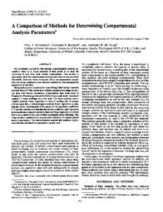
Reddy et al., 1991). Preliminary evidence indicates that XNF7 is involved in dorsal ventral patterning of the embryo (W.Shou and L.Etkin, unpublished observations). Interestingly, XNF7 has been detected in association with the mitotic spindle and condensed chromosomes during mitosis (Li et al., 1994b), a process which is functionally important. XNF7 belongs to a family of proteins which contain two novel zinc finger domains named RING (Freemont et al., 1991; Freemont, 1993; Lovering et al., 1993) and B-box (Reddy and Etkin, 1991) followed closely (5-8 amino acids) by a predicted a-helical coiled-coil domain forming a tripartite motif (Figure la; Kastner et al., 1992; Reddy et al., 1992). The spacing between the three elements of the motif is highly conserved among family members, suggesting that the relative positions of the domains is of functional importance. There are a number of proto-oncoproteins within the B-box family, including the ret finger protein (RFP) (Takahashi et al., 1988), PML (de The et al., 1991; Goddard et al., 1991; Kakizuki et al., 1991; Kastner et al., 1992) and TIFI (T18; Miki et al., 1991; Kastner et al., 1992; Le Douarin et al., 1995). RFP, PML and T18 (TIF1) are oncogenic in humans and mice when found as translocations that include the RING, B-box and a-helical coiled-coil domains recombined with other genes. PML is of particular importance owing to its direct association (a) RING finger
Coled-moil DLiZH±.241111111111ll_llilii_llM B2
Introduction
B2 (184-232) ATDC Bl (168-216) EFP Bl (102-148)
PM0L
TIF1 Bl (157-204) PML Bl (124-166)
SEE
30
20
10
1
RPLEKC KPKEKC QKVNIC SS-A/Ro (87-128) TQGERC GEMGVC RFP (101-142) ATDC B2 (220-260) FEARXC EFP B2 (152-191) LLRRXC TIF1 B2 (217-258) QRPVFC
finger/Xenopus XNF7
K Oxford University Press
Coied-coil iiiiiiiiiiiiiii1111n1111111111111111111111
B-box
B L. fiZ RI-
XNF7 (219-260) PwA33 (233-274) RPT-1 (91-132)
Xenopus nuclear factor, XNF7 (609 amino acids), is a maternally expressed protein that is retained in the cytoplasm until the mid-blastula transition, at which time it enters the nucleus (Dreyer et al., 1983; Miller et al., 1989, 1991; Reddy et al., 1991; Li et al., 1994a). Based on its primary structure, subcellular localization, nucleic acid binding properties and the ability of the N-terminal domain to transactivate a reporter gene, XNF7 is thought to function as a transcription factor (Miller et al., 1989;
a
DERLKLYCKDDGTLSCVICRDSLK
H
40 ASHNFLPI
SFLPI KDGTLACVICRDSXL HH RGHQTALI AQO GEXLRLFCRXDMQVICWLCERSQE RDEAMVPL GERLHLFCEKDGKALCWVCAQSRX HH AVE RGHSVLPL EXB REPLKLYCEEDQMPICVVCDRSRE H XNSTVTV PVH GKTMEiFCQTDQTCICYLCMFQE H KTCSPASL SQH NRLREFFCPEHSECICHICLVE H KENRYQFI PFH KKEQLKLYCETCDKLTCRDCQLLE SELKCDISAE TNNIFCSNPNH RTPTLTSIYCRGCSKPLCCSCALIDSSH HLEGAAFRDHQLLEP SEEVLC DSCIGNKQKAVKSCLVCQASFCELHLKP NAQVAC DHC LKEAAVKTCLVCMASFCQEELQP HFDSPAFQDHPLQPP KSNQVC TSC EDNAEANGFCVECVEWLCKTCIRA RQRVKFTKDHTVRQK DAQAVC TRC KESADFWCFECEQLLCAXCFEA H* QWFL]3EARPL * SSS *
DEH
S* *
DER
S$SS*
*
SS*
Fig. 1. The B-box family. (a) Schematic representation of the tripartite motif for XNF7. The RING and B-box are separated by 39 amino acids. The B-box and a-helical coiled-coil domains are adjacent. (b) Sequence alignment of the B-box family. The putative conserved metal ligands are shown in bold and are labelled with a *. Conserved hydrophobic residues are labelled with a $. Each sequence is referenced in the text and the top numbering refers to the XNF7 B-box peptide (residues 1-42). The actual protein numbering is given in parentheses.
5947
K.L.B.Borden et aL Table I. 'H NMR assignments of the B-box peptide Residue
ArgI Pro2 Leu3 Glu4 Lys5 Cys6 Ser7 Glu8 His9 AsplO Glu 1l Argl2 Leul3 Lysl4 Leu15 TyrI6 Cysl7 Lysl8 Aspl9 Asp 20 Gly2l Thr22 Leu23 Ser24 Cys25 Val26 Ile27 Cys28 Arg29 Asp3O Ser3l Leu32 Lys33 His34 Ala35 Ser36 His37 Asn38 Phe39 Leu4O Pro4l Ile42
Chemical shift (p.p.m.) NH
aH
8.53 9.14 8.28 7.74 6.86 8.33 9.18 8.54 7.74 7.56 7.74 8.63 9.78 9.58 7.07 7.01 8.13 8.34 7.31 8.49
4.35 4.51 5.00 3.78 3.86 4.14 4.55 4.84 3.96 4.28 4.14 4.35 4.13 4.56 5.33 5.00 5.18 3.48 4.45 4.77 3.78/4.00 4.56 4.34
8.82 9.29 8.94 8.23 8.48 8.20
5.46 3.58 3.68 4.00 4.25 4.88
2.77/3.67
1.85 3.36/3.58
(y),1.16 (yMe), 0.73 1.65 (y), 3.22 (6)
7.78
4.09 3.90 4.02 4.30
1.30/1.75 1.45/1.65 3.05/3.46 1.42
1.61 (y), 0.91/0.95 (6/6') 1.01/1.06 (y/y'), 1.60 (6), 2.89 8.24 (H2), 7.47 (H4)
4.07 5.06 5.45 5.08 4.86
3.70 2.71 2.73/2.95 1.76 2.18/2.38
8.18 (H2), 7.49 (H4) 7.14,7.82 (NH2A)/7.02,7.88 (NH2B)a 6.85 (4), 7.45 (3,5), 7.18 (2,6)
7.40 7.37 8.85 8.92 9.61
P/P'H
Others
1.94
1.78 (y), 3.25(6) 1.92/2.00 (y/y'), 3.62/3.78 1.54 (y), 1.04
2.22/2.35 1.95 2.25/2.89 1.82 4.16/4.29 2.28/2.36 3.19/3.49 3.06/3.19 1.45/1.85 1.94 1.81/1.97 1.42/1.66 1.35/1.42 2.73/2.85 3.25/3.81 1.55
2.71(Y) 1.51(y),
(6)
1.71
(6), 2.96
(6/6')
(E)
8.27(H2), 6.86 (H4) 1.70 1.44
3.25 (6) (y), 0.74/0.84 (y), (6/6') 1.11 (y), 1.51 (6), 2.85 (E) 1.27 (y), 0.33/0.79 (6/6') 6.85 6.95
(3,5),
(2,6)
0.78/0.99
(y/y'), 2.98 (E)
4.04 1.68
1.21 (y) 0.91/0.96
(6/6')
2.69/3.58 2.12 1.08
0.28
2.42/2.95 2.71/2.90
0.92/0.98
1.67 2.04
(y), (y),
(y/y')
0.99/1.02 3.75/3.88
(6)
(E)
(6/6')
(6/6')
Assignments were carried out using standard assignment methods as described in the text. Data were collected at pH 7.5 and 30°C. aThere were two cross-peaks in the 'H20 TOCSY which corresponded to the amide side-chain of Asn38. The presence of two cross-peaks indicated that these protons were undergoing slow exchange on the NMR time-scale. with acute promyelocytic leukaemia as a fusion protein with retinoic acid receptor alpha (e.g. see Goddard et al., 1991). TIFI has been shown to interact with several nuclear receptors in vivo and is proposed to mediate the ligand-dependent transcriptional activation function of nuclear receptors (Le Douarin et al., 1995). EFP, an oestrogen-responsive gene product, is another family member thought to represent an oestrogen-responsive transcription factor mediating phenotypic expression due to oestrogen action (Inoue et al., 1993). PML, TIFI and EFP all possess two B-box-like domains (B1 and B2) which appear to form a subgrouping of the B-box family as highlighted by the presence of an extra potential metal ligand (Figure Ib; Reddy et al., 1992; Inoue et al., 1993). The other members of the family include RPT-1 (Patarca et al., 1988), SS-A/Ro-52 (Chan et al., 1991) and PwA33 (Bellini et al., 1993). RPT-1 (mouse) has been implicated in the down-regulation of the interleukin-2 receptor, acting 5948
putative transcription factor (Patarca et al., 1988). SS-A/Ro-52 is a human auto-antigen and forms part of a ribonucleoprotein complex, although no direct interaction between SS-A/Ro-52 and RNA has been observed (Chan et al., 1991; Slobbe et al., 1992). PwA33 (Pleurodeles) may also function as a component of RNP complexes (Bellini et al., 1993). To date, two gene products have been reported as possessing a B-box and coiled-coil domain but with no RING finger. These include a candidate gene for ataxia-telangiectasia group D (ATDC) which possesses two B-box domains and a coiled-coil domain similar to PML, TIFI and EFP (Leonhardt et al., 1994). The other is a gene found at the breast cancer locus, 1 A1.3B (Campbell et al., 1994) which has one B-box and a coiled-coil domain, and appears tightly linked to the BRCA 1 gene which interestingly possesses a RING finger but no B-box or coiled-coil domains (Brown et al., 1994; Miki et al., 1994). as a
XNF7 B-box structure
A
L15
F2
(ppm): 8. 7-
C25
N3QL.
8. 88. g
9.0.
F39
9. I-
C
9.2-
+ 4i 40
9 4--
L40
30 -
9.6-
x+
+4* + IxO
4+
C17
9.5-
0 S
ax0 .o ta t:;
S^x
0
Y16
9.7-
0
9.8-
20-
S
tx x.
9.9g 10
B
Of
40 -
9. 3-
a . Soo0*
+0
4D
F2
^
-
o -T-
(ppm
0
10
20
40
30
8.7 residue
8.8
8.9
D
9.0-:
40-
N38a, F39NH-
9.19.2 9.3
30
_ _S P C25a, V26NH
9.4
a
20-
9.5
0
9.6 9.7
F39a,
L40NH
Y16a, C17NH
Il
10
9.89.9
LI 5a, Y16NH
Rs
oJM I 5.6 5.5 5.4 5.3 5.2 5.1 5.0 4.9
F1 (ppm)
.I.I.I.
.
R
.
i
i
i
I i
iI
I
residue
Fig. 2. NMR data used in XNF7 B-box structure calculations. (A) A section of the amide alpha region of a TOCSY and (B) a 290 ms NOESY in IH20. In (A) and (B) the ajNj+I connectivities observed in the P-strands are traced out and the corresponding positions of the intra-residue connectivities are indicated in the TOCSY spectrum. Most NOEs occur between the a-carbon proton from the preceding residue to the amide proton of the next residue. For example, Ll5aY16NH indicates a NOE between Leul5 a-carbon proton and Tyrl6 amide proton. See text for further details. (C) A distance map summarizing the NOEs observed for the XNF7 B-box domain. Reading the residue from the x-axis indicates the proton type. Symbols: 0, a-carbon proton; *, side chain proton; X, amide proton; +, ring proton. (D) Distance restraints per residue are shown with long-range (open bars), medium-range (cross-hatched bars) and short-range (solid bars).
In order to determine the molecular function of the B-box domain a number of studies have been initiated. Deletions of the XNF7 B-box domain result in a loss of binding to mitotic chromosomes (Li et al., 1994b) while deletions and point mutations in the B-box of PwA33 results in the loss of association with the lampbrush loops of chromosomes in the oocyte nucleus in Pleurodeles (Bellini et al., 1993; M.Bellini, personal communication). In addition, results of studies of the B-box family member RFP, which is closely related to XNF7 (Figure lb), showed that the B-box and coiled-coil domains are both involved in homodimerization (T.Cao and L.Etkin, unpublished observations). Together, these data suggest that the B-box domain may mediate protein-protein interactions and in the case of XNF7 and PwA33 is important for the association of these proteins
i1
i IEEE-
residueNN NNNC'CtCC
with subcellular structures during Xenopus and Pleurodeles development. In order to understand the functions of the RING, B-box and coiled-coil domains in molecular detail, we have initiated a number of structural and biophysical studies of these domains (Borden et al., 1993, 1995; Lovering et al., 1993). Previously, we reported a biophysical characterization, including metal binding and secondary structure analysis, of a 42-residue synthetic peptide corresponding to the XNF7 B-box domain (Borden et al., 1993). Here, we extend these earlier studies and now present the three-dimensional solution structure of the XNF7 B-box, representing the first structure, topology and metal ligation system for a B-box domain. The structure now provides a basis for investigating the molecular role of the B-box domain in XNF7 function 5949
K.L.B.Borden et al.
and Xenopus development and also provides insights into the function of the B-box domain within the B-box family.
Results and discussion NMR structure determination The three-dimensional structure of the XNF7 B-box domain (XNF7 residues 219-260) was determined using standard
A
5950
two-dimensional NMR techniques (see Materials and methods). It should be noted that experiments were carried out at physiological pH of 7.5 as the peptide-although soluble-was unstructured at lower pHs as assayed both by CD and NMR. 'H chemical shift assignments are given in Table I. The NOESY footprint region (Figure 2B) shows connectivities from Leul5 to Cysl7 and Asn38 to Leu4O which form part of the two ,B-strands in the B-box structure.
XNF7 B-box structure
Also shown are the alignments to the corresponding region of the TOCSY spectra (Figure 2A). It should be noted that in the NOESY (Figure 2B) the amide proton (i) to a-carbon proton (i) intra-residue cross-peaks are missing whereas the a-carbon proton (i) to amide proton (i+1) inter-residue cross-peaks are very intense. This pattern of cross-peak intensity is characteristic of phi and psi angles which occur in extended conformation, such as 1-strands (Wuithrich, 1986). In addition there are several tertiary NOEs between the strands (see Figure 2C). A summary of the observed sequential and long-range NOEs is given in Figure 2C in the distance map format. A total of 154 non-intra-residue distance constraints and 27 dihedral angle constraints (13 Phi, 7 Psi and 7 Chi 1), were used as input into the program XPLOR (Briinger, 1992). Of the distance constraints, 64 were short-range (i to i+2), 12 medium (i+3 to i+5) and 78 long-range (i to > i+5). A summary of the constraints per residue is given in Figure 2D. It is notable that there are a number of areas within the peptide where there are no constraints. These regions generally correspond to positions where amide protons could not be assigned due to either overlap or exchange with bulk solvent. Of the 50 structures calculated, 39 converged with the remaining 11 as mirror images. The r.m.s. deviations for all 39 converged structures (residues 5-17; 25-41) are 1.39 A for the backbone and 2.13 A for all atoms. From the XPLOR calculations, 18 structures had NOE violations