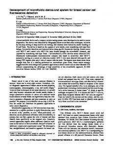
vices that exploit the advantages of each material in a single device. (Fig 2). Fig 2. ... Further, custom software has been developed to objectively characterise ...
DEVELOPMENT OF A MICROFLUIDIC PLATFORM AND DETECTION SYSTEM FOR PLATELET FUNCTION ANALYSIS 1,2
1
1,3
1
Nigel J. Kent , Geradene Meade , Lourdes Basabe-Desmonts , 1 1,3 1 Bryan Lincoln , Dermot Kenny ,Antonio J. Ricco , 1 2 Brian D. MacCraith , Brian G. Corcoran .
Biomedical Diagnostics Institute, Dublin City University, Glasnevin, Dublin 9, IRELAND 2 School of Mechanical and Manufacturing Engineering, Dublin City University, Glasnevin, Dublin 9, IRELAND 3 Molecular and Cellular Therapeutics, Royal College of Surgeons in Ireland, Dublin 2, IRELAND ABSTRACT We have developed a prototype point-of-care diagnostic tool for early warning of platelet related disorders. The system objectively quantifies early-stage, shearmediated platelet-surface interactions for well characterized protein surfaces. The hybrid microfluidic platform significantly reduces sample volume (from ~ 5000 µL to ~150 μL) relative to current laboratory systems, while the detection system utilises low-cost optical components for fluorescence detection of platelet-surface interactions in whole blood. KEYWORDS: Platelets, Microfluidics, Polymer Fabrication, Point of Care INTRODUCTION Platelets (thrombocytes) are small discoid (2-4 μm by 0.5 μm) cell fragments circulating in the blood stream and are involved in the cellular mechanisms of haemostasis, leading to the formation of blood clots [1]. When a platelet is activated, e.g. by exposure to a damaged vessel wall, various proteins are expressed, mediating platelet-wall and platelet-platelet interactions. Abnormalities in these interactions may increase risk of bleeding and are linked to atherosclerosis, coronary artery disease, myocardial infarction, stroke and cancer. While the mechanisms of platelet activation are complex, with multiple simultaneous interactions, its initial stages are a function of the shear rate of the platelet-carrying medium [2]. THEORY The standard platform for characterization of shear-mediated cellular interactions is the parallel-plate flow chamber [3]. Its low aspect ratio ensures a uniform shear rate over most of the channel width. This configuration, however, is sampleinefficient, requiring much of the sample to pass through the device undetected. Our design uniquely combines two existing microfluidic technologies: parallel plate confinement and a variation on hydrodynamic focusing [4].
Twelfth International Conference on Miniaturized Systems for Chemistry and Life Sciences October 12 - 16, 2008, San Diego, California, USA 978-0-9798064-1-4/µTAS2008/$20©2008CBMS
1048
Fig 1a. CFD model Fig 1b. Modelled results based on a buffer sample reduction as a revolume flow rate of 0.4 sult of increasing buffer ml/min. flow rate to 0.55 ml/min. Fig 1. Schematic showing the principles of hydrodynamic (HD) shaping. Flow chamber dimensions are 100 µm high and 4 mm wide. Fig 1a and Fig 1b show the numerically modelled effect, on the sample (in centre/red), of increasing the inlet volume flow rate of the buffer (surrounding stream/yellow). This ‘hydrodynamic-shaping’ (Fig 1) is achieved by fixing the outlet flow rate of the device (0.6 ml/min to achieve a shear rate of 1500/sec in whole blood) and tuning the relative flow rate of the buffer inlet thus enabling control of fluidic shear rate independent of sample flow rate and resulting in a significant reduction in required sample volume. The fabrication strategy for the device allows rapid prototyping of multilayer, hybrid polymer-glass devices that exploit the advantages of each material in a single device (Fig 2).
Fig 2. Schematic and image of HD shaping platform. Channel geometries are defined by laser patterning, 50 µm thick, polymer laminate adhesive layers. A 6 mm thick PMMA lid is manufactured using conventional mchining and a glass cover slip used as a base layer.
EXPERIMENTAL In order to observe platelet surface interactions, a miniature epi-fluorescent microscope (Fig 3) was built to implement bright-field and fluorescence microscopy (Fig 4). The sample medium, whole human blood, is fluorescently stained with dihexaoxacarbocyanine iodide (DiOC6(3)), a lipophilic dye commonly used to study blood platelets. The sample passes though the device at controlled shear rates (~ 1500/sec) typical of vascular flow and under conditions conducive to surface contact (i.e. a fluid layer < 10 platelet diameters thick) with immobilised von Willebrand Factor (VWF), an activating protein prominent in the initial stages of platelet activation. The sample is excited with a high-brightness blue LED. Fluorescent images are captured via CCD camera over defined ranges of time and frame rates.
Twelfth International Conference on Miniaturized Systems for Chemistry and Life Sciences October 12 - 16, 2008, San Diego, California, USA
1049
Readout/Data Analysis
Electronics Temp ctrl Holder
Camera Stages Microscope
Fig 3. Image of miniaturised detection Fig 3. Image of miniaturied detection and acquisition system. and acquisition system.
Fig 4. Fluorescence and bright field Fig 4. Fluorescence and bright images using HD shaping platform field images using HD shaping platand miniaturised detection system. form and miniaturised detection system.
RESULTS AND DISCUSSION In agreement with previous results [1,2], we find that the shear-activated response of platelets in the presence of VWF surfaces manifests itself as translocation of the platelets along the surface at rates largely independent of sample flow velocity. Further, custom software has been developed to objectively characterise these translocation velocities as a measure of platelet function (Fig 5). Fig 5. Platelet trajectories determined from a sequence of fifty images. Experiment carried out in whole blood using VWF and a vascular mimetic shear rate of 1500/sec. REFERENCES [1] P. Harrison. Platelet function analysis. Blood reviews, 19(2):111–123, Mar 2005. [2] Schmugge, M., Rand, M.L. & Freedman, J. 2003, "Platelets and von Willebrand factor", Transfus.Apher.Sci., vol. 28, no. 3, pp. 269-277. [3] H. Lu, L. Y. Koo, W. M. Wang, D. A. Lauffenburger, L. G. Griffith, and K. F.Jensen. Microfluidic shear devices for quantitative analysis of cell adhesion. Analytical Chemistry, 76(18):5257–5264, Sep 15 2004. [4] S. Chung, S. J. Park, J. K. Kim, C. Chung, D. C. Han, and J. K. Chang. Plastic microchip flow cytometer based on 2- and 3-dimensional hydrodynamic flow focusing. Microsystem Technologies, 9(8):525–533, 10/ 2003.
Twelfth International Conference on Miniaturized Systems for Chemistry and Life Sciences October 12 - 16, 2008, San Diego, California, USA
1050