MOPEA062
Proceedings of IPAC’10, Kyoto, Japan
DEVELOPMENT OF ADVANCED QUANTUM RADIATION SOURCE BASED ON S-BAND COMPACT ELECTRON LINAC R. Kuroda*, H. Toyokawa, M. Yasumoto, H. Ikeura-Sekiguchi, N. Sei, H. Ogawa, M. Koike, K. Yamada, AIST, Ibaraki, Japan Abstract We have developed the advanced quantum radiation sources such as a laser Compton scattering (LCS) X-ray and high intense THz sources based on S-band compact electron linac at AIST. In case of the LCS X-ray source, its biological and medical applications have been started using the in-line phase-contrast imaging. And now, the upgrade plan to increase X-ray yields has been executed for these applications. In case of the THz source, coherent THz radiation has been obtained using an ultra-short electron bunch and the THz scanning transmission imaging system has been developed for the investigation of unknown materials.
INTRODUCTION Advanced quantum radiation sources such as a laser Compton scattering X-ray and a high intense THz radiation sources have been developed based on an Sband compact electron linac at AIST in Japan. All of system is built in one research room about 10 meters square including an electron injector, an electron linac, quadrupole magnets, bending magnets, an rf source and a high power laser system [1-2]. Figure 1 shows a top view of our conceptual diagram for the advanced quantum radiation sources. Figure 2 shows a top view photograph of the S-band compact electron linac. The injector consists of a laser photo-cathode rf gun which has the BNL type S-band 1.6 cell cavity with a Cs2Te photocathode load-lock system and a solenoid magnet for emittance compensation. The linac has two 1.5-m-long accelerator structures which is a 1/2 π mode standing wave structure. The electron beam can be accelerated up to about 42 MeV using the rf source of a 20 MW klystron. The laser Compton scattering X-ray source using a TW Ti:Sapphire laser can generate a hard X-ray pulse which
has variable energy of 10 keV - 40 keV with narrow bandwidth by changing electron energy and collision angle for medical and biological applications [3]. The high intense THz radiation source based on the electron linac has been also developed instead of a conventional laser based THz source. The designed THz pulse has high peak power more than 1 kW in frequency range between 0.1 - 2 THz. The THz pulse will be generated with coherent radiation such as synchrotron radiation and transition radiation using an ultra-short electron bunch with bunch length of less than 0.5 ps (rms). The coherent synchrotron radiation in the THz region has been already generated and it will be applied to the THz time domain spectroscopy (TDS) and THz scanning transmission imaging systems [4]. In this conference, we will report present status of our advanced quantum radiation sources.
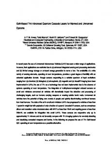
Figure 2: Top view photograph of S-band compact electron linac.
LASER COMPTON SCATTERING X-RAY SOURCE
In case of the laser Compton scattering (LCS) X-ray source, two mode-locked laser systems was operated in 10 Hz, whose mode-lock frequencies are synchronized to 36th sub-harmonic frequency (79.3 MHz) of the linac accelerating frequency (2856 MHz). An all-solid state UV laser (266 nm) was used for the photocathode rf gun. The other laser system was a femtosecond Ti:Sapphire laser with a chirped-pulse amplification (CPA), which is used as collision laser for LCS. It consists of a mode-locked oscillator, a pulse stretcher, a regenerative amplifier, a multi-pass pre-amplifier, a multi-pass main amplifier and a pulse compressor. The pulse width and energy per pulse were 100 fs (FWHM) and 140 mJ, respectively. Figure 1: Top view of conceptual diagram for advanced Pulse width of the LCS X-rays is 150 fs – 3 ps quantum radiation source. depending on a beam sizes of the laser and electron, ___________________________________________ * beam-beam crossing-angle, and pulse widths. The shortest E-mail:
[email protected] 08 Applications of Accelerators, Technology Transfer and Industrial Relations 220
U05 Applications, Other
Proceedings of IPAC’10, Kyoto, Japan
X-ray pulse width is obtained at 90-degree collision angle. The maximum energy of the LCS X-rays can be tuned 10 – 40 keV in about 5 % energy spread, and total photon yields are about 107 photons/s at 165-degree collision angle. Overall system specifications are summarized in Table 1. Table 1: Specification of laser Compton X-ray source
Electron beam
Ti:Sapphire laser
X-ray
Energy Bunch charge Energy spread Bunch length Beam size at collision point Repetition rate Wavelength Pulse width Repetition rate Pulse energy Spot size Energy Yield at 165 deg Yield at 90 deg Stability
~42 MeV ~1 nC 0.2% 3 ps (rms) 43 μm x 30 μm (rms) 10 Hz 800 nm 100 fs (FWHM) 10 Hz 140 mJ 28 μm (rms) ~40 keV 107 photons/s 106 photons/s ~6% (15 min)
We have applied the LCS X-ray to medical and biological imaging using a in-line phase-contrast imaging [5], which is to enhance the contrast of the difference of densities in bone and soft-tissues of living specimens. Figure 6 shows the imaging results with 26.4-keV LCS X-rays obtained for the human bone when the distance between the specimen and the IP is 0 cm and 20 cm. The images obtained at distances of 0 cm and 20 cm are the absorption image (Top figure) and the phase-contrast image (Bottom figure), respectively [6]. The enlarged areas in Fig. 3 show the close up of a part of the fractured bone. And a bone trabecula is also clearly visible in Fig. 3. Even though the quasi-monochromatic LCS X-ray is very useful for the biological and medical imaging,, the photon yields are not enough for the real-time imaging and the much higher resolution imaging. The upgrade plan to increase the X-ray yields several orders of magnitude has been executed with multi-pulse LCS, that is to generate a train of X-ray pulses using laser and electron pulse-trains [7].
MOPEA062
INTENCE COHERENT THZ SOURCE Coherent THz generation The coherent synchrotron radiation (CSR) of the THz region was generated from the ultra-short and high charge electron bunch at the 90 degree bending magnet located at end of the beam line downstream from the bunch compressor [8]. The THz CSR pulse is extracted from a zcut quartz window for THz applications. Typical electron beam parameters for the THz CSR generation and our expected THz specification were described in Table 2. The observed temporal width of the CSR pulse depends on the frequency response of the measurement system. In this system with Electro-Optic (EO) sampling method, it is supposed to be about 700 fs (rms) because of the frequency response of the quartz window, the EO crystal and the detector [9]. Table 2: Design values of the coherent THz source Electron beam Max. Energy 30 ~ 42 MeV Charge per bunch 1 nC – 2 nC Energy Spread ~ 5% for compression Bunch length 300 fs (rms) (after compression) Bunch number 1 – 100 Rep. rate 10 Hz ~ 50 Hz THz pulse Frequency 0.1 – 2 THz Pulse energy 65 nJ Peak power 25 kW Rep. rate 10 – 50 Hz Pulse width 700 fs (rms)
Figure 4: Achromatic arc section for bunch compression
THz scanning imaging system
Figure 3: Results of the in-line phase contrast imaging
The THz scanning transmission imaging as the preliminary experiment was demonstrated with an IC (integrated circuit) card in the previous article [9]. Figure 3 shows the setup of the imaging experiment with the THz CSR pulse. In this work, the THz pulse was extracted from the quarts window and collected by the
08 Applications of Accelerators, Technology Transfer and Industrial Relations U05 Applications, Other
221
MOPEA062
Proceedings of IPAC’10, Kyoto, Japan
parabolic antenna. It passed through a W-band waveguide (WR-10) with an E-bend waveguide and it was guided to a W-band rf detector (WiseWave FAS-10SF-01) whose sensitive area is 1mm × 2mm with sensitive range of 0.075 - 0.11 THz. Its signal of 500 mV corresponds to 1 mW. In this work, a vegetable leaf with about 50 mm length was selected for the imaging sample. The THz transmission imaging has been performed with a scanning step of 500 μm. Although the aperture size of the W-band waveguide is about 1 mm × 2 mm, its resolution is less than 1mm limited by a pin-hole located at the end of the waveguide due to the near field effect. Figure 7 shows results of the THz imaging of the vegetable leaf with the passage of the time from 0 hour to 14 hours after plucking it [10]. The image indicates the water distribution in corresponding to the freshness of the vegetable because the THz radiation is strongly absorbed by water in the fresh leaf. In case of the fresh leaf, it is found that sufficient water is observed along veins of the leaf. However, after 14 hours later, the freshness was quickly lost. As a result, it is found that the THz imaging is a powerful tool for the freshness measurement of the vegetable with the passage of the time.
Figure 5: Setup of THz transmission imaging using THz CSR pulse with W-band waveguide, detector and X-Y sample stage.
Figure 7: THz imaging of a fresh vegetable leaf.
SUMMARY We have developed the advanced quantum radiation sources such as a laser Compton Scattering (LCS) X-ray and high intense THz sources based on S-band compact electron linac at AIST. In case of the LCS X-ray source, the total number of generated photons and maximum X-ray energy were 107 photons/pulse and approximately 40 keV, respectively, at a crossing angle of 165°. We have successfully demonstrated the use of our in-line phase-contrast imaging system with 26.4-keV LCS X-rays for the observation of a fractured human bone. In this study, we observed good contrast enhancement in the absorption and phase-contrast images of the bone fracture and bone trabecula. In case of the coherent THz radiation source, the generation of the THz coherent synchrotron radiation (CSR) in the 90 degree bending magnet has been successfully performed using the ultra-short electron bunch. The THz scanning transmission imaging of the vegetable leaf has been also successfully demonstrated with the W-band rf detector. As a result, it is found that the intense THz radiation source is a powerful tool for imaging of THz absorption with the passage of the time such as the freshness measurement of the vegetable. These works were partially supported by Grant-in-Aid for Scientific Research (B) No. 20360043 and No. 22360040 from MEXT Japan.
REF ERENCES
Figure 6: Setup of THz transmission imaging using THz CSR pulse with W-band waveguide, detector and X-Y sample stage.
[1] H. Toyokawa, R. Kuroda et al., Proc. PAC07, 121 (2007). [2] R. Kuroda et al., Infrared Physics & Technology, 51, 390 (2008). [3] K. Yamada, R. Kuroda et al., Nuc. Inst. Meth. A 608, S7 (2009). [4] R. Kuroda et al., Nucl. Inst. Meth. A 593, 91 (2008). [5] H. Ikeura-Sekiguchi, R. Kuroda et al., Appl. Phys. Let., 92, 131107 (2008) [6] R. Kuroda et al., Nucl. Inst. Meth. A (2010) in press. [7] R. Kuroda et al., Nucl. Inst. Meth. A 608, S28 (2009). [8] N. Sei, R. Kuroda et al., J. Appl. Phys. 104, 114908 (2008). [9] R.Kuroda et al., Rad. Phys. Chem. 78, 1102 (2009). [10] R. Kuroda et al., Nucl. Inst. Meth. A (2010) in press.
08 Applications of Accelerators, Technology Transfer and Industrial Relations 222
U05 Applications, Other