Sensing and Bio-Sensing Research 20 (2018) 16–21
Contents lists available at ScienceDirect
Sensing and Bio-Sensing Research journal homepage: www.elsevier.com/locate/sbsr
Development of an optical sensor based on surface plasmon resonance phenomenon for diagnosis of dengue virus E-protein
T
⁎
Nur Alia Sheh Omara, Yap Wing Fena,b, , Jaafar Abdullahc, Che Engku Noramalina Che Engku Chikd, Mohd Adzir Mahdie a
Functional Devices Laboratory, Institute of Advanced Technology, Universiti Putra Malaysia, 43400 UPM Serdang, Selangor, Malaysia Department of Physics, Faculty of Science, Universiti Putra Malaysia, 43400 UPM Serdang, Selangor, Malaysia c Department of Chemistry, Faculty of Science, Universiti Putra Malaysia, 43400 UPM Serdang, Selangor, Malaysia d Department of Bioprocess Technology, Faculty of Biotechnology and Biomolecular Sciences, Universiti Putra Malaysia, 43400 UPM Serdang, Selangor, Malaysia e Wireless and Photonics Network Research Centre, Faculty of Engineering, Universiti Putra Malaysia, 43400 UPM Serdang, Selangor, Malaysia b
A R T I C LE I N FO
A B S T R A C T
Keywords: Optical sensor Surface plasmon resonance Sensing Dengue virus
Application of surface plasmon resonance (SPR) optical sensor in the diagnosis of dengue virus (DENV) emerged over recent years. Immobilized monoclonal antibody (IgM) on gold/Fe-MPA-NCC-CTAB/EDC-NHS thin film was prepared using a spin coating technique. DENV E-protein can be detected by measuring the SPR signal when IgM immobilized gold/Fe-MPA-NCC-CTAB/EDC-NHS thin film is exposed to the DENV E-protein solution, by varying the concentration ranging from 0.0001 nM to 10 nM. A linear relationship between the shift of SPR angle and concentration of DENV E-protein up to 0.01 nM, with sensitivity of 39.96° nM−1 has been observed. The surface morphology of sensor chip was also recorded using atomic force microscopy to confirm the presence of bound DENV E-protein into IgM surface.
1. Introduction Early diagnosis of dengue is crucial to decrease the fatal illnesses known as dengue hemorrhagic fever (DHF) and dengue shock syndrome (DSS). Understanding the structure of dengue virus and the host immune response is highly desirable to develop an effective diagnostic tests in wide range. There are four serotypes of dengue virus, i.e. DENV1, -2, -3, -4, and each serotypes consist 3 structural proteins (capsid, membrane, envelope (E) protein) and 7 non-structural proteins (NS1, NS2a, NS2b, NS3, NS4a, NS4b, NS5) [1–5]. Over the past years, the diagnosis of dengue non-structural 1 (NS1) antigen reactive IgM have been identified and proven as a robust diagnostic method. However, there have some limitations that could not provide an early detection since antibodies (IgM) is released in response to NS1 product on the 5th day of infection. To overcome these limitations, it is of interest to enhance the rapid detection by combining detection of DENV E-protein antigen and dengue-specific IgM antibody. The E-protein is one of the structural protein that forms the coat of the host virus itself, therefore it is enough to mount sufficient immune response by producing detectable antibodies in patients for diagnosis [6, 7]. Currently, optical biosensor has provided a good performance in diagnosing for high sensitivity and selectivity, handling facility and
⁎
rapid detection. Surface plasmon resonance (SPR) sensor is an effective optical technique to detect the changes of the refractive index of the dielectric close to the metal layer [8]. This sensor has also been exploited for DENV detection by specifically monitor biomolecular interactions between antigen and antibody. In addition, the use of metal oxide nanoparticles in biosensor fabrication is essential to improve the efficiency of protein immobilization. Of these nanoparticles, magnetite (Fe3O4) nanoparticles have many advantages in biological and biomedical applications [9], such as separation of biochemical products, magnetic resonance imaging, targeted drug delivery [10, 11] and biosensing, due to their superior biocompatibility, super paramagnetic property [12] and more importantly, the intimate contact among the nanoparticles, biocatalyst and substrates [13, 14]. These nanoparticles were prepared in basic aqueous solution in the presence of 3-mercaptopropionic acid (MPA) as an effective capping agent to produce highly dispersed coated magnetite nanoparticles. Nanocellulose crystalline (NCC), extracted from renewable plant resources, is one of the outstanding material in biomedical applications because of its remarkable physical properties, special surface chemistry and excellent biological properties (biocompatibility, biodegradability and low toxicity) [15]. Apart from that, NCC is capable to be functionalized with antibody for rapid agglutination and accurate detection
Corresponding author at: Functional Devices Laboratory, Institute of Advanced Technology, Universiti Putra Malaysia, 43400 UPM Serdang, Selangor, Malaysia. E-mail address:
[email protected] (Y.W. Fen).
https://doi.org/10.1016/j.sbsr.2018.06.001 Received 24 October 2017; Received in revised form 12 May 2018; Accepted 1 June 2018 2214-1804/ © 2018 The Authors. Published by Elsevier B.V. This is an open access article under the CC BY license (http://creativecommons.org/licenses/BY/4.0/).
Sensing and Bio-Sensing Research 20 (2018) 16–21
N.A.S. Omar et al.
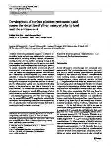
Scheme 1. (A) Illustration of possible mechanism for the preparation of Fe-MPA-NCC-CTAB composite, (B) The sensor functionalization. (a) Surface activation of gold/Fe-MPA-NCC-CTAB layer, (b) Immobilize the IgM antibody via EDC-NHS cross-linker, (d) Injection of DENV E-protein solution.
and 1 mg/ml, respectively, were purchased from Meridian Life Science. A prism with refractive index, n = 1.7786 at 632.8 nm and the substrate, glass cover slips 24 × 24 mm with thickness 0.13–0.16 mm were purchased from Menzel-Glaser. Phosphate buffered saline (PBS, pH 7.4) was prepared by compounding the solution of Na2HPO4 and NaH2PO4 to be used in a dilution of DENV E-protein and IgM. All chemicals were of analytical reagent grade.
[16]. To enhance the sensitivity of biosensor, the modification of commercial NCC by hexadecyltrimethylammonium bromide (CTAB), a cationic surfactant with an alkyl chain and quaternary ammonium group, has been employed [17]. The use of CTAB also help to stabilize and locate the magnetite nanoparticles on the NCC surface by noncovalent interactions with hydroxyl groups. The aim of this study is to propose a technique for the early detection of DENV E-protein by immobilized the ligand IgM antibody as the sensing layer in SPR. The preparation of Au-Fe3O4-MPA-NCCCTAB/EDC-NHS thin films and the studies on its sensing properties in an optical system for identification of IgM antibodies to the DENV antigen is explored.
2.2. Preparation of Fe3O4-MPA 1 wt% of Fe3O4 was capped with MPA using a mol ratio of 1:30 in deionized water and was shaked for overnight. The mixture was separated by centrifugation at 4000 rpm for 5 min, and afterwards, it was washed twice with deionized water and ethanol. The suspended mixture allow to dry at 100 °C for 2 h under vacuum condition [18].
2. Experimental section 2.1. Reagent and materials The magnetic (Fe3O4) powder was supplied by the Institute of Advanced Technology, Malaysia. 3-Mercaptopropionic (MPA) acid, hexadecyltrimethylammonium bromide (CTAB) and N-hydroxysuccinimide (NHS) were purchased from Sigma Aldrich, Germany. NEthyl-N-3-(dimethylaminopropyl) carbodiimide (EDC) was bought from Fluka, Switzerland. The freeze dried nanocellulose crystalline (NCC) with a features fibers 5–20 nm wide and 150–200 nm in length was purchased from the University of Maine, United States. The standard powder of recombinant dengue type 2 envelope protein (DENV E-protein) and its monoclonal antibody (IgM) with concentration 2.85 mg/ml
2.3. Preparation of NCC-CTAB The preparation of NCC-CTAB was based on previous work [17], which begin by preparing a 1 wt% suspension of NCCs at adjusted pH 10 through the addition of 1 M NaOH and a 1 wt% aqueous CTAB solution (2:1 CTAB to sulfur mol ratio). The suspension was slowly added to the CTAB solution, and then the mixture was stirred thoroughly for 3 h at 60 °C. After 3 h of stirring, the reaction mixture was left to stir at room temperature overnight. 17
Sensing and Bio-Sensing Research 20 (2018) 16–21
N.A.S. Omar et al.
2.4. Preparation of Fe3O4-MPA-NCC-CTAB solution The Fe3O4-MPA solution was prepared by dissolving 5 mg of Fe3O4MPA powders in 1 ml deionized water. The obtained solution was then mixed with 1 ml NCC-CTAB by shaking or vortex at room temperature for 1 min to ensure proper mixing. This sample will be referred as Fe3O4-MPA-NCC-CTAB in future discussion. 2.5. Preparation of DENV and IgM solution The DENV E-protein and IgM standard powder were diluted in PBS (pH 7.4) using dilution formula (M1V1 = M2V2) to produce DENV and IgM solutions with concentration 0.0001, 0.001, 0.01, 0.1, 1, 10 nM and 10 nM, respectively. 2.6. Preparation of thin films The glass cover slips were cleaned using acetone to cleanse off the dirt and fingerprint marks laid on the surface of glass slides. The glass slides were then deposited with a thin gold layer using SC7640 Sputter Coater. Approximately 0.55 ml of the Fe3O4-MPA-NCC-CTAB solution was first dropped on the top glass cover slip covering the surface. They were spun at 6000 rev/min for 30 s using Spin Coating System, P6708D. The formation of Fe3O4-MPA-NCC-CTAB composite can be explained through the electrostatic interaction of positively charged of NCC-CTAB and negatively charge to carboxyl group of Fe3O4-MPA. After that, a cross link solution containing EDC (2 mM) and NHS (5 mM) was coated to the sensor surface, forming a stable amide bond via carboxylic functions of Au/Fe-MPA-NCC-CTAB sensor surfaces. The sensor surface was further activated through covalent binding of terminal amine groups of IgM antibody immobilization. DENV E-protein solution was then introduced as a target antigen. The schematic representation of the entire surface of thin film modifying steps is illustrated in Scheme 1.
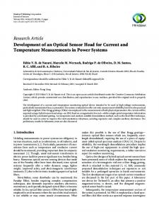
Fig. 1. Experimental set up for angle scan SPR technique.
2.7. Instrumental The thin films were imaged using an AFM in Scan Asyst mode. The Scan Asyst mode is a Peak Force Tapping technique that enables to observe the surface morphology in the high resolution images using single-touch scanning of the thin film. An optical spectroscopy was designed in the laboratory to test the capability of the thin film to diagnosis of DENV based on surface plasmon resonance principle (Fig. 1). The SPR measurement had been carried out by measuring the reflected He-Ne laser beam (632.8 nm, 5 mW) as a function of incident angle. The optical setup consists of a He-Ne laser, an optical stage driven by a stepper motor with a resolution of 0.001° (Newport MM 3000), a polarizer and an optical chopper (SR 540). The He-Ne laser beam was incident on to the prism (refractive index of 1.77861), passed through the sample (derivative thin film), and the reflected beam was detected by a large area photodiode. The signal was then processed by the lockin amplifier (SR 530) [19].
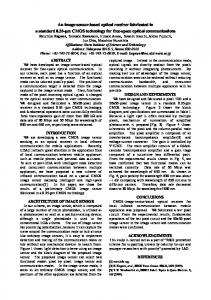
Fig. 2. SPR reflectivity curves for Au/IgM film in contact with different concentration of DENV E-protein solution ranged from 0.0001 nM to 10 nM.
It was probably due to the less amount of DENV E-protein existed in these low concentration solutions to bind to immobilized IgM antibody. While for high DENV E-protein concentration (1 nM and 10 nM), the resonance angles were being shifted to the smaller angle. The reason for this shift was probably due to the increment number of analyte adsorb to the gold surface, which consequently increased the shift of resonance angle, such as in the works of Fen et al. (2011) [20]. Based on the above results, the researchers were interested to increase the sensitivity of SPR technique in the DENV E-protein detection below 10 nM.
3. Results and discussion 3.1. SPR reflectivity for DENV E-protein on Au/IgM thin film Prior to the measurement, the IgM antibodies were immobilized on gold film using spin coating technique in order to prepare the Au/IgM layer. The SPR test was then carried out for different concentration of DENV E-protein (ranging from 0.0001 to 10 nM) solution, which were injected one after another into the cell to be in contact with immobilized IgM on gold surface. The SPR curve for DENV E-protein concentrations in contact with Au/IgM film is shown in Fig. 2. The result showed that the resonance angle did not show any shifting, i.e. 53.471° when the target antigen concentration was varied below 1 nM.
3.2. SPR signal for DENV E-protein on Au/Fe-MPA-NCC-CTAB/EDCNHS/IgM thin film 3.2.1. Optimization of SPR sensor A primarily test was conducted with three different sensing materials layered on gold film after being exposed to the 0.01 nM DENV E18
Sensing and Bio-Sensing Research 20 (2018) 16–21
N.A.S. Omar et al.
Table 1 The SPR resonance angle and shift of resonance angle for different concentrations of DENV E-protein solution in contact with Au/Fe-MPA-NCC-CTAB/EDCNHS/IgM (0 nM represents PBS solution). Concentration of DENV Eprotein (nM)
Resonance angle, θ (degree)
Shift of resonance angle, Δθ (degree)
0 0.0001 0.001 0.01 0.1 1 10
54.404 54.226 54.543 54.792 54.788 54.538 54.489
0 0.178 0.139 0.388 0.384 0.134 0.085
Fig. 3. SPR reflectivity curves for different sensing film in contact with concentration of 0.01 nM DENV E-protein solution.
protein solution for 10 min as shown in Fig. 3. The first sensing layer consists of Fe-MPA/IgM, followed by Fe-MPA-NCC-CTAB/IgM and finally Fe-MPA-NCC-CTAB/EDC-NHS/IgM. The purpose of this procedure is to determine the sensitivity of the sensor layer in detecting DENV E-protein. From the result, the resonance angle for Au/Fe-MPA/ IgM in contact with DENV is 54.321° and then slightly decreased to 54.217° when NCC-CTAB layer is added. This may due to the less immunospecific binding of IgM onto the Fe-MPA-NCC-CTAB surface. To enhance the attachment of protein binding, a cross-linker of EDC-NHS has been introduced above Fe-MPA-NCC-CTAB layer. It was found that the resonance angle became narrowed and shifted obviously to the 54.641°. Therefore, it was concluded that Fe-MPA-NCC-CTAB/EDCNHS/IgM thin film is more sensitive for the detection of DENV E-protein solution.
Fig. 5. Langmuir isotherm model of the SPR angle shift.
angle for different concentrations of DENV E-protein in contact with FeMPA-NCC-CTAB/EDC-NHS/IgM thin film is shown in Table 1. The shift in the resonance angle (Δθ) was determined by taking the difference between the resonance angle of the sample and the PBS solution as a reference (54.404°). The SPR signal shifts mostly depended on the number of binding dengue onto Au/Fe-MPA-NCC-CTAB/EDC-NHS immobilized IgM antibodies surface. In other words, the more attached dengue antigens are in the IgM surface area, the greater the shift can be
3.2.2. SPR reflectivity The analysis of SPR reflectivity curves for Au/Fe-MPA-NCC-CTAB/ EDC-NHS/IgM thin film in contact with different concentration of DENV b-protein solution ranged from 0.0001 nM to 10 nM are presented in Fig. 4 (a) and (b). The resonance angle and shift of resonance
Fig. 4. SPR reflectivity curves for Au/Fe-MPA-NCC-CTAB/EDC-NHS/IgM thin film in contact with different concentration of DENV E-protein solution ranged from (a) 0.0001 nM to 0.01 nM and (b) 0.01 nM to 10 nM. 19
Sensing and Bio-Sensing Research 20 (2018) 16–21
N.A.S. Omar et al.
concentrations of 0.0001 nM to 0.1 nM were used as a target antigen detection in future measurement. By fitting the data from Table 1 using Langmuir isotherm model [22], the binding affinity of the DENV E-protein towards the Fe-MPANCC-CTAB/EDC-NHS/IgM sensing layer can be obtained as follows the expressed equation
Δθ =
Δθmax C 1 K
+C
(1)
where Δθmax is the maximum SPR shift at the saturation, C is the DENV E-protein concentration and K is the affinity constant. The plots which are fitted in the Langmuir model through the MATLAB fitting tool are shown in Fig. 5. The fitting of the SPR curve for DENV E-protein using Langmuir isotherm model yielded an adjusted R2 value of 0.70 and Δθmax is approximately of 0.399° which is about 0.011° higher than an experimental maximum shift of the SPR angle, i.e., 0.388°. From this model, the binding affinity constant (K) for DENV E-protein to the Au/ Fe-MPA-NCC-CTAB/EDC-NHS/IgM was 938.41 nM−1, while −1 0.09 nM to the Au/IgM layer. It was observed that a higher affinity constant value was attributed to Au/Fe-MPA-NCC-CTAB/EDC-NHS/ IgM sensing layer, which was envisaged to have high potential sensitive and stronger binding towards DENV E-protein. The sensitivity of Fe-MPA-NCC-CTAB/EDC-NHS/IgM sensing layer within the range of DENV E-protein concentration was also determined by plotting the linear regression graph for concentration up to 0.01 nM by neglecting the data of 0.1 nM (Fig. 6). The linear regression analysis yielded the gradient of 39.96° nM−1 with its adjusted correlation coefficients, R2, of 0.73. The gradient of the SPR curve is defined as the sensitivity of sensing layer towards antigen detection. Therefore, it can be concluded that the sensitivity of the Fe-MPA-NCC-CTAB/EDC-NHS/ IgM towards DENV E-protein concentration, i.e., 0.0001 nM to 0.01 nM is 39.96° nM−1.
Fig. 6. Change in SPR resonance angle (Δθ) versus DENV E-protein concentration.
observed. Overall, it was observed that the shift in the resonance angle was slightly increased from 0.178° to 0.384° when the DENV E-protein concentration was increased up to 0.1 nM. This finding can be attributed to the increment number of binding between analyte-ligand, which led to an increase in the refractive index of the sensing layer. The result is also in agreement with Shankaran et al. [21], who found the increased in resonance angle is the effect of the increasing binding event of antigen-antibody immunoreaction. While for the high DENV Eprotein concentrations of 1 nM and 10 nM, the shift in the resonance angle were dropped into 0.085°. It is because the highly antigen concentration will lead the antibody immobilized sensor surface was fully covered and congested, and for this reason, the DENV E-protein
Fig. 7. (a) AFM image of Au/Fe-MPA-NCC-CTAB/EDC-NHS surface with immobilized antibody. (b) AFM image of Au/Fe-MPA-NCC-CTAB/EDC-NHS/IgM surface in contact to DENV E-protein. 20
Sensing and Bio-Sensing Research 20 (2018) 16–21
N.A.S. Omar et al.
3.3. AFM analysis
[3] P.R. Barrero, A.S. Mistchenko, Complete genome sequencing of dengue virus type 1 isolated in Buenos Aires, Argentina, Virus Res. 101 (2004) 135–145. [4] M.G. Guzmán, G. Kouri, Dengue diagnosis, advances and challenges, Int. J. Infect. Dis. 8 (2004) 69–80. [5] P. Chawla, A. Yadav, V. Chawla, Clinical implications and treatment of dengue, Asian Pac J Trop Med 7 (2014) 169–178. [6] E. Pokidysheva, Y. Zhang, A.J. Battisti, C.M. Bator-Kelly, P.R. Chipman, C. Xiao, G.G. Gregorio, W.A. Hendrickson, R.J. Kuhn, M.G. Rossmann, Cryo-EM reconstruction of dengue virus in complex with the carbohydrate recognition domain of DC-SIGN, Cell 124 (2006) 485–493. [7] T.C. Pierson, D.H. Fremont, R.J. Kuhn, M.S. Diamond, Structural insights into the mechanism of antibody-mediated neutralization of flavivirus infection: implications for vaccine development, Cell Host Microbe 4 (2008) 229–238. [8] H.H. Nguyen, J. Park, S. Kang, M. Kim, Surface plasmon resonance: A versatile technique for biosensor applications, Sensors 15 (2015) 10481–10510. [9] H. Labiadh, T.B. Chaabane, R. Silille, L. Balan, R. Schneider, A facile method for the preparation of bifunctional Mn: ZnS/ZnS/Fe3O4 magnetic and fluorescent nanocrystals, Beilstein J. Nanotechnol. 6 (2015) 1743–1751. [10] Z.P. Xu, Q.H. Zeng, G.Q. Lu, A.B. Yu, Inorganic nanoparticles as carriers for efficient cellular delivery, Chem. Eng. Sci. 61 (2006) 1027–1040. [11] A.S. Lubbe, C. Bergemann, J. Brock, D.G. McClure, Physiological aspects in magnetic drug-targeting, J. Magn. Magn. Mater. 194 (1999) 149–155. [12] C. Wang, J. Yang, X. Cui, H. Wang, Synthesis of raspberry-like monodisperse magnetic hollow hybrid nanospheres by coating polystyrene template with Fe3O4@ SiO2 particles, J. Colloid Interf. Sci. 354 (2011) 94–99. [13] S.M. Zhu, J.J. Guo, J.P. Dong, Z.W. Cui, T. Lu, C.L. Zhu, D. Zhang, J. Ma, Sonochemical fabrication of Fe3O4 nanoparticles on reduced graphene oxide for biosensors, Ultrason. Sonochem. 20 (2013) 872–880. [14] J.J. Hong, J.S. Fu, Y. Wei, L.C. Yue, Preparation and application of magnetic Fe3O4 nano-crystalline, Prog. Chem. 22 (2010) 1566–1574. [15] N. Lin, A. Dufresne, Nanocellulose in biomedicine: current status and future prospect, Eur. Polym. J. 59 (2014) 302–325. [16] I. Oke, Nanoscience in nature: cellulose nanocrystals, Surg. J. 3 (2010) 77–80. [17] T. Abitbol, H. Marway, E.D. Cranston, Surface modification of cellulose nanocrystals with cetyltrimethyliammonium bromide, Nord. Pulp Pap. Res. J. 29 (2014) 46–57. [18] S. Sathish, S. Balakumar, Influence of physichochemical interactions of capping agent on magnetic properties of magnetite nanoparticles, Mater. Chem. Phys. 173 (2016) 364–371. [19] Y.W. Fen, W.M.M. Yunus, Z.A. Talib, Analysis of Pb(II) ion sensing by crosslinked chitosan thin film using surface plasmon resonance spectroscopy, Optik 124 (2013) 126–133. [20] Y.W. Fen, W.M.M. Yunus, N.A. Yusof, Optical properties of cross-linked chitosan thin film for copper ion detection using surface plasmon resonance technique, Opt. Appl. 41 (2011) 999–1013. [21] D.R. Shankaran, K.V. Gobi, T. Sakai, K. Matsumoto, T. Imato, K. Toko, N. Miura, A novel surface plasmon resonance immunosensor for 2,4,6-trinitrotoluene (TNT) based on indirect competitive immunoreaction: a promising approach for on-site landmine detection, IEEE Sensors J. 5 (2005) 616–621. [22] N.H. Kamaruddin, A.A.A. Bakar, N.N. Mobarak, M.S.D. Zan, N. Arsad, Binding affinity of a highly sensitive Au/Ag/Au/chitosan-graphene oxide sensor based on direct detection of Pb2+ and Hg2+ ions, Sensors 17 (2017) 1–16. [23] P. Jahanshahi, E. Zalnezhad, S.D. Sekaran, F.R.M. Adikan, Rapid immunoglobulin M-based dengue diagnostic test using surface plasmon resonance biosensor, Sci. Rep. 4 (2014) 1–7.
Furthermore, the images of the antibody immobilization site were studied using atomic force microscopy (AFM) and are shown in Fig. 7 (a). According to the 3D image, high sporadic hills were created by immobilized antibody on the sensor surface, which anchored well to the amine groups on the Au layer. The above finding was also consistent with the studies conducted by Jahanshahi [23]. After being injected with the DENV E-protein as shown in Fig. 7 (b), the clear asymmetric shapes and also the addition of sporadic hills on the surface were observed in 2D and 3D images, respectively, confirming the DENV attachment to the antibody surface. 4. Conclusion In the present work, immobilization of IgM in gold/Fe-MPA-NCCCTAB/EDC-NHS for detection of DENV using SPR was developed. By introducing IgM immobilized Fe-MPA-NCC-CTAB/EDC-NHS on a gold surface, DENV concentration range of 0.0001 nM to 10 nM can be determined. The sensitivity found by optical sensor in contact with DENV is 39.96° nM−1. Further studies on the modification of the active layer to enhance sensitivity and selectivity for the early detection of DENV using SPR method is important. Hence, it is envisaged that the future directions in the diagnosis of dengue virus will focus on the improvement of the sensitivity and the specificity for a particular dengue virus protein. Acknowledgements The authors gratefully acknowledged the financial support for this study from the Universiti Putra Malaysia (UPM) through the Putra Grant (9531500 and 9520600). The laboratory facilities provided by the Functional Devices Laboratory, Institute of Advanced Technology, Universiti Putra Malaysia, were also acknowledged. References [1] K. Navakul, C. Warakulwit, P. Yenchitsomanus, A. Panya, P. Lieberzeit, C. Sangma, A novel method for dengue virus detection and antibody screening using a grapheme-polymer based electrochemical biosensor, Nanomedicine 13 (2017) 549–557. [2] C.M. Rice, E.M. Lenches, S.R. Eddy, S.J. Shin, R.L. Sheets, J.H. Strauss, Nucleotide sequence of yellow fever virus: implications for flavivirus gene expression and evolution, Science 229 (1985) 726–733.
21