Jun 17, 2016 - Oxford College of Pharmacy, Ghaziabad, India. 3. Meerut Institute of Engineering & Technology, Meerut. ABSTRACT. An attempt has been ...
WORLD JOURNAL OF PHARMACY AND PHARMACEUTICAL SCIENCES Priya et al.
World Journal of Pharmacy and Pharmaceutical Sciences
SJIF Impact Factor 6.041
Volume 5, Issue 8, 992-1024
Research Article
ISSN 2278 – 4357
DEVELOPMENT OF COLON TARGETING DRUG DELIVERY SYSTEM USING PLANT POLYSACCHARIDE Bhanu Priya1*, Shubham Verma2 and Nitin Sharma3 1
IIMT College of Pharmacy, Greater Noida, India.
2 3
Article Received on 28 May 2016,
Oxford College of Pharmacy, Ghaziabad, India.
Meerut Institute of Engineering & Technology, Meerut. ABSTRACT An attempt has been made to formulate colon targeted tablet dosage
Revised on 17 June 2016, Accepted on 07 July 2016
form of metronidazole as model drug using natural polysaccharide
DOI: 10.20959/wjpps20168-7342
extracted from Abelmoschus moschatus (Mushkadana, familyMalvaceae). In preliminary study, extracted polysaccharide was
*Corresponding Author
characterized in terms of physicochemical properties. Swelling study at
Bhanu Priya
different pH confirmed, utility of plant polysaccharide as a colon
IIMT College of
targeting carrier. Core tablets were prepared by changing the
Pharmacy, Greater Noida,
concentration of polysaccharide (10 to 30 mg) and using wet and dry
India.
granulation method. Optimized core tablets were coated by compression coating with 350 mg polysaccharide (Abelmoschus moschatus). The amount of metronidazole released from tablets at different time intervals was estimated by UVSpectroscopy method. In-vitro dissolution studies revealed that natural polysaccharide based colon targeted tablet was successfully following the release pattern specific to colon delivery. It was observed that release of drug was become rapid by decreasing the concentration of extracted polysaccharide. The result of the studies shows that compression coated formulation of metronidazole tablets with 350 mg of Abelmoschus moschatus polysaccharide as coating material is most likely to provide targeting of drug for local action in the colon owing to its minimal release of the drug in the first 5 hr. KEYWORDS: Colon, Abelmoschus moschatus, Metronidazole, Natural Polymer, Anti Amoebic.
www.wjpps.com
Vol 5, Issue 8, 2016.
992
Priya et al.
World Journal of Pharmacy and Pharmaceutical Sciences
INTRODUCTION An ideal targeted drug delivery system is the one which delivers the drug at a predetermined rate locally or systemically for a specified period of time. Oral route has been the most preferred and suitable route for the delivery of drug. Oral route of drug management has gained consideration in the pharmaceutical field, because of more adaptability in the designing of dosage form as compared to other dosage form for other routes. Drug delivery by oral route depends on different variables such as patient, type of delivery system, disease being treated, duration of therapy and drug properties of the majority of oral controlled drug delivery system. Oral drug delivery for colon targeting is the most preferred route by a majority of patients due to its ease of administration. By definition, colonic delivery refers to targeted delivery of drugs into the lower GI tract, which occurs primarily in the large intestine (i.e. colon). Targeted drug delivery to the colon would therefore ensure direct treatment at the disease site, lower dosing and fewer systemic side effects. In addition to local therapy, the colon can also be utilized as a portal for the entry of drugs into the systemic circulation. Colon drug delivery is beneficial not only for oral delivery of peptide and protein drugs but also delivery of those compounds whose molecular weight is very low and used to treat the colon diseases.[1] ANATOMY OF COLON The gastro intestine is divided into three parts- stomach, small intestine, and large intestine. The large intestine in human is about 1.5 m long. The colon is upper part (five feet) of large intestine situated mainly in the abdomen. The large intestine extending from the ileocecal junction to the anus is divided in to three main parts- colon, rectum and anal canal. The colon is cylindrical tube which is lined by moist, soft pink lining called mucosa and the pathway is called the lumen. The cecum forms the first part of the colon and leads to the right colon (just under the liver) followed by the descending colon, sigmoid colon, rectum and anal cana.[2]
Figure 1: Structure of human intestine www.wjpps.com
Vol 5, Issue 8, 2016.
993
Priya et al.
World Journal of Pharmacy and Pharmaceutical Sciences
Table 1: Anatomical Properties of Gastrointestinal Tract. Region of gastrointestinal tract Duodenum Small intestine Jejunum Ileum Cecum Ascending colon Descending colon Large intestine Transverse Colon Sigmoid colon Rectum Anal canal pH of colon pH of stomach and small intestine is given below.[3]
Length (cm) 20- 30 150- 250 200-350 6- 9 20- 25 10- 15 40- 45 35- 40 12 3
Table 2: pH of Stomach and Small Intestine. Region Stomach Small intestine Jejunum Illeum Large intestine Colon
pH 1- 1.5 (Fasted) 2- 3 (fed) 5.0-7.5 6.0-7.3 6.4-7.0
Advantages of colon targeting 1. Direct treatment at the disease site, lower dosing and fewer systemic side effects. 2. Drug absorption may be delay by using colon targeting. 3. Colon targeting specific formulation allow oral administration of protein and peptides drugs that could be used to prolong the drug delivery. 4. In the treatment of colon diseases. 5. To reduce incidence of adverse effects. Limitations and challenges in colon targeted drug delivery.[4] Multiple manufacturing steps. Delivery of colon specific drug delivery system requires well established dissolution testing method. Drug should be in solution form before it arrives in the colon or alternatively. Metabolic degradation of drug and colonic performances are also effected by
resident
microflora.
www.wjpps.com
Vol 5, Issue 8, 2016.
994
Priya et al.
World Journal of Pharmacy and Pharmaceutical Sciences
The tight junction and lower surface area in colon can also restrict the drug transport across the mucosa and into the systemic circulation. Before the designing of colon delivery system, the stability of the drug should be known. The drug may potentially bind in non specific way to dietary residues, mucus or feacal matter and intestinal secretion. MATERIALS AND METHOD Albemoschus moschatus (AM) was procured from the local market of Greater Noida, India. It was authenticated by the Department of Botany, Banaras Hindu University, Varanasi, India. Metronidazole drug was obtained from Zee Laboratories, Himachal Pradesh, India. Isolation of Polysaccharide The stems of the plant AM were collected and washed with distilled water to remove the dirt particles and crushed. The crushed stems were soaked in double distilled water at room temperature and kept overnight to settle down undissolved material. The supernatant was separated by decantation. Supernatant was concentrated to its 1/3rd volume under vacuum by using rotatory evaporator (Yamato) and was cooled. The concentrated liquid was precipitated with acetone using 1:3 mucilage: acetone ratio. Green precipitates were obtained after repeated washing, which were then dried at 50-60°C. The dried material was powdered and kept in desiccator till further use. After extraction of polysaccharide by above procedure the percentage yield of the polysaccharide was found to be 8%.w/w. Preliminary Chemical Tests for Characterization of Isolated Albemoschus moschatus Polysaccharide Preparation of test solution of Albemoschus moschatus (AM). Pinches of the above dried precipitate were dissolved in distilled waterhence forth referred as test solution. TEST FOR CARBOHYDRATE a) Molish’s Test: The Molish’s reagent was prepared by dissolving 10 g of alpha naphthol in 100 ml of 95% alcohol. A few ml of test solution was placed in test tube, and it was mixed with 2 drops of Molish’s reagent. To this solution, 1mL of concentrated sulphuric acid was added from the side of the inclined test tube, so that the acid formed a layer beneath the aqueous solution
www.wjpps.com
Vol 5, Issue 8, 2016.
995
Priya et al.
World Journal of Pharmacy and Pharmaceutical Sciences
without mixing with it. Development of red brown ring at the junction of two layers of the liquids indicates the presence of carbohydrates. b) Fehling’s Solution Test The two solutions (Fehling‘s solution A + Fehling‘s solution B) were mixed in equal volumes immediately before use. A little of test solution was taken in the above test tube and then this mixture was warmed. Red precipitates of cuprous oxide formation confirmed the presence of carbohydrates. TEST FOR STEROLS a) Salkowaski Reaction Few mg of dried precipitates were taken in 2 mL of chloroform and 2 mL of conc. sulphuric acid was added from the side of test tube. The test sample was shaken for few minutes. The presence of red colour in the chloroform layer will indicate the presence of sterols. b) Libermann-Burchard Reaction Few mg of dried precipitates were dissolve in chloroform and few drops of acetic anhydride were added to it. Then few drops of conc. sulphuric acid were added from the side of the test tube. If the transient colour develops from red to blue and finally green, then indicates the presence of sterol. TEST FOR ALKALOIDS Few mg of dried precipitates were taken separately in 5 mL of 1.5% v/v hydrochloric acid and filtered. These filtrates were then used for testing alkaloids with following reagents. a) Dragendroff’s reagent In the presence of dragendroff’s reagent (potassium bismuth iodide solution) the above filtrates give raddish brown precipitate then it indicates the presence of alkaloids. b) Mayer’s Reagent In the presence of alkaloids it gives cream colour precipitate with mayer’s reagent (potassium iodide solution).
www.wjpps.com
Vol 5, Issue 8, 2016.
996
Priya et al.
World Journal of Pharmacy and Pharmaceutical Sciences
c) Wagner’s reagent 1.27 g of iodine and 2 g of potassium iodide were dissolved in 5 mL of water and the solution was diluted to 100 mL with water. When few drops of this reagent were added to the test filtrate, If a brown flocculent precipitate was formed indicating the presence of alkaloids. d) Hanger’s Reagent A saturated aqueous solution of picric acid was employed for this test. When the test filtrate was treated with this reagent. If it gives orange yellow precipitate then it will indicate the presence of alkaloids. TEST FOR SAPONINS (Foam Test) A few mg of the dried precipitate was taken in a test tube and shaken vigorously with a small amount of sodium bicarbonate and water. If stable, characteristic honeycomb like froth is obtained, then saponins are presents. TEST FOR TANNINS The dried precipitate was taken separately in water, warmed and filtered. Tests were carried out with the filtrate using following reagents. a) Ferric chloride reagent A 5% w/v solution of ferric chloride in 90% alcohol was prepared. Few drops of this solution were added to the filtrate. The presence of Dark green or deep blue colour will indicate the presence of tannins. b) Lead acetate test A 10% w/v solution of basic lead acetate in distilled water was added to the test filtrate if the formation of precipitate then it indicates the presence of tannins. c) Bromine water test Bromine solution was added to the test filtrate. Decolourization of bromine water indicates the presence of tannins. PROTEIN TEST a) Xanthoproteic test A few mg of dried precipitate was taken with 2mL of water and 0.5 mL of concentrated nitric acid was added to it. If the yellow colour produced it will indicate the presence of proteins.
www.wjpps.com
Vol 5, Issue 8, 2016.
997
Priya et al.
World Journal of Pharmacy and Pharmaceutical Sciences
b) Million’s Test (Mercuric Nitrate Solution) Million’s reagent was prepared by dissolving 3 mL of mercury in 27 mL of fuming nitric acid keeping the mixture well cooled; this solution was then diluted with equal quantity of distilled water. Aqueous solution of the dried precipitate was taken and 2 mLor 3 mL of million’s was added. In the presence of proteins it will gives white precipitate, which slowly turns to pink. c) Warming test Test solution was heated in a boiling water bath, proteins get coagulated. TEST FOR AMINO ACIDS a) Ninhydrin Test The Ninhydrin reagent is 0.1% w/v solution of ninhydrine in n-butanol. A little of this reagent was added to the extract. A violet or purple colour is developed if the amino acids are present. TEST FOR GLYCOSIDES About 2mL of dried precipitate were taken and subjected to following tests. a) Killer - killani Test glacial acetic acid (1 mL) containing traces of ferric chloride and 1mL of concentrated sulphuric acid was added to the dried precipitate and observed for formation of reddish brown colour at the junction of two layers formation of the bluish green colour at the upper layer,will indicates the presence of glycoside. b) Borntrager’s Test One mL of benzene and 0.5mL of dilute ammonia solution were added to the extracted dried precipitate and observed it will produce the reddish pink colour in the presence of glycosides. c) Legal Test The above test solution was made alkaline with few drops of 10% w/v of sodium hydroxide solution and then freshly prepared sodium nitroprusside solution was added to the solution and observed for the formation of blue colour.
www.wjpps.com
Vol 5, Issue 8, 2016.
998
Priya et al.
World Journal of Pharmacy and Pharmaceutical Sciences
d) Baljet Test To the concentrated extracts sodium picrate reagent were added and observed for the formation of orange and yellow colour. TEST FOR PHENOLIC COMPOUNDS a) Ferric Chloride Solution The test sample was taken and warmed add 2mL of ferric chloride solution and observed for the formation of green and blue colour in the presence of phenolic compounds. b) Lead Acetate Solution The test sample was taken and add the 2mL of lead acetate solution if precipitate were observed then phenolic compounds are present. c) Gelatine Solution A few mL of Gelatine Solution was added to the test solution and observed the formation of precipitate or turbidity will indicate the presence of phenolic compounds. TEST FOR LIPIDS AND OIL a) Rub a small quantity of dried precipitate on a paper in the presence of lipids or oil it will produced a permanent translucent stain. b) To 2 mL of test solution, 2-3 drops of N/50 iodine solution was added and the colour of the particles was noted. TEST FOR STARCH a) Iodine test Mix 3 mL test solution and few drops of dilute iodine solution. In the presence of starch blue colour appear, it disappear on boiling and reappear on cooling. b) Tannic acid test for starch The test sample was mixed with 20% tannic acid; if it gives precipitate then indicates the presence of starch. TEST FOR MUCILAGE a) Ruthenium red test: The dried precipitate was mounted on a slide with ruthenium red solution and covered with a cover slip. After a few seconds, it was irrigated with lead acetate and the excess stain was sucked off with a blotting paper. Lead acetate solution was added to
www.wjpps.com
Vol 5, Issue 8, 2016.
999
Priya et al.
World Journal of Pharmacy and Pharmaceutical Sciences
prevent undue swelling of the test solution. The pink colour of the particles shows the presence of mucilage. b) The test sample was heated for some time and then cooled. Formation of gelatinous mass indicates presence of mucilage. PREFORMULATION STUDIES Preformulation study is an important step for determination of physical and chemical properties of the drug before incorporating it in formulation development. The nature of the drug highly affects the processing parameters like method of preparation, selection of solvents, compatibility and pharmacokinetic response of the formulation. Preformulation studies are indispensable protocol for development of safe, effective and stable dosage form. Thus, in order to ensure optimum condition for clinically beneficial delivery system, preformulation studies were carried out. A thorough understanding of these properties, ultimately provide a rational for formulation design. Drug identification test and drug excipients compatibility studies were done in this phase to provide a useful support in development of dosage form.[5,6] Performulation Study of Drug Preformulation is defined as the application of pharmaceutical principles to the physicochemical parameters of drug substance and other excipients characterized with the aim of designing optimum drug delivery system. Drug Identification Tests The selected drug Metronidazole was identified by various methods like physical characterization, melting point determination, UV-spectrophotometric study, and infrared (IR) spectroscopy. Physical Characterization Metronidazole was physically characterized on the basis of appearance, colour, odour and taste. All these organoleptic parameters were recorded and compared with standard data. Melting Point Apparatus Capillary melting point apparatus was used to determine melting point of the drug. The temperature at which a solid melts becomes a liquid is the melting point. The melting point of a drug can be determined by introducing a tiny amount of drug into a small capillary tube
www.wjpps.com
Vol 5, Issue 8, 2016.
1000
Priya et al.
World Journal of Pharmacy and Pharmaceutical Sciences
(one side fused by heat), attaching this to the stem of a thermometer centered in a heating bath, bath was heated slowly, and temperatures was observed at which melting begins and is completed. The melting point was recorded in triplicate and compared with literature value. UV- Spectrophotometer Study UV spectrophotometric study was carried out in order to determine the λmax of metronidazole in phosphate buffer solution of pH 6.8 as per IP 1996. A standard stock solution of metronidazolewas prepared by dissolving 100 mg of drug in a 100 ml volumetric flask and the volume was made upto 100 ml by using phosphate buffer solution of pH 6.8 to get the concentration 1000 µg/ml of standardmetronidazole. From the standard stock solution, 1 ml was pipette out into 10 ml volumetric flask and the volume was made upto 10 ml with phosphate buffer solution of pH 6.8 to get the concentration. Maximum wavelength (λmax) was obtained by scanning the resulting solution (10 µg/ml) in the wavelength region between 200 nm to 400 nm by using UV-VIS spectrophotometer (UV1700 Pharma Spec, Shimadzu, Japan). Same procedure was repeated with pH 1.2 and maximum wavelength (λmax) was obtained by scanning the resulting solution (10 µg/ml) in the wavelength region between 200 nm to 400 nm by using UV-VIS spectrophotometer. Infrared (IR) Spectroscopy IR spectroscopy was done using Shimadzu Fourier Transform Infrared (FTIR) spectrophotometer. Perfectly dried metronidazole (5mg) was mixed with powder, which have no absorption in IR region such as potassium bromide (KBr). Pellet of this mixture was then prepared by using KBr press at 15 tons and IR spectrum was taken by at frequency of 4000400 cm
-1
. Obtained IR spectrum was compared with reported spectra to match the
characteristic peaks. Drug Excipients Compatibility Screening Drug (Metronidazole), polysaccharide of Albemoschus moschatus and all excipient were taken in ratio similar to that to be used in formulation. The prepared physical mixture was placed in a petri-dish and kept for 15th days at room temperature. Compatibility of this mixture was examined after 15th days via observation for physical changes. Physical mixture was also subjected to FTIR spectroscopy to find out any interaction at molecular level.
www.wjpps.com
Vol 5, Issue 8, 2016.
1001
Priya et al.
World Journal of Pharmacy and Pharmaceutical Sciences
Calibration Curve Calibration curve of metronidazole was prepared by using U.V. (UV 1700 PharmaSpec, Shimadzu, Japan).
Selection of Media pH values was selected on the basis of pH value in stomach and intestine (Colon) for preparation of calibration curves. The media selected was USP phosphate buffer, pH 1.2 and pH 6.8. Preparation of Buffers and Reagents a) Sodium hydroxide solution, 0.2 M: 8 gm of sodium hydroxide was dissolved in distilled water and diluted to1000 mL with distilled water. b) Potassium dihydrogen phosphate solution, 0.2 M: Potassium dihydrogen phosphate (27.218 g) was dissolved in distilled water and diluted to 1000 mL. c) Hydrochloric acid solution, 0.1 N: Concentrated hydrochloric acid (8.5 mL) acid was diluted with distilled water and volume was made up to 1000 mL with distilled water. pH (1.2) was adjusted with dilute hydrochloric acid. Preparation of Calibration Curve of Metronidazole at pH 1.2 A standard stock solution of metronidazole was prepared by dissolving 100 mg of drug in a 100 mL volumetric flask and the volume was made upto 100 ml by using pH 1.2 to get the concentration 1000 μg/mL of standard metronidazole. From the standard stock solution, 1 mL was pipette out into 10 mL volumetric flask and the volume was made upto 10 mLwith pH1.2 buffer solution to get the concentration 100 μg/mL. From the above prepared stock solution, ten dilutions were made by using buffer solution pH 1.2 which has ultimate concentrations of 1, 2,3,4,5,6,7,8,9, and 10 µg/mL. The absorbance was measured at λmax 277 nm by using UV-VIS spectrophotometer (UV1700 PharmaSpec, Shimadzu, Japan). Preparation of Calibration Curve of Metronidazole at pH 6.8 A standard stock solution of metronidazole was prepared by dissolving 100 mg of drug in a 100 mL volumetric flask and the volume was made upto 100 mLby using phosphate buffer pH 6.8 to get the concentration 1000 μg/mL of standard metronidazole. From the standard
www.wjpps.com
Vol 5, Issue 8, 2016.
1002
Priya et al.
World Journal of Pharmacy and Pharmaceutical Sciences
stock solution, 1 mL was pipette out into 10 ml volumetric flask and the volume was made upto 10 ml with phosphate buffer pH 6.8 to get the concentration 100 μg/ml. From the above prepared stock solution, ten dilutions were made by using pH 6.8 which has ultimate concentrations of 1, 2,3,4,5,6,7,8,9, and 10 µg/mL. The absorbance was measured at λmax 319 nm by using UV-VIS spectrophotometer (UV1700 PharmaSpec, Shimadzu, Japan). Preparation of Rat Cecal Content Medium Albino rat weighing 150-200 gm and kept in fasting condition over night. Forty- five minutes before the commencement of drug release studies, seven rats were killed. The abdomen were opened, the cecae were traced, ligated at both the ends, dissected, and immediately transferred into pH 6.8 buffer. The cecal bages were opened, their contents were individually pooled and suspended in the buffer.[7] RESULT AND DISCUSSIONS Chemical Characterization of Isolated Polysaccharide Chemical characterization of isolated polysaccharide was done as per the reported models test and obtained results are given in Table 3. There was only carbohydrate tests (ruthenium red test, and molish’s test) found to be positive that indicated polysaccharide contain mucilage and reducing sugar. Table 3: Chemical Characterization of Isolated Polysaccharide. Test for Carbohydrate: Molish’s test Fehling solution A & B Benedict’s test Steroids and terpenoids: Salwoski reaction Libernann- Burchared reaction Alkaloid: Dragendroff test Mayer’s test Wagner test Saponins: Froth formation test
www.wjpps.com
Positive if Present
Albemoschus moschatus polysaccharide
Reddish brown layer Brick red Red precipitate
+ve +ve +ve
Red colour layer Green colour
-ve -ve
Orange brown colour Cream-colored precipitate Reddish-brown precipitate
-ve -ve -ve
Froth is formed
-ve
Vol 5, Issue 8, 2016.
1003
Priya et al.
World Journal of Pharmacy and Pharmaceutical Sciences
Tannins: Potassium dichromate test Bromine water Protien: Biuret test Xanthoprotien test Warming test Amino acid: Ninhydrin test Glycoside: Killer killani test Baljet test Legal test Phenolic compound: Ferric chloride test Lead acetate test Acetic acid solution Starch: N/50 iodine solution Mucilage test Ruthenium test
Red precipitate Discolourisation
-ve -ve
Violet or pink colour White precipitate Protein coagulates
-ve -ve -ve
Violet purple colour
-ve
Reddish brown ring Orange colour Pink to red colour
ve -ve -ve
Green or brown colour Precipitate formation Red colour solution
-ve -ve -ve
Blue colour
-ve
Pink colour
+ve
Organoleptic Evaluation of Polysaccharide Organoleptic evaluation of the extracted polysaccharidewas done and obtained results are given in Table 4. The surface morphology of polysaccharide was found to be slightly rough, determined by scanning electron microscopy. Table 4: Organoleptic Properties of Polysaccharide. Colour Brown
Odour Odourless
Taste Texture Shape Characteristic Rough Irregular
Solubility Profile of Polysaccharide The solubility profile is given in Table 5. From the solubility data it was found that polysaccharide forms viscous solution in hot and cold water and insoluble in organic solvents. Table 5: Solubility Profile of polysaccharide Solvents Cold water Hot water Ethanol Alcohol Acetone Phosphate buffer(6.8pH)
www.wjpps.com
Solubility Form viscous solution Form viscous solution Insoluble Insoluble Insoluble Form viscous solution
Vol 5, Issue 8, 2016.
1004
Priya et al.
World Journal of Pharmacy and Pharmaceutical Sciences
pH of Polysaccharide Solution pH of polysaccharide was investigated to check the acceptability with the biological mucosa. pH study showed that the polysaccharide have pH within physiological range so they are non – irritating to the biological mucosa. The result of are summarized in Table 6. Loss on Drying Loss on drying was done to identify the amount of volatile content or moisture which was entrapped within the particles. The result polysaccharide AM was represented in Table 6. Swelling Index Swelling index of polysaccharide was examined to find out the water holding capacity of the polysaccharide. Swelling index of AM was found 323.1±233. The result polysaccharide AM was represented in Table 6. Table 6: pH, Swelling Index and Loss on Drying of AM Polysaccharide. polysaccharide AM
pH of 1% w/v solution 7.86±0.03
Swelling index (%) 323.1 ± 2.33
Loss on drying (%w/v) 13±0.70
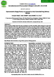
*Average of three readings ±S.D. Determination of swelling index of extracted polysaccharide Albemoschus moschatus (AM) in different pH media The swelling behaviour of extracted polysaccharide was examined in different pH media such as 1.2, 5.8, 7.8, and 8. The swelling behaviour of polysaccharide was found to be 82.1±0.1 and 72.1±0.015 in pH8 and 7.8 respectively. Henceits indicate this polysaccharide shows higher swelling behaviour in alkaline medium and may be used as colon delivery of drug. Table 7: Determination of swelling index of extracted polysaccharide (Albemoschus moschatus) in different pH. pH 8 7.8 5.8 1.2
www.wjpps.com
Swelling index (%) 82.1±0.1 72.1±0.15 53.2±0.18 30.1±0.20
Vol 5, Issue 8, 2016.
1005
Priya et al.
World Journal of Pharmacy and Pharmaceutical Sciences
Figure 2: swelling index of Polysaccharide at different pH. Ash Value Ash value was determined to identify the quality and purity of both the extracted polysaccharide. Ash value of both the polysaccharide was calculated by three ways and shown in Table 8. Table 8: Ash Value of AM Polysaccharide. Total Acid soluble ash (%) ash (%) 4±0.35 1±0.15 AM *Average of three readings ±S.D. Polysaccharide
Water soluble ash (%) 6±0.02
Scanning Electron Microscopy (SEM) From the SEM photograph (Fig 3), itis clear that polysaccharide(AM) possess rough surface and may be used for various pharmaceutical operations.
Figure 3: Scanning electron microscopy of extracted polysaccharide of Abelmoschus moschatus(AM).
www.wjpps.com
Vol 5, Issue 8, 2016.
1006
Priya et al.
World Journal of Pharmacy and Pharmaceutical Sciences
Micromeritics properties The result of the AM were found as represented in Table 9. Table 9: Physical Characterization of Polysaccharide. Parameters
Value
Bulk density(g/mL)
0.510±0.03
Tapped density(g/mL)
0.717±0.21
Carr’s index (%)
29.87±0.346
Hausner’s ratio
1.405±0.12
Angle of repose
28.52±0.09
Physical Characterization of Drug Metronidazole was evaluated for its physical properties and it was observed that metronidazole was a pale‐yellow crystal and odourless that was slightly soluble in water and alcohol. Metronodazole is an antiprotozoal, anti parasitic agent, very effective in the treatment of amoebiasis, trichomoniasis, giardiasis and many other parasitic diseases. The physical properties were found similar to that reported in. (IP 2007).This proves the identity of metronidazole. Melting Point Determination The melting point of drug was determined in triplicate and their mean values with standard deviation. The melting point of metronidazole was found to be 156 ±2ºC, which corresponds to the literature value of 158ºC to 160ºC that proves the identity and purity of drug. UV- Spectrophotometer Study Scannedλmax value of metronidazole in phosphate buffer (pH 6.8) U-V Spectroscopy study was carried out in order to determine the λmax of metronidazole in pH 6.8 phosphate buffer. Scanned λmax for metronidazole was shown in Figure 4.
www.wjpps.com
Vol 5, Issue 8, 2016.
1007
Priya et al.
World Journal of Pharmacy and Pharmaceutical Sciences
Figure: 4 Scannedλmax value of metronidazole in phosphate buffer (pH 6.8). Table 10: Scanned λmax and the absorbance value of sample of metronidazole prepared in 6.8 phosphate buffer. S.No. Strength Scanned λmax Absorbance 1 10 µg/mL 319 0.084 The scanned λmax found to be 319 nm and absorbance value for 10 to be 0.084. Scanned λmax value of metronidazole in phosphate buffer (pH 1.2) The absorbance and concentration data of metronidazole in pH 1.2 were given in Table 11. These absorbance were plotted on Y-axis against concentration on X-axis, and slope of the standard curves was obtained, shown in Figure 5.
Figure 5: Scannedλmax value of metronidazole in buffer solution pH 1.2. www.wjpps.com
Vol 5, Issue 8, 2016.
1008
Priya et al.
World Journal of Pharmacy and Pharmaceutical Sciences
Table 11: Scanned λmax and the absorbance value of sample of metronidazole prepared in 1.2 pH phosphate buffers. S.No. 1
Strength 10 µg/mL
Scanned λmax 277
Absorbance 2.660
FTIR Spectroscopy of Drug FTIR spectra of Metronidazole are given in Figure 6 and the interpretation in shown in Table 12.
Figure 6: FTIR Spectra of Metronidazole. Table 12: Interperation of FTIR of Metronidazole. Functional group
Reported frequency
(cm-1)
Observed frequency
OH stretching
3330
3409.91
C=C ,C=CH stretching
3105
3099.39
1538 -1375
1537.16
C-OH, C-O stretching
1078
1074.28
C-NO2,C-N stretching
700
763.76
NO2,N-O stretching
(cm-1)
Drug Excipient Compatibility Studies Drug excipient compatibility studies were carried out by FTIR spectroscopy method. Preformulation studies were carried out to study the compatibility of pure drug metronidazole with the extracted polysaccharide Albemoschus moschatus. The individual IR spectra and of
www.wjpps.com
Vol 5, Issue 8, 2016.
1009
Priya et al.
World Journal of Pharmacy and Pharmaceutical Sciences
the combination of the drug and excipients are shown in figure 7, 8 and 9 that indicates no interaction between drug and polymer when compared with infrared spectrum of pure drug.
Figure 7: FTIR Spectra of polysaccharide (Albemoschus moschatus) From the above IR spectrum of AM polysaccharide(Fig 7) it was interpreted that AM polysaccharide have characteristic peak at 3382.91 cm-1 for O-H stretching, 2927.74 cm-1 for methyl C-H stretching, 1596.38 cm-1 for O-H bending and 1384.79 cm-1 for C-O-C absorption band.
Figure 8: FTIR Spectra of mixture of drug and polysaccharide(Albemoschus moschatus)
www.wjpps.com
Vol 5, Issue 8, 2016.
1010
Priya et al.
World Journal of Pharmacy and Pharmaceutical Sciences
Figure 9: FTIR Spectra of physical mixture ofdrug, polysaccharide(AM) and excipients. Standard Curve of metronidazole in buffer solution pH 1.2 Table 13: Standard curve data of metronidazolein buffer at 1.2 pH
.
S. No. 0 1 2 3 4 5 6 7 8 9 10
Concentration (µg/ml) 0 1 2 3 4 5 6 7 8 9 10
Absorbance(nm) 0.000 0.056 0.117 0.191 0.251 0.307 0.368 0.415 0.472 0.513 0.606
Figure 10: Standard curve of metronidazole in pH 1.2.
www.wjpps.com
Vol 5, Issue 8, 2016.
1011
Priya et al.
World Journal of Pharmacy and Pharmaceutical Sciences
2.2.5.6 Standarad curve of metronidazole in phosphate buffer of pH 6.8 The absorbance and concentration data of metronidazole in phosphate buffer of pH 6.8 were given in Table 14. These absorbances were plotted on Y-axis against concentration on Xaxis, shown in Figure 11. Table 14: Standard curve data of metronidazole in phosphate buffer at pH 6.8.
Figure 11: Standard curve of metronidazole at 6.8 pH phosphate buffer.
www.wjpps.com
Vol 5, Issue 8, 2016.
1012
Priya et al.
World Journal of Pharmacy and Pharmaceutical Sciences
Formulation Development Preparation of metronidazole tablets were carried out by wet granulation and dry granulation method by using natural polysaccharide(AM).Twelve different batches of metronidazole were prepared using natural polysaccharide as binder in core tablets and coating material. The present investigation is aimed using the inexpensive, naturally and abundantly available polysaccharide(Albemoschus moschatus) for colon targeted delivery of metronidazole. Preparation of metronidazole tablets by wet granulation method The most widely used and most general method of tablet preparation is wet granulation method. Tablets formulation were blended and granulated with 10% starch paste (corn)and albemoschus moschatus and mix the microcrystalline cellulose. The wet mass was passed through a mesh (20µm) sieve and the granules were dried at 50˚C for 2-3 hour. The dried granules were sieved (22µm), lubricated with magnesium sterarate and talc, and compressed on a tablet punching machine by using 10 mm round slightly concave punches. Preparation of metronidazole tablets by dry granulation method In dry granulation method all powder ingredients are mix and sieved (22µm), blended and mix talc, magnesium stearate and compressed using 10 mm round concave punches. Preparation of compression coated tablets The core tablets (average weight 398 mg) of metronidazole were prepared by wet granulationand dry granulation method.The coating of core tablets was done by using 350 mg of Albemoshus moschatus (polysaccharide) in six batches i.e. from F1 to F6.For compression coating, 175 mg of coat weight was placed in the die cavity followed by carefully placing the core tablet in center and addition of the reminder 175 mg of coat weight. The coating material was compressed around the core tablet by using 12 mm of round concave punch by on 16station tablet punching machine. Quantity of polysaccharide (AM) was replaced by MCC in coating material from batch F7 to F12prepared by wet granulation and drygranulation method. Composition of metronidazole (200 mg) core tablet formulations prepared by wet granulation and dry granulation methods (total wt 398 mg).
www.wjpps.com
Vol 5, Issue 8, 2016.
1013
Priya et al.
World Journal of Pharmacy and Pharmaceutical Sciences
Table 15: Composition of prepared tablet with coating material. Technique and Formulation
Ingredients (mg) Coating material Starch AM Corn Magnesium Talc Paste MCC Polysaccharide (polysaccharide) starch Stearate 1% (10%) AM MCC
Wet granulation F1 F2 F3 Dry granulation F4 F5 F6 Wet granulation F7 F8 F9 Dry granulation F10 F11 F12
30 20 10
100! 100 100
10 20 30
50 50 50
4 4 4
4 4 4
350 350 350
0 0 0
30 20 10
_ _ _
10 20 30
150 150 150
4 4 4
4 4 4
350 350 350
0 0 0
10 10 10
100 100 100
30 30 30
50 50 50
4 4 4
4 4 4
325 300 275
25 50 75
10 10 10
_ _ _
30 30 30
150 150 150
4 4 4
4 4 4
325 300 275
25 50 75
RESULTS AND DISCUSSION The colon specific tablets of metronidazole were prepared, evaluated and the best formulation was selected for their use in colon targeted drug delivery. In the present work total 12 formulations were prepared. These formulations were subjected to evaluation studies. Evaluation of all these formulation batches showed promising results and confirmed the utility of extracted polysaccharide as pharmaceutical excipient for colonic drug delivery. Evaluation of Granules Granules were prepared bywet granulation and dry granuulation method by using extracted polysaccharide as binder in core tablet. Prepared granules were evaluated for bulk density, tapped density, compressibility index (carr’s index), angle of repose and hausner’s ratio and results are presented in Table 16. Bulk density and Tapped density The result of bulk density and tapped density are found to be0.36±0.00 to0.63 ±0.00 and 0.47±0.00 to 0.83±0.00 respectively. It was observed that density data of granules prepared by wet granulation was less than the data of granules prepared by dry granulation method. The data of density characterstic of prepared granules is given in Table no 16.
www.wjpps.com
Vol 5, Issue 8, 2016.
1014
Priya et al.
World Journal of Pharmacy and Pharmaceutical Sciences
Angle of repose Although all prepared batches were showing good floability but angle of repose of batch F3 and F6 were below 30º which shows excellent flow properties in comparison to other batches. The angle of repose for the formulation blends was carried out after lubrication and result were reported in Table no. 16 Which ranges between 28.33 to 33.66. It concluded that, all the formulation blends have good flow property. Compressibility index The data of compressibility was found in between 15.47±0.46 to21.88±0.57.Generally compressibility index values up to 15% result in good to excellent flow properties. The compressibility index of optimize batch F3 and F6 had lower than other F1, F2, F4 and F5 because of F3 and F6 exhibit lower concentration of polysaccharide than other batches then others having comparatively poor flow properties. Data of compressibility index is reported in Table no. 16. Hausner’s ratio: Batch F1, F2, F4, F5 had higher hausner’s ratio than other batches because of higher concentration of polysaccharide hence, it may be concluded that these batchespossess comparatively poor flow properties. The data of Hausner’s ratioof prepared granules is givenin Table 16. Table 16: Precompression results for all batches prepared by wet and dry granulation method. Formulation Code F1 F2 F3 F4 F5 F6
Bulk density 0.42±0.00 0.42±0.00 0.36±0.00 0.64±0.00 0.63±0.00 0.51±0.00
Tapped density 0.51±0.00 0.55±0.00 0.47±0.00 0.77±0.00 0.92±0.00 0.55±0.00
Angle of repose 33.66±0.57 33.33±0.57 28.33±0.57 32.66±0.57 33.66±0.57 27.31±0.57
Carr’s Index 21.88±0.57 20.71±0.51 15.47±0.46 23.66±0.57 20.77±0.68 16.33±0.57
Haursner’s Ratio 1.27±0.00 1.24±0.00 1.10±0.00 1.32±0.00 1.22±0.00 1.06±0.00
Evaluation of Uncoated Core Tablets The in-process quality control parameters like average diameter, thickness, weight variation, hardness, friability and disintegration were evaluated and obtained data is summerized in Table 17. The average length, width, and thickness of core tablets were found within limit.
www.wjpps.com
Vol 5, Issue 8, 2016.
1015
Priya et al.
World Journal of Pharmacy and Pharmaceutical Sciences
Weight Variation According to the USP limit of weight variation the formulated tablets fall within limit of ± 5. All the formulations show748 ±0.01 to748± 0.03 % of weight variation (Table no.17). Hardness The hardness of different trial batches was determined with Monsanto hardness tester and results showed that all the formulations possessed good hardness.The hardness of all the tablets were maintained within 7.2±0.00 to 7.5±0.05 (Table no 17). Friability: According to the IP limit friability of the formulated batches showed be less than 1% w/w. The results of friability test indicates that all formulations showed 0.042% to 0.048% friability which was within the range of IP limit (Table no 17). Disintegration Time According to IP limit of disintegration time, the formulated tablets showed disintegration within 15 minutes (Table no. 17). Thickness The thickness of all formulation ranged between 3.64±0.00 to 36.64±0.01 mm (Table no 17). Drug content The percentage drug content of both tablet prepared by different granulation methods was found to be within limits.A percentage drug content value of metronidazole was foundin the range from 97.22±0.00 to 98.56±0.00 (Table no 17). Table 17: Tablet parameters of tablets prepared by different granulation method.
F1 F2 F3 F4 F5 F6
Hardness
Friability
Thickness
7.5±0.05 7.5±0.01 7.3±0.05 7.5±0.01 7.4±0.05 7.2±0.00
0.048±0.00 0.047±0.00 0.047±0.00 0.042±0.00 0.046±0.00 0.048±0.00
3.64±0.01 3.64±0.00 3.64±0.00 3.64±0.00 3.64±0.01 3.64±0.01
Weight variation 748±0.01 748±0.03 748±0.01 758+0.02 748±0.03 748±0.02
Disintegration Time 7.06±0.05 7.03±0.05 7.01±0.01 7.01±0.01 7.06±0.05 7.02±0.01
Drug content 98.56±0.00 98.23±0.00 98.23±0.01 97.31±0.00 97.23±0.01 98.46±0.01
In Vitro Drug Release of compression coated tablets Cumulative percentage drug release of compression coated tablets were estimated for the all 12 batches at different time intervals. Data of percentage drug release are given in Table 18
www.wjpps.com
Vol 5, Issue 8, 2016.
1016
Priya et al.
World Journal of Pharmacy and Pharmaceutical Sciences
and 19.The present study was aimed at developing oral colon targeting formulation for metronidazole using Albemoschus moschatus as binder in core tablets and as a tablet coating material. Metronidazole tablets were prepared by wet and dry granulation method. The tablets were subjected to in-vitro drug release studies in pH1.2 (2h), pH 6.8 phosphate buffer (3 h), and simulated colonic fluid contain rat cacecal medium at the end of 24 hours. In order to determine the effect of granulation method and concentration of extracted polysaccharide over drug release first six formulations (F1 to F6) was subjected to release study. Obtained data are summarized in Table 18 and illustrated in Figure 12 and 13. It was observed polysaccharide concentration in prepared tablets play crucial role to release the drug in both types of tablets prepared by changing the granulation method. Obtained data revealed that percentage drug release of metronidazole was increased by decreasing the concentration of extracted polysaccharide as binder. In the tabletsprepared by wet granulation method, the least drug release was found in batch F1 with highest concentration of polysaccharide (70.16%) whereas, batch F3 possess highest drug release with lowest concentration of extracted polysaccharide in tablet as binder. Reason may be attributed that increased concentration of polysaccharide in the tablets may alter the permeation of drug through highly branched network of polysaccharide. Hydration of polysaccharide cause conversion in to a thick gel network which retard the drug release from polymer matrix. Table 18: In-vitro drug release of metronidazole tablets of prepared by different granulation method. Time(hr) 0 1 2 3 4 5 6 7 8 9 10 11 12 16 20 24
F1 0 1.3±0.00 3.16±0.00 6.53±0.01 7.58±0.00 8.45±0.01 24.38±0.00 26.38±0.00 33.87±0.01 39.51±0.01 42.15±0.00 44.85±0.01 46.25±0.00 52.75±0.02 54.95±0.01 70.16±0.07
www.wjpps.com
F2 0 3.72±0.02 4.67±0.05 7.30±0.05 8.36±0.01 10.44±0.01 20.70±0.00 19.26±0.11 22.41±0.05 39.54±0.07 45.72±0.03 48.72±0.00 53.71±0.01 58.72±0.05 60.31±0.00 72.81±0.05
F3 0 4.15±0.00 5.80±0.00 8.01±0.05 11.63±0.00 13.47±0.01 25.06±0.00 27.17±0.00 36.52±0.05 41.68±0.00 52.52±0.01 54.44±0.05 60.81±0.06 65.64±0.00 69.54±0.01 73.54±0.01
F4 0 3.33±0.00 4.80±0.05 6.56±0.00 7.56±0.00 10.58±0.01 11.85±0.02 20.89±0.05 25.48±0.07 30.16±0.00 41.07±0.00 46.39±0.00 51.46±0.01 56.18±0.00 60.18±0.00 73.30±0.01
Vol 5, Issue 8, 2016.
F5 0 4.33±0.00 5.80±0.01 7.38±0.00 20.07±0.06 24.81±0.05 31.47±0.00 40.63±0.01 41.69±0.00 44.79±0.01 49.07±0.03 51.06±0.00 52.77±0.00 54.23±0.00 63.96±0.01 75.30±0.00
F6 0 5.87±0.00 6.87±0.05 9.32±0.00 22.44±0.00 28.13±0.01 35.44±0.01 45.61±0.05 49.68±0.01 50.15±0.01 52.69±0.01 58.12±0.01 60.69±0.01 63.12±0.00 65.18±0.05 76.13±0.00
1017
Priya et al.
World Journal of Pharmacy and Pharmaceutical Sciences
Figure 12:Comparative cumulative drug releases prepared by wet granulation method
Figure 13: comparative cumulative drug releases prepared by dry granulation method. Similarly, in order to investigate the effect of concentration of extracted polysaccharide as coating material six more batches (F7-F12) were prepared by replacing the polysaccharide concentration with microcrystalline cellulose (MCC). Although, the core tablets content was same in (F7-F9) and (F10-12). Data of percentage release of drug from the tablets prepared by changing the coating material are summerized in Table 19 and illustrated in Figure 14 and 15. Obtained data indicates that concentration of extracted polysaccharide again play an crucial role during drug release. It was observed that the whole drug was come out within 7h in both cases (F9 and F12) with least content of polysaccharide and higest content of MCC as coating material. Drug release pattern was found proportional to the concentration of extracted polysaccharide as coating material. Reason may be attributed that higher amount of polysaccharide make highest viscous polymeric network cause to alter the drug from it.
www.wjpps.com
Vol 5, Issue 8, 2016.
1018
Priya et al.
World Journal of Pharmacy and Pharmaceutical Sciences
Table 19: In-vitro release of tablets prepared by changing the content of polysaccharide as coating material. Time(hr) F7 F8 F9 F10 F11 F12 0 0 0 0 0 0 0 1 5.93±0.01 6.72±0.00 9.12±0.01 7.88±0.01 10.01±0.00 9.52±0.01 2 6.18±0.00 7.52±0.05 10.34±0.11 8.80±0.00 12.31±0.00 11.21±0.00 3 7.63±0.05 14.14±0.05 20.16±0.05 22.09±0.05 32.33±0.00 23.09±0.00 4 12.53±0.00 20.17±0.01 31.33±0.03 31.04±0.03 40.11±0.05 39.12±0.00 5 17.85±0.00 28.75±0.11 52.47±0.01 31.71±0.01 42.18±0.01 53.06±0.00 6 23.58±0.00 30.87±0.11 62.11±0.05 39.81±0.00 56.99±0.01 67.15±0.01 7 25.28±0.00 49.96±0.11 71.12±0.00 49.68±0.01 61.01±0.05 72.25±0.00 8 36.58±0.00 57.47±0.00 52.08±0.05 63.11±0.01 9 42.32±0.00 70.47±0.01 62.39±0.05 72.11±0.05 10 57.06±0.01 70.13±0.01 11 58.19±0.01 72.02±0.01 12 74.54±0.01 75.06±0.00 -
Figure 14: Cumulative drug releases prepared by wet granulation method and polysaccharide concentration as coating material.
Figure 15: Cumulative drug releases prepared by dry granulation method
and
polysaccharide concentration as coating material. www.wjpps.com
Vol 5, Issue 8, 2016.
1019
Priya et al.
World Journal of Pharmacy and Pharmaceutical Sciences
Drug Release Kinetics The correlation coefficients for the different drug release kinetic models are shown in Table 20 Models with the highest correlation coefficient were judged to be the most appropriate model for the dissolution data. The results of in vitro drug release studies were treated with zero order, first order kinetics, Higuchi and Korsemeyer‐Peppas model. The dissolution data of the various batches of tablets were fitted into the various kinetic models and their regression values used to assess the best fit. The higher the R2 value (i.e. the more linear the graph), the better the fit of the dissolution profile to that kinetic model. From the results obtained from the study, higher R2 values were obtained for the Higuchi model than the other kinetic models. This happened especially in the batches that passed the dissolution test (i.e. batches 1 to 12). Higuchi describes drug release as a diffusion process based on the Fick’s law, square root time dependent (Kalam et al. 2007). This model can be used to describe the drug dissolution from several types of modified release in pharmaceutical dosage forms. (Suvakanta et al. 2010).
The dissolution data was fitted into the Korsmeyer-Peppas model to
determine the exact mechanism of drug release. This model is generally used to analyze the release of polymeric dosage form, where the release mechanism is not well known or when more than one type of release phenomenon could be involved (Kalam et al. 2007). The graphs of log cumulative percent release against log time was plotted and the release rate constant, k, and the release exponent, n, determined.
Table 20: Drug release kinetics model of different batches. S. No 1 2 3 4 5 6 7 8 9 10 11 12
Formulation Code F1 F2 F3 F4 F5 F6 F7 F8 F9 F10 F11 F12
Formulation/model and parameter Zero order First order Higuchi (r2) 2 (r ) (r2) 0.8953 0.9461 0.9581 0.9429 0.9755 0.9546 0.918 0.9776 0.9722 0.9595 0.9114 0.9642 0.8484 0.9843 0.9414 0.8116 0.9114 0.9582 0.9766 0.9773 0.9316 0.9634 0.9657 0.9185 0.9572 0.9878 0.8305 0.8343 0.9456 0.954 0.8055 0.9289 0.9209 0.959 0.9653 0.8531
Korsemeyer Peppas (r2)/n 0.9241/0.0217 0.9696/0.0197 0.9673/0.0234 0.9842/0.0206 0.9536/0.0207 0.9582/0.0218 0.9615/0.025 0.9578/0.0354 0.9236/0.0495 0.9067/0.0518 0.9155/0.0373 0.9336/0.0518
Table 21: Release mechanism with variation of n* values ‘n’ 1
www.wjpps.com
Mechanism Fickian diffusion Non Fickian diffusion or anomalous release Case II Transport
Vol 5, Issue 8, 2016.
1020
Priya et al.
World Journal of Pharmacy and Pharmaceutical Sciences
All formulation shows high linearity (r2=0.9289 to 0.9878) in case of first order release profile this shows that all formulation follow first order release kinetic. This is indicates that the amount of drug release is dependent on the amount of drug loaded. (Concentration dependent). In our experiments, the in‐vitro release profiles of drug from all the formulations could be best expressed by Higuchi’s equation, as the plots showed high linearity (r2= 0.8305 to 0.9722 ). To confirm the diffusion mechanism, the data were fitted into Korsemeyer‐Peppas model. All formulations F1 to F12 showed high linearity (r2= 0.9067 to 0.9842), with slope (n) values ranging from 0.0197 to 0.0518. This indicates that F1 to F12 shows purely fickian diffusion. This means drug release mechanism involving swelling of the tablets. It might be concluded that the drug release is controlled by one mechanism i.e. diffusion mechanism.
Figure 16: Log cumulative % drug retain vs. Time.
Figure 17: Cumulative % drug releases vs. Time (hr). www.wjpps.com
Vol 5, Issue 8, 2016.
1021
Priya et al.
World Journal of Pharmacy and Pharmaceutical Sciences
Figure 18: Cumulative % drug release vs. √T.
Figure19: Log cumulative % drug retain vs.log Time. Evaluation of Stability Study Coated colon targeted tablet of metronidazole (F3) was subjected to stability study at room temperature and relative humidity (250C ± 20C/ 60% ± 5%) and evaluated for physical appearance, drug content, average weight. The formulation showed no significant change in the physical appearance at room temperature (250C ± 20C/ 60% ± 5%). Table 21: Room Temperature stability study data. Tests Description Odour Weight (Avg.) Drug content
www.wjpps.com
Specification Brown round biconvex coated tablets Odourless 748mg±0.01 mg 98.59±0.00%
After 15 days Brown round biconvex coated tablets Odourless 748 ±0.05mg 98.37±0.01%
Vol 5, Issue 8, 2016.
After 30 days Brown round biconvex coated tablets Odourless 748±0.01 mg 98.61±0.00%
1022
Priya et al.
World Journal of Pharmacy and Pharmaceutical Sciences
SUMMARY AND CONCLUSION In present study, oral colon targeting drug delivery system was developed by using natural polysaccharide
extracted
froml
plant
Abelmoschus
moschatus(AM).
Chemical
characterization of the polysaccharide showed that AMwere of mucilaginous in nature as ruthenium red test result was found to be positive. Micromeritics study showed that AM powder has good flow property as data ofcarr’s index, hausner’s ratio and angle of repose was found within the prescribed limit. Organoleptic evaluation of the polysaccharide dried powder showed that powder possess rough surface as observed in SEM photographs. From the swelling study as function of pH it can be conclude that AM polysaccharide powder has more capability to use as colon targeting excipient since,swelling behavior of polysaccharide was found to be more in alkaline medium. The result of the study show that compression coated formulation of metronidazole tablets with 350 mg of Albemoschus moschatus (polysaccharide) coat is most likely to provide targeting of drug for local action in the colon owing to its minimal release of the drug in the first 5 hr. The comparison between corresponding concentrations different ratio of the same polysaccharide was found it was more effective at low concentration. Stability study was conducted on tablets of Batch F3 stored at 25°C ± 2°C/60% RH ± 5% RH (Room temperature). After one month no significant changes were observed in any of the studied parameters during the study period, thus it could be concluded that formulation was stable at room temperature. Thus a stable and effective colon targeting tablet (metronidazole) containing natural polysaccharide was successfully developed. ACKNOWLEDGEMENT Authors are highly thankful to Department of Pharmacy, IIMT College of Pharmacy, Greater Noida and Oxford College of Pharmacy, Ghaziabad, India for providing necessary facilities. Conflict of Interest: There is no conflict of Interest.
www.wjpps.com
Vol 5, Issue 8, 2016.
1023
Priya et al.
World Journal of Pharmacy and Pharmaceutical Sciences
REFERENCES 1. Vinaykumar KV, Sivakumar T., et al. Colon Targeting Drug Delivery system, A review on recent approaches. Int Journal of Pharm Biomed Sci., 2011; 2(1): 1-19. 2. Patel A, Bhatt N, et al. Colon Targeted Drug Delivery System A Revie. Int Journal of Pharmaceutical Science and Bioscientific Research., 2011; 1: 1-49. 3. Malik K, Goswami L, et al. A review on Colon Targeting Drug Delivery System, Novel Approaches, Anatomy and Evaluation. The Pharma Innovation., 2012; 9: 1-12. 4. Sonasaniya B, Patel M R, .Patel K R, Patel N M. A Review on colon targeted drug delivery system. International Journal of Universal Pharmacy and Bio Sciences., 2013; 2(1): 20-34. 5. Sanket DG, Priyanka RP, Girish KJ, Nishant NU, Upendra N. Formulation development and evaluation of colon targeted tablets of secnidazole for the treatment of amoebiasis. Int J Pharm Sci Rev Res., 2010; 5(3): 64-71. 6. Chetan SC, Pushpendra SS, Naruka, Rajendrapal SR, Viral KB. Formulation and evaluation of prednisolone tablet for colon targeted drug delivery sytem. J Chem Pharm Res., 2010; 2(4): 993-8. 7. Sinha V R, Mittal B R, Kumria Rachna. In vivo evaluation of time and site of disintegration of polysaccharide tablet prepared for colon-specific drug delivery. International journal of Pharmaceutics., 2005; 289: 79-85.
www.wjpps.com
Vol 5, Issue 8, 2016.
1024