must be well defined so that the desired results are achieved [18,19]. ..... A monoclonal antibody for FRα (Farletuzumab) has been developed and may be an.
Journal of Drug Targeting
ISSN: 1061-186X (Print) 1029-2330 (Online) Journal homepage: http://www.tandfonline.com/loi/idrt20
Development of nanoparticulate systems with action in breast and ovarian cancer: nanotheragnostics Fabiana de Sousa Cunha, Laise Nayra dos Santos Pereira, Thâmara Pryscilla de Costa e Silva, Roberto Alves de Sousa Luz & Anderson Nogueira Mendes To cite this article: Fabiana de Sousa Cunha, Laise Nayra dos Santos Pereira, Thâmara Pryscilla de Costa e Silva, Roberto Alves de Sousa Luz & Anderson Nogueira Mendes (2018): Development of nanoparticulate systems with action in breast and ovarian cancer: nanotheragnostics, Journal of Drug Targeting, DOI: 10.1080/1061186X.2018.1523418 To link to this article: https://doi.org/10.1080/1061186X.2018.1523418
Accepted author version posted online: 12 Sep 2018.
Submit your article to this journal
View Crossmark data
Full Terms & Conditions of access and use can be found at http://www.tandfonline.com/action/journalInformation?journalCode=idrt20
Development of nanoparticulate systems with action in breast and ovarian cancer: nanotheragnostics Fabiana de Sousa Cunha2, Laise Nayra dos Santos Pereira1, Thâmara Pryscilla de Costa e Silva1, Roberto Alves de Sousa Luz1, Anderson Nogueira Mendes1,3*
1
t
Departamento de Química, Centro de Ciências da Natureza, Universidade Federal do Piauí, Campus Universitário Ministro Petrônio Portella, Ininga, Teresina, PI 64049-550, Brazil 2
cr
ip
Departamento de Química, Campus Poeta Torquato Neto, Universidade Estadual do Piauí, Rua João Cabral, Pirajá, Teresina, PI, 64002-150, Brazil 3
Ac
ce
pt
ed
M
an
us
Departamento de Biofísica e Fisiologia, Centro de Ciências em Saúde, Universidade Federal do Piauí, Campus Universitário Ministro Petrônio Portella, Ininga, Teresina, PI 64049-550, Brazil
Abstract The use of nanoparticulate systems with action in breast and ovarian cancer has been highlighted in recent years as an alternative to increasing the therapeutic index of conventional anticancer drugs. Thus, nanoparticles have advantageous characteristics in the treatment of cancer. Several nanocarriers of drugs and nanoparticles are described in the literature. The pharmacokinetics of the drugs can be modified by the use of nanocarriers, which in turn facilitate the specific delivery of the drug to the tumor cell. Therefore, the present work is a review that examines some nanosystems with nanoparticles for action in the treatment of breast cancer and ovarian cancer.
Ac
ce
pt
ed
M
an
us
cr
ip
t
Keywords: Nanomedicine; Theranostics; Breast cancer; Ovarian cancer; Nanocarriers.
Introduction Recent developments in nanotechnology, research opportunities to significantly transform cancer therapy. This technology allowed manipulation of the biological and physicochemical properties of nanomaterials to facilitate more efficient focusing and delivery of drugs. Chemotherapeutic drugs are toxic against cancer cells, as well as their low specificity and high toxicity, these drugs are also toxic to healthy cells, in addition to the side effects associated with medication [1]. One possible strategy that can improve such a problem is the development of
t
nanoparticulate systems. As these drugs are encapsulated within nanoparticles of 50-800 nm,
ip
it is not possible to cross the vascular wall of healthy regions of the body (the space between
cr
these cells is only 15-30nm). This is different from what occurs in inflamed regions or even in regions where tumors are located, where endothelial cells are less packed together than in
us
healthy regions, resulting in an accumulation of nanoparticles in the tumor tissue near the blood vessel [2–5].
an
Clinical studies suggest that therapeutic nanoparticles may increase efficacy and reduced side effects compared to conventional cancer therapeutic drugs [6]. Encouraged by
M
rapid and promising progress in cancer nanotechnology, researchers continue to develop new and effective nanoparticles for drug delivery [2]. The use of therapeutic nanoparticles as
breast and ovarian cancer.
ed
unique drug delivery systems is a significant addition to current cancer therapy, especially for
pt
A strategy could be to associate antitumor drugs with colloidal nanoparticles, with the aim to overcome non-cellular and cellular-based mechanisms of leaky, resistance and to
ce
increase the selectivity of drugs towards cancer cells while reducing their toxicity towards normal tissues. Considering the above, this work is a review of the specialized literature,
Ac
aiming to contribute to the academic training and updating of health professionals on the main advances of nanomedicine in several areas, especially in the area of cancerology, in the development of nanoparticulated systems with cancer action breast and ovary.
Nanotechnology in medicine Nanomedicine in breast and ovarian cancer Cancer is one of the leading causes of morbidity and lethality around the world, with recent advances that result in modest impacts on patient survival [7]. Nanomedicine has emerged as an apparatus that represents an enormous potential for improving the treatment of cancer [8–10]. Currently, several nanoparticles have been approved by the US Food and Drug Administration (FDA) [11–13]. Among those approved, Cisplatin® the first drug based on
t
nanotechnology approved by the FDA [14]. Another example is liposomal doxorubicin,
ip
Doxil®, Myocet®, Abraxane® and Paclitaxel®. Thus, the literature reports the main
Ac
ce
pt
ed
M
an
us
cr
nanotechnological events used in the treatment of breast and ovary as shown in Figure 1.
Figure 1. Timeline with some milestones of nanotechnology for medicine especially for treatment of breast and ovarian cancer.
Actually, drug delivery has been one of the first areas to grow in such scenario, and one of the most developed ones over the years, the field of nanomedicine is generating a new wave of nanoscale drug delivery strategies, opening trends that involve functionalization of these constructs with portions that improve site-specific delivery and tailor-made release [15– 17]. For the development of nanomaterials with high specificity, whatever the applications,
characteristics such as stability, size dispersion, morphology, surface charge, and toxicity must be well defined so that the desired results are achieved [18,19]. Currently, the use of nanomaterials in medicine makes traditional instrumentation and methodologies of analysis better with each new discovery. Researchers in the field say that nanomedicine can be accustomed to innovations for a refinement of molecular medicine that is, studying case by case, from early diagnosis to point treatment minimizing the risks to the patient's body. Therefore, the use of diagnostic techniques based on nanoparticles offers a high sensitivity, as in the case of the diagnosis of cancers in the initial stages [20,21]. Nanotechnology is proven to provide certain benefits in drug administration,
ip
t
improving solubility, increasing uptake to target sites, and changing traditional drug pharmacokinetic patterns. The use of nanocarriers as drug delivery systems for therapeutic or
cr
imaging agents may enhance the pharmacological properties of the compounds normally used
us
in the treatment and diagnosis of cancer [2,5,22,23].
Breast cancer is the most common neoplasm among women where every year 1,5
an
million people are diagnosed with cancer worldwide [7,24]. According to WHO records, 570,000 women died of breast cancer, in 2015. This accounted for about 15% of all cancer deaths among women [24]. In the United Kingdom, where age-standardized incidence and
M
mortality is the highest in the world, the incidence among 50-year-old women approaches two per 1,000 women per year and the disease is the only cause of death among women of 40 to
ed
50 years. There are more than 14,000 deaths per year, and the incidence is increasing particularly among women aged 50-64, probably due to breast screening in this age group
pt
[25,26].
ce
Ovarian cancer accounts for about 152,000 deaths worldwide annually [27]. Ovarian cancer represents a great clinical challenge in gynecological oncology and varies widely in the
Ac
frequency between different geographical regions and ethnic groups, with a high incidence in northern Europe and the United States and a low incidence in Japan. Most cases are sporadic, and only 5% to 10% of ovarian cancers are familial [27–30]. The etiology of ovarian cancer is poorly understood. Less than 1% of epithelial ovarian cancer occurs before age 20 and twothirds of ovary malignity in these young patients are germ cell tumors [28,29]. The maximum incidence of invasive epithelial ovarian cancer occurs at 60 years. Exposure of the ovaries to pelvic and carcinogenic contaminants may play a role in the pathogenesis of ovarian cancer. Epidemiological and molecular genetic studies identify numerous risk factors and protection. The most important risk factor is a family history of the disease [31,32].
Nanoscience using nanoparticles for the treatment of cancer is gaining prominence due to its numerous applications. In addition to the amount of drug released in the body of the individual diagnosed with cancer to be less than that used in a normal treatment, nanocarriers have receptors that must bind only to the cancer cell to produce cellular apoptosis through endocytosis [33]. This new era of nanoscience has come to minimize the side effects caused by the treatment of diseases and generate a new expectation for the individuals who have this cancerous pathology. Nanomedicine demonstrated the possibility of targeting drugs to tumor cells. Nanoparticles are widely explored to create drug delivery platforms and ensure safety in
cr
ip
t
healthy tissues [34].
us
Nanomaterials for cancer diagnosis and treatment Theragnostics nanomaterials to breast and ovarian cancer
an
In recent years, with the advancement of nanotechnology, nanoparticles have been developed for different applications due to the great potential in the diagnosis and therapy of
M
cancer [35–37]. Nanoparticles can generally be defined as submicron colloidal systems and may be made of different polymeric materials. When properly developed, the nanoparticles
ed
can effectively perform the role of drug transport to tumor tissues or cells, affecting the protection of the drug from the inactivation of the temperature during its transport.
pt
With their size from 4 to 100 nm, the nanoparticles are 10,000 times smaller than normal human cells. They exhibit good interactions with biomolecules such as enzymes,
ce
receptors, and antibodies both inside and on the cell surface. Nevertheless, they can be coated, functionalized and integrated with several conjugates for selective detection and treatment of
Ac
cancer. Depending on the process that can be used to prepare the nanoparticles, it is possible to obtain nanoparticles in the form of nanospheres or nanocapsules [38]. Nanoparticles in the form of nanospheres are systems in which the drug is distributed
in all nanoparticles, whereas nanoparticles in the form of nanocapsules are vesicular systems in which the drug is confined in an aqueous or oily environment. Such nanocapsules can be evaluated as a reservoir system [17,39]. Thus, studies evaluate how nanoparticles carry the anticancer drug effectively by increasing the concentration of this drug in cancerous tissues and enhancing the effectiveness of the antitumor [40].
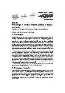
The nanoparticles have characteristics that instigate great promise for therapeutic and diagnostic consistency. In the preparation of therapeutic nanoparticle formulations, it is possible to improve the accumulation and release of pharmacologically active agents at the pathological site, allowing their application as monitoring biodistribution and accumulation of target sites, visualizing and quantifying drug release, longitudinally evaluating the efficacy reducing the incidence and intensity of side effects [35,41–43]. Among the formulations of nanoparticles that are reported, formulations such as liposomes, polymers, micelles, proteins, antibodies, gold nanoparticles, iron oxide nanoparticles among others, also aim to efficiently deliver a therapeutic drug to the
ip
t
pathological site, a as it will prevent accumulation in healthy organs and tissues of the body
ce
pt
ed
M
an
us
cr
[44] (Fig. 2).
Ac
Figure 2. Nanocarriers for cancer treatment of breast cancer and ovary cancer. The application of nanotechnology to the drug delivery in cancer has been widely
explored in the hope of improving the efficacy of chemotherapy and reducing the side effects [45]. Nanomaterials must be developed to interact with proteins and cells without intervening in their biological activities. Their physical properties should be maintained after undergoing surface modifications as well as being non-toxic [46]. The use of nanomaterials in the study of medicine favors an efficiency in the improvement of conventional instruments and methodologies of analysis with each new discovery [22]. In this sense, the use of diagnostic techniques based on the study of
nanoparticles provides a high sensitivity, as in the diagnosis of cancers in the early stages [10,23]. Although advances in diagnostics are being worked on rapidly, it is important that treatments through nanomaterials be investigated. That is, by improving the sensitivity of the initial diagnosis, as well as the types of treatment and the reduction of the toxicity of nanomaterials [47]. In order to obtain nanomaterials with good and high specificity, independent of the possible applications and characteristics such as stability, dispersion of size, morphology, surface load and toxicity, these should present a definition of quality to obtain good results [21].
ip
t
The rapid advance of nanomedicine is closely related to some properties of nanomaterials which will allow applications in diagnostics and therapies [10,15-16]. The
cr
following are the most studied and reported nanomaterials in the literature.
us
Metallic nanomaterials have excellent optical, electronic and catalytic properties, due to their structuring at the nanometric scale. Specifically, gold-based nanomaterials have
an
been the most extensively studied because of the high stability and efficient absorption of light [48,49]. In studies with gold nanoparticles, these have unique physical properties such as large surface area, high surface reactivity, and biocompatibility, thus establishing their use in
M
cancer therapy [50,51].
These nanoparticles also present as a method for photothermal applications,
ed
establishing the role of drug carriers [47] because of their physicochemical properties depending on the size and shape, that is, modifying the shape of the nanoparticle material for
pt
nanorods, stick-shaped nanomaterials. They are nanoparticles that can also be covered by
ce
silica particles with a gold layer forming core-shell structures [48]. Moreover, studies with carbon nanotubes, which are nanoparticles of one or several
Ac
layers of cylindrical carbons [52], have shown that the functionalization of this material allows its action as drug loading molecules for applications in drug delivery and has been successfully employed in the treatment of cancer [53]. Carbon nanotubes functionalized with polyethylene glycol and conjugated with the
chemotherapeutic provided greater efficacy in the suppression of the growth of breast cancer, due to the greater accumulation of the chemotherapeutic in the region of cancer when conjugated with the nanotubes [54]. Nanotechnology also presents the preparation of dual-purpose nanomaterials, diagnosis, and simultaneous therapy. They are the so-called theragnostics, these are agents characterized by being different nanoparticles used as theragnostics in cancer, where it is
possible to combine different active agents [55]. For example, iron oxide nanoparticles (IONPs) can be used in magnetite or hematite forms, as contrast agents due to their superparamagnetic activity, biocompatibility, and low cost. Modifying the surface with different inorganic molecules as well as linking polymeric and non-polymeric stabilizers provide the opportunity to use IONPs based agents for different applications [56]. Another interesting study is the evaluation of the effects of nanoparticles on the blood coagulation system. In this study, nanoparticles can be manipulated to avoid compromising coagulant processes, thus maintaining homeostasis. By this manipulation, it is
ip
factors or platelets interacting specifically with the nanomaterial [57].
t
possible to prevent the components of coagulation systems such as blood flow, coagulation
Several studies also present the use of nanoparticles as tumor markers in vitro or in
cr
vitro qualitative detection. In this type of process, the nanoparticles act by concentrating and
us
protecting a marker of degradation in order to make the analysis more sensitive, as is the case of fluorescent nanospheres. Such nanoparticles carry contrast agents, which will allow
an
redirection and concentration of the marker at the desired location. For this, the size, surface charge, surface coating, nanomaterial stability is of paramount importance [40]. Investigations have also been conducted with the aim of showing how nanoparticle
form of trapping in a colloidal system.
M
systems can control the distribution profiles of tissues and cells of anticancer drugs in the
ed
The result of this type of study is that the efficacy of the antitumor is increased, significantly reducing the side effects. Thus nanoparticles perform well for the selective
pt
delivery of oligonucleotides to tumor cells, and some types of nanoparticles may provide an
ce
interesting ability to maintain resistance to many drugs, which in fact remains a challenge in chemotherapy [40].
Ac
There are also reported works using magnetic nanoparticles which are generally modified and function through a polymer or liposome and are conjugated to an anticancer drug to try to improve stability and affinity with the tumor cell or impart a specific cleavage ability to the tumor [2,4,58]. The diseases caused by cancer are diverse, but when the picture is breast cancer and ovarian cancer, the nanoparticles cited in the course of this Review gain support for the diagnosis and treatment of these pathologies.
Advances and use of nanocarriers for breast and ovarian cancer Currently, the study materials that are being used as a transport module from one substance to another, favoring a good selectivity [34,59,60] are the nanocarriers. These are being studied for use in drug delivery, especially anticancer drugs, because of the small size that allows them to escape into the vasculature of the leaked tumor and accumulate at the site of the tumor through a permeability and retention effect (EPR) [61–64]. These nanomaterials have unique characteristics that demonstrate their great potential in chemotherapy [2], such as drug delivery systems for therapeutic or imaging
t
agents to improve the pharmacological properties of compounds commonly used in the
ip
diagnosis and treatment of cancer. In addition, they offer important advantages in the passive
cr
biodistribution at the site of the tumor due to increased permeation and the retention effect. In the functionalization of drug delivery systems with chemical fragments, which
us
are capable of recognizing molecular elements expressed specifically by cancer cells or structures involved in the development of tumors, these nanomaterials can increase the
an
biodistribution to the cancer sites, improving the therapeutic efficiency.
Generally, nanocarriers range in size from 1 to 1000 nm [2,9], however, because of
M
the width of 200 nm microcapillaries, nanomedicine generally refers to devices < 200 nm [2]. Because of their small size, nanocarriers can deliver drugs to inaccessible sites around the
ed
body where it is often difficult to deliver large doses of drugs. Much of the research on nanocarriers is being applied to their great potential in drug
pt
administration, which makes them useful in the drug delivery process because they are able to transport the drug to a specific destination (receiver, active site) to exercise their therapeutic
ce
activity with maximum safety [65].
Site specificity is an important therapeutic benefit as it prevents drugs from being
Ac
delivered to the wrong places [54,66,67], and nanocarriers show promise of being used especially in chemotherapy [31] because they can help minimize the broader adverse toxicity in cells healthy and rapid growth throughout the body [68]. Since chemotherapy drugs can be extremely toxic to human cells, it is important that they are delivered to the tumor without being released to other parts of the body [69–71].
t ip cr
an
us
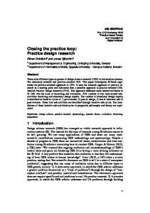
Figure 3. Nanocarriers surface ligands and therapeutic agents used for cancer diagnosis or treatment: A (nanosphere), B (solid lipid nanoparticle), C (nanocapsule), D (dendrimers), E (nanodiamond), F (liposomes), G (quantum dot), H (gold nanoparticles) I (virus-mediated nanocarriers) and J (nanotubes). The most commonly used nanocarriers include conjugated polymers [72,73],
M
polymer nanoparticles [74–76], micelles [16], liposomes and lipid-based transporters [3,77], dendrimers [8,78], carbon nanotubes [53,54,69] and nanoparticles [43,79]. Lipid-based transporters include liposomes and micelles. Examples of gold nanoparticles are nanoshells
ed
and gold nanocages [74]. (Fig. 3)
A major potential problem with nanocarriers is the undesired toxicity of the type of
pt
nanomaterial being used, that is, it can be toxic to the human body if it accumulates in certain
ce
cellular organelles [80]. Thus, new research is being conducted to invent more effective and safer nanocarriers, such as protein-based nanocarriers which according to studies, are
Ac
promising for therapeutic use because they occur naturally and generally demonstrate less cytotoxicity than synthetic molecules [81]. In the literature, there are four methods in which nanocarriers can deliver drugs that
include passive cleavage, active cleavage, pH specificity and temperature specificity. Passive cleavage facilitates the deposition of nanovectors within the tumor microenvironment due to distinctive features inherent to the tumor environment, not normally present in healthy tissues [82,83]. This accumulation is caused by increased permeability and retention effect [74,84]. Also in this context, the nanocarriers carrying the medicament remain sufficiently long in the circulation to accumulate in the desired tissue due to the size and surface properties as well as the properties of the tissue [85].
In active targeting, the antibodies are attached to the surface of the nanocarrier to be recognized by the receptors expressed by the target cell surfaces [86]. Thus, the encapsulated drug is released, or internalized with the nanocarrier, at the site of action [64]. The nanocarriers have such a high ratio of surface area to volume, allowing the incorporation of multiple antibodies on their surfaces [68]. These targeting modules allow the incorporation of nanocarriers in the cells, but they present some disadvantages, such as toxicity, which can decrease the efficiency of drug delivery cells [44]. Some nanocarriers have only released the drugs they contain in specific pH ranges. The specificity of pH allows nanocarriers to deliver drugs directly to the tumor site
ip
t
[87,88]. Tumors are known to be generally more acidic than normal human cells, with lower pH value (~6.7) [89]. Normal tissue has a pH of about 7.4 [49,87]. Nanocarriers that only
cr
release drugs at certain pH ranges can, therefore, be used to release the drug only in acidic
us
tumor environments [87,89,90]. Environments with high acidity cause the release of the drug due to the acid environment that degrades the structure of the nanocarrier.
an
These nanocarriers do not release drugs in neutral or basic environments, effectively targeting the acidic environments of tumors, leaving normal body cells intact [87,91]. This pH sensitivity can also be induced in micelle systems by adding chains of copolymers to micelles
M
that have been determined to act in a pH-independent mansion [87]. These complexes of micelles and polymers also help prevent cancer cells from developing resistance to various
ed
drugs.
The low pH environment triggers a rapid release of the micelle polymers, causing
pt
most drugs to be released at the same time, rather than gradually, as other drug treatments
ce
[18,92]. This rapid-release mechanism significantly decreases the time it takes for anticancer drugs to kill a tumor, effectively preventing the tumor from having time to undergo mutations
Ac
that would make it resistant to drugs [18]. Some nanocarriers provide drugs more effectively at certain temperatures. Since
tumor temperatures are generally higher than temperatures throughout the rest of the body at about 40 º C, this temperature gradient helps to act as a safeguard for delivery of the tumor site-specific [9].
Nanocarriers for breast cancer Although developments in the treatment and diagnosis of breast cancer have been advanced over time, delays in diagnosis make this pathology increasingly fatal. Conventional
treatment approaches include hormone therapy, immunotherapy, chemotherapy, among others [18,34]. Immunotherapy against cancer involves the use of antibodies that bind to receptors and inactivate signaling pathways for the growth of breast cancer cells. Commercial use of trastuzumab antibodies against human epidermal growth factor receptor 2 (Her2) is an example of successful use of cancer immunotherapy [93,94]. In hormone therapy, the hormone-stimulating hormone, estrogen, is reduced in the body or estrogen (ER) receptors are blocked. For example, the analgesic analog tamoxifen (TMX) is used to block estrogen from binding to ER and activate the estrogen-responsive
ip
t
genes that stimulate the growth of breast cancer cells. Both of the above-mentioned therapies can, however, be used only in the case of breast cancer cells expressing estrogen,
cr
progesterone or Her2 receptors [23,58,95].
us
However, breast cancer cells expressing none of these receptors, that is, triple negative breast cancer (TN) cells are very aggressive and difficult to treat. Chemotherapy has
an
been used to treat TN breast cancer, which has serious side effects due to non-specificity. However, due to the various limitations and side effects associated with these therapies, new treatment strategies are being developed [96].
M
Some clinical trials are comparing the efficacy of various chemotherapeutic drugs, used in different combinations, for the treatment of triple negative breast cancer. Among
ed
them, it highlights the folate alpha-receptor (FRα), membrane receptor protein that facilitates the transport of folate, employed in several types of cancer, including lung, ovary and breast
pt
cancers [97,98]. FRα is a central mediator of cell growth regulation that can serve as an important target for cancer therapy. Studies have shown that FRα overexpression in breast
ce
cancer is strongly correlated with early relapse and decreased median survival; therefore, FRα emerged as a potentially promising therapeutic target in breast cancer [99–101].
Ac
A monoclonal antibody for FRα (Farletuzumab) has been developed and may be an
attractive treatment strategy, either alone or in combination with chemotherapy. Therefore, an antibody suitable for immunohistochemistry (IHC) FRα segmentation may have significant significance in the future, particularly in identifying patients appropriate for treatment with folate therapy [98]. FRα-targeted therapies, alone or in combination with cytotoxic chemotherapeutics, may represent a novel approach to the treatment of TNBC, or therapies with anti-folate alpha-receptor directed antibodies to restrict the growth of triple breast cancer negative [102].
Nanoparticles in breast cancer therapy are showing promising results [44,77,103]. The use of nanosystems presents as favorable properties high biocompatibility, low immunogenicity, maintenance of plasma levels in constant concentrations and efficient drug protection. Among the nanocarriers, liposomes are considered excellent platforms for the release of hydrophobic drugs (carried along the lipid bilayer) and hydrophilic (delivered inside the aqueous vesicle). In particular, they present high blood time to facilitate drug delivery efficiently to their target-specific site [6,94]. They are good carriers of drugs since they are biocompatible, with low toxicity, and can release hydrophilic and lipophilic drugs,
biological membranes until reaching the target-specific organ [21,62].
ip
t
protect their degradation charges by plasma enzymes and transport their charge through the
cr
For purposes of improving blood circulation and targeting specifically to the cell,
us
the surface of the liposomes may be modified primarily by the inclusion of polymers, peptides or antibodies [104]. Although these advantages are significant, there are disadvantages of a
an
possible toxicity, absence of biocompatibility of the materials used and the high cost of
Nanocarriers for ovarian cancer
M
obtaining nanotransporters compared to conventional pharmaceutical formulations.
ed
Ovarian cancer is the leading cause of death from gynecological malignancies worldwide. Although most tumors initially respond to standard treatments combining surgery
pt
and chemotherapy with platinum-based chemotherapy, frequent recurrence and subsequently acquired chemoradiation are responsible for the therapeutic failure, leading to a 5% overall
ce
survival rate of 30% [94].
Considering the usual initial sensitivity of ovarian tumors to chemotherapy over the
Ac
past ten years, efforts have focused on the last decade to cure ovarian cancer using currently available chemotherapeutic agents in various combinations, doses, schedules (durations and/ or routes of administration). However, with such a systemic chemotherapeutic approach, there are considerable limitations, including toxicities for healthy tissues and low concentrations of drug achievable at tumor sites [105]. Considerable efforts are made to engineer systems capable of transporting large doses of cytotoxic agents specifically in targeted malignant cells while sparing healthy cells. These nanocarriers will be analyzed by citing examples of their use in preclinical development [106].
Epithelial ovarian cancers account for 90% of ovarian cancers. They could derive from cells of the epithelium of the surface of the ovary, or from cells of the fallopian tube epithelium. Compared with other types of solid tumors, ovarian carcinomas present peculiar and varied processes of dissemination and development [59,60]. Two major types of development are observed: If tumors grow slowly and remain confined to the ovary, ovarian carcinomas are referred to as Type I. They include serous, mucinous, endometrioid, and clear cell carcinomas. However, if the tumors are more aggressive, they are said to be type II, where they include high-grade serous carcinomas, malignant mixed tumors, and undifferentiated carcinomas [34].
ip
t
The criteria for effective nanocarriers for the delivery of chemotherapy drugs against ovarian cancer are: non-toxic carrier, stable within the bloodstream with minimal premature
cr
drug release, low absorption in all normal organs and reticuloendothelial system, high tumor
us
uptake and prolonged retention within the tumor, ability to be absorbed by tumor cells, inherent mechanisms for drug delivery at the site of the tumor or within the tumor cells,
an
ability to release the loaded drug on demand, and convenient formulation protocols which can be readily performed by clinical pharmacists at the clinic. Several innovative nanocarrier systems with telodendrimer have recently been developed to meet most of the above criteria
M
[107].
Numerous active operators have been developed, with potential applications of
ed
interest in the treatment of ovarian cancer. Studies focused on the various therapeutic drug delivery systems currently evaluated for the treatment of ovarian cancer.
pt
CA-125 is a large antigen (2500 kDa), absent in normal ovarian tissues, but
ce
expressed in more than 80% in patients with ovarian cancer [108]. CA-125 is the most widely used serum biomarker in ovarian cancer and the sensitivity of serum CA-125 correlates with
Ac
the clinical stage of the disease [109]. As it is also expressed on the surface of ovarian cancer cells, it can also be used specifically for ovarian cancer cells. In vitro and in vivo preclinical studies reported in the literature on nanocarrier
evaluation are dealing with different ovarian cell models. The most used models are ovarian cancer cells SKOV3, A2780, and OVCAR-3. In order to improve image sensitivity and detection of intraperitoneal ovary tumors, the use of dendrimers as a targeted diagnostic tool is being explored. Thus, the development of G4-PAMAM folate-dendrimers capable of specifically targeting the folate receptor is reported [109,110]. When given in vivo, folatedendrimer conjugated to gadolinium paramilitary contrast agents have been shown to
accumulate in xenotransplanted ovarian tumors (OVCA432 cells), resulting in a significant increase in contrast compared to a nonspecific extracellular gadolinium complex [111]. Other avidin-based ovarian tumor probes based on avidin-biotin-dendrimer complexes are also reported in the literature for scintigraphy images of intraperitoneal tumors [112], magnetic resonance imaging of two modalities and fluorescence imaging [113]. Several micelle carrier systems have been developed and evaluated as a potential drug delivery system for chemotherapy in ovarian cancer. Thus, it has been repeatedly demonstrated in preclinical studies that micelles of various anticancer drugs (eg. paclitaxel [114], docetaxel [115], doxorubicin [116], camptothecin [117]) have shown a better
ip
t
accumulation in tumors than free drugs, thus exhibiting superior cytotoxicity against human ovarian carcinoma cells and minimizing undesired drug toxicity over normal tissues.
cr
Polymer micelle formulations of paclitaxel were investigated in clinical trials
us
[118,119]. For example, Paclical®, a micellar formulation of paclitaxel in the intravenous infusion Ringer's acetate solution currently evaluated in phase III initiated in the fall of 2008
an
for platinum-sensitive ovarian cancer. Genexol-PM ®, a micellar formulation of paclitaxel consisting of PEG and PLA, was generally well tolerated and demonstrated sufficient
M
antitumor activity to ensure further development. Conclusions
ed
This review presents a revolution that comes to contribute to scientific advances, a nanoscience, and nanomedicine, in general, are proposing a new targeting without treatment
pt
of disease that is diagnosed with cancer. About 10 million people each year are diagnosed
ce
with this pathology and breast and ovarian cancer are more present types of cancer in women's lives. Nanocarriers and nanoparticles indicate how a vehicle to specifically deliver
Ac
the cancer drug as well as cancerous cells would be destroyed. Thus, the treatment of cancer would not be so excruciating and that would entail in
the minimization of the side effects. The contribution of nanoparticles to cancer chemotherapy will certainly grow as long as more efficient tumor targeting strategies are developed. Future research is being segmented with folic acid due to the strategy of biocompatibility and cellular internalization. However, active targeting generally encompasses numerous chemical reactions and the challenge will, therefore, be to avoid a complex synthesis of constructs. However, new approaches to associating drugs with nanoparticles must appear and contribute to progress in science.
As the use of nanocarriers, there has been an improvement in the transport of drugs for the treatment of cancer. However, they suggest new challenges that are directly linked to nanoparticle efficiency, stability, and interactions between receptor and ligand. To minimize the problems associated with the transport of nanocarriers, strategies will be developed combining chemotherapeutics, nanocarriers and target molecules to optimize the treatment of cancer, where there will be a reduction of costs for therapy and diagnosis of cancer. Finally, to further promote the clinical translation of nanoparticle formulations for
cr
ip
t
diagnostic and therapeutic purposes and to further improve their implementation for personalized medicine, especially in the field of oncology, it is essential to resolve the main regulatory challenges of a controlled nanoparticle synthesis, uniformity, batch-to-batch reproducibility and upscaling of nanoparticle production. The specific combinations of nanocarriers and target molecules - similar to the strategies of chemotherapy combination that
an
us
can be personalized to improve treatment against cancer – that will contribute to improve therapeutic results and reduce costs. These combinations will represent an important modality for cancer diagnosis and treatment.
Acknowledgments
M
The authors acknowledge the financial support of the Brazilian National Council for Research and Development (CNPq), Coordination for the Improvement of Higher Education Personnel
ed
(CAPES) and the Foundation for Research Support of the State of Piauí (FAPEPI).
pt
Conflict of interest statement
ce
The authors declare the research was conducted in the absence of any commercial relationship
Ac
and there are no potential conflicts of interest.
References [1] [2] [3] [4]
[5]
ip
t
[6]
Wang X, Wang Y, Chen ZG, et al. Advances of Cancer Therapy by Nanotechnology. Cancer Res Treat. 2009;41:1–11. Peer D, Karp JM, Hong S, et al. Nanocarriers as an emerging platform for cancer therapy. Nat. Nanotechnol. 2007;2:751–760. Allen TM, Cullis PR. Drug Delivery Systems: Entering the Mainstream. Science. 2004;303:1818–1822. Matsumura Y, Maeda H. A New Concept for Macromolecular Therapeutics in Cancer Chemotherapy: Mechanism of Tumoritropic Accumulation of Proteins and the Antitumor Agent Smancs. Cancer Res. 1986;46:6387–6392. Khawar IA, Kim JH, Kuh H-JJ. Improving drug delivery to solid tumors: Priming the tumor microenvironment. J. Control. Release. 2015;201:78–89. Tomasina J, Lheureux S, Gauduchon P, et al. Nanocarriers for the targeted treatment of ovarian cancers. Biomaterials. 2013;34:1073–1101. Plummer M, de Martel C, Vignat J, et al. Global burden of cancers attributable to infections in 2012: a synthetic analysis. Lancet Glob. Heal. 2016;4:e609–e616. Perinotto AC, Caseli L, Hayasaka CO, et al. Dendrimer-assisted immobilization of alcohol dehydrogenase in nanostructured films for biosensing: Ethanol detection using electrical capacitance measurements. Thin Solid Films. 2008;516:9002–9005. Blanco E, Hsiao A, Mann AP, et al. Nanomedicine in cancer therapy: Innovative trends and prospects. Cancer Sci. 2011;102:1247–1252. Caruso F, Hyeon T, Rotello VM. Nanomedicine. Chem. Soc. Rev. 2012;41:2537–2538. Gradishar WJ. Albumin-bound paclitaxel: a next-generation taxane. Expert Opin. Pharmacother. 2006;7:1041–1053. Saif MW. U.S. Food and Drug Administration approves paclitaxel protein-bound particles (Abraxane(R)) in combination with gemcitabine as first-line treatment of patients with metastatic pancreatic cancer. JOP. 2013;14:686–688. Northfelt DW, Martin FJ, Working P, et al. Doxorubicin encapsulated in liposomes containing surface-bound polyethylene glycol: pharmacokinetics, tumor localization, and safety in patients with AIDS-related Kaposi’s sarcoma. J. Clin. Pharmacol. 1996;36:55–63. Monneret C. Platinum anticancer drugs. From serendipity to rational design. Ann. Pharm. Fr. 2011;69:286–295. Mura S, Couvreur P. Nanotheranostics for personalized medicine. Adv Drug Deliv Rev. 2012;64:1394–1416. Torchilin VP. Micellar nanocarriers: Pharmaceutical perspectives. Pharm. Res. 2007;24:1–16. Farokhzad OC, Langer R. Impact of nanotechnology on drug delivery. ACS Nano. 2009;3:16–20. Fan Z, Fu PP, Yu H, et al. Theranostic nanomedicine for cancer detection and treatment. J. Food Drug Anal. 2014;22:3–17. Boulaiz H, Alvarez PJ, Ramirez A, et al. Nanomedicine: Application areas and development prospects. Int. J. Mol. Sci. 2011;12:3303–3321. Kranz C, Eaton DC, Mizaikoff B. Analytical challenges in nanomedicine. Anal. Bioanal. Chem. 2011;399:2309–2311. Venditto VJ, Szoka FC. Cancer nanomedicines: So many papers and so few drugs! Adv. Drug Deliv. Rev. 2013;65:80–88. Korsmeyer R. Critical questions in development of targeted nanoparticle therapeutics. Regen. Biomater. 2016;3:143–147.
[13]
[14] [15]
an
Ac
[16]
M
[12]
ed
[10] [11]
pt
[9]
ce
[8]
us
cr
[7]
[17] [18] [19] [20] [21] [22]
Ac
ce
pt
ed
M
an
us
cr
ip
t
[23] Zhan C, Li C, Wei X, et al. Toxins and derivatives in molecular pharmaceutics: Drug delivery and targeted therapy. Adv. Drug Deliv. Rev. 2015;90:101–118. [24] WHO. WHO | Breast cancer [Internet]. WHO. World Health Organization; 2018 [cited 2018 Aug 21]. Available from: http://www.who.int/cancer/prevention/diagnosisscreening/breast-cancer/en/#.W3v30DwIPRA.mendeley. [25] Cedolini C, Bertozzi S, Londero AP, et al. Type of breast cancer diagnosis, screening, and survival. Clin. Breast Cancer. 2014;14:235–240. [26] Juhascik MP, Negrusz A, Faugno D, et al. An estimate of the proportion of drugfacilitation of sexual assault in four U.S. localities. J. Forensic Sci. 2007;52:1396– 1400. [27] Reid BM, Permuth JB, Sellers TA. Epidemiology of ovarian cancer: a review. Cancer Biol. Med. 2017;14:9–32. [28] Kujawa KA, Lisowska KM. [Ovarian cancer--from biology to clinic]. Postepy Hig. Med. Dosw. (Online). 2015;69:1275–1290. [29] Dong X, Men X, Zhang W, et al. Advances in tumor markers of ovarian cancer for early diagnosis. Indian J. Cancer. 2014;51 Suppl 3:e72-6. [30] Meinhold-Heerlein I, Fotopoulou C, Harter P, et al. The new WHO classification of ovarian, fallopian tube, and primary peritoneal cancer and its clinical implications. Arch. Gynecol. Obstet. 2016;293:695–700. [31] Bhoola S, Hoskins WJ. Diagnosis and management of epithelial ovarian cancer. Obstet. Gynecol. 2006;107:1399–1410. [32] Goldberg JM, Piver MS, Jishi MF, et al. Age at onset of ovarian cancer in women with a strong family history of ovarian cancer. Gynecol. Oncol. 1997;66:3–9. [33] Gharpure KM, Wu SY, Li C, et al. Nanotechnology: Future of oncotherapy. Clin. Cancer Res. 2015;21:3121–3130. [34] Vieira DB, Gamarra LF. Advances in the use of nanocarriers for cancer diagnosis and treatment. Einstein (São Paulo). 2016;14:99–103. [35] Baetke SC, Lammers T, Kiessling F. Applications of nanoparticles for diagnosis and therapy of cancer. Br. J. Radiol. 2015;88:1–12. [36] He C, Lin W. Hybrid nanoparticles for cancer imaging and therapy. Cancer Treat. Res. 2015;166:173–192. [37] Zhao J, Lee P, Wallace MJ, et al. Gold Nanoparticles in Cancer Therapy: Efficacy, Biodistribution, and Toxicity. Curr. Pharm. Des. 2015;21:4240–4251. [38] Moghimi SM, Hunter AC, Murray JC. Long-circulating and target-specific nanoparticles: theory to practice. Pharmacol. Rev. 2001;53:283–318. [39] Yu MK, Park J, Jon S. Targeting strategies for multifunctional nanoparticles in cancer imaging and therapy. Theranostics. 2012;2:3–44. [40] Duncan R, Gaspar R. Nanomedicine(s) under the microscope. Mol. Pharm. 2011;8:2101–2141. [41] Ahmed N, Fessi H, Elaissari A. Theranostic applications of nanoparticles in cancer. Drug Discov. Today. 2012;17:928–934. [42] Rizzo LY, Theek B, Storm G, et al. Recent progress in nanomedicine: Therapeutic, diagnostic and theranostic applications. Curr. Opin. Biotechnol. 2013;24:1159–1166. [43] Mohanraj V, Chen Y, Chen M&. Nanoparticles – A Review. Trop. J. Pharm. Res. Trop J Pharm Res. 2006;5:561–573. [44] Solanki A, Kim JD, Lee K-B. Nanotechnology for regenerative medicine: nanomaterials for stem cell imaging. Nanomedicine (Lond). 2008;3:567–578. [45] Cancino J, Marangoni VS, Zucolotto V. Nanotechnology in medicine: concepts and concerns. Quim. Nova. 2014;37:521–526. [46] Jain PK, ElSayed IH, El-Sayed MA. Au nanoparticles target cancer. Nano Today.
[47] [48] [49] [50] [51] [52]
[59] [60]
[61] [62]
cr
us
Ac
[63]
an
[58]
M
[57]
ed
[56]
pt
[55]
ce
[54]
ip
t
[53]
2007;2:18–29. Anikeeva P, Deisseroth K. Photothermal genetic engineering. ACS Nano. 2012;6:7548–7552. Lee J, Chatterjee DK, Lee MH, et al. Gold nanoparticles in breast cancer treatment: Promise and potential pitfalls. Cancer Lett. 2014;347:46–53. Sperling RA, Rivera Gil P, Zhang F, et al. Biological applications of gold nanoparticles. Chem. Soc. Rev. 2008;37:1896. Xu Y, Wang J, Li X, et al. Selective inhibition of breast cancer stem cells by gold nanorods mediated plasmonic hyperthermia. Biomaterials. 2014;35:4667–4677. Oldenburg S., Averitt R., Westcott S., et al. Nanoengineering of optical resonances. Chem. Phys. Lett. 1998;288:243–247. Madani SY, Naderi N, Dissanayake O, et al. A new era of cancer treatment: carbon nanotubes as drug delivery tools. Int. J. Nanomedicine. 2011;6:2963–2979. Shi Kam NW, O’Connell M, Wisdom JA, et al. Carbon nanotubes as multifunctional biological transporters and near-infrared agents for selective cancer cell destruction. Proc. Natl. Acad. Sci. 2005;102:11600–11605. Liu Z, Chen K, Davis C, et al. Drug delivery with carbon nanotubes for in vivo cancer treatment. Cancer Res. 2008;68:6652–6660. Ilinskaya AN, Dobrovolskaia MA. Nanoparticles and the blood coagulation system. Part I: benefits of nanotechnology. Nanomedicine. 2013;8:773–784. Li Z, Kawashita M, Araki N, et al. Magnetite nanoparticles with high heating efficiencies for application in the hyperthermia of cancer. Mater. Sci. Eng. C. 2010;30:990–996. Jabir NR, Tabrez S, Ashraf GM, et al. Nanotechnology-based approaches in anticancer research. Int. J. Nanomedicine. 2012;7:4391–4408. Sutradhar KB, Amin ML. Nanotechnology in Cancer Drug Delivery and Selective Targeting. ISRN Nanotechnol. 2014;2014:1–12. Singh R, Lillard JW. Nanoparticle-based targeted drug delivery. Exp. Mol. Pathol. 2009;86:215–223. Chen Z, Zhang L, Song Y, et al. Hierarchical targeted hepatocyte mitochondrial multifunctional chitosan nanoparticles for anticancer drug delivery. Biomaterials. 2015;52:240–250. Basile L, Pignatello R, Passirani C. Active targeting strategies for anticancer drug nanocarriers. Curr. Drug Deliv. 2012;9:255–268. Thierry B. Drug Nanocarriers and Functional Nanoparticles: Applications in Cancer Therapy. Curr. Drug Deliv. 2009;6:13. Shi J, Kantoff PW, Wooster R, et al. Cancer nanomedicine: Progress, challenges and opportunities. Nat. Rev. Cancer. 2017;17:20–37. Torchilin V. Tumor delivery of macromolecular drugs based on the EPR effect. Adv. Drug Deliv. Rev. 2011;63:131–135. Chen YC, Lo CL, Lin YF, et al. Rapamycin encapsulated in dual-responsive micelles for cancer therapy. Biomaterials. 2013;34:1115–1127. Tabatabaei Rezaei SJ, Nabid MR, Niknejad H, et al. Multifunctional and thermoresponsive unimolecular micelles for tumor-targeted delivery and sitespecifically release of anticancer drugs. Polym. (United Kingdom). 2012;53:3485– 3497. Pérez-Herrero E, Fernández-Medarde A. Advanced targeted therapies in cancer: Drug nanocarriers, the future of chemotherapy. Eur. J. Pharm. Biopharm. 2015;93:52–79. R. Khan D. The use of Nanocarriers for Drug Delivery in Cancer Therapy David R. Khan. J. Cancer Sci. Ther. 2010;02:058–062.
[64] [65] [66]
[67] [68]
Ac
ce
pt
ed
M
an
us
cr
ip
t
[69] Qian WY, Sun DM, Zhu RR, et al. pH-sensitive strontium carbonate nanoparticles as new anticancer vehicles for controlled etoposide release. Int. J. Nanomedicine. 2012;7:5781–5792. [70] Kaur S, Prasad C, Balakrishnan B, et al. Trigger responsive polymeric nanocarriers for cancer therapy. Biomater. Sci. 2015;3:955–987. [71] Duncan R. Polymer conjugates as anticancer nanomedicines. Nat. Rev. Cancer. 2006;6:688–701. [72] Luo Y, Prestwich G. Cancer-Targeted Polymeric Drugs. Curr. Cancer Drug Targets. 2002;2:209–226. [73] Kwon GS. Polymeric Micelles for Delivery of Poorly Water-Soluble Compounds. Crit. Rev. Ther. Drug Carrier Syst. 2003;20:357–403. [74] Yavuz MS, Cheng Y, Chen J, et al. Gold nanocages covered by smart polymers for controlled release with near-infrared light. Nat. Mater. 2009;8:935–939. [75] Sun Y, Li Y, Huang H, et al. PH-Sensitive Poly(itaconic acid)-poly(ethylene glycol)poly(L-histidine) Micelles for Drug Delivery. J. Macromol. Sci. Part A Pure Appl. Chem. 2015;52:925–933. [76] Mendes ANAN, Hubber I, Siqueira M, et al. Preparation and Cytotoxicity of Poly(Methyl Methacrylate) Nanoparticles for Drug Encapsulation. Macromol. Symp. 2012;319:34–40. [77] Zhu RR, Qin LL, Wang M, et al. Preparation, characterization, and anti-tumor property of podophyllotoxin-loaded solid lipid nanoparticles. Nanotechnology. 2009;20:055702. [78] Wu LP, Ficker M, Christensen JB, et al. Dendrimers in Medicine: Therapeutic Concepts and Pharmaceutical Challenges. Bioconjug. Chem. 2015;26:1198–1211. [79] Brigger I, Dubernet C, Couvreur P. Nanoparticles in cancer therapy and diagnosis. Adv. Drug Deliv. Rev. 2002;54:631–651. [80] Wang J, Fang X, Liang W. Pegylated phospholipid micelles induce endoplasmic reticulum-dependent apoptosis of cancer cells but not normal cells. ACS Nano. 2012;6:5018–5030. [81] Elzoghby AO, Samy WM, Elgindy NA. Protein-based nanocarriers as promising drug and gene delivery systems. J. Control. Release. 2012;161:38–49. [82] Kreuter J. Nanoparticles-a historical perspective. Int. J. Pharm. 2007;331:1–10. [83] Cajot S, Van Butsele K, Paillard A, et al. Smart nanocarriers for pH-triggered targeting and release of hydrophobic drugs. Acta Biomater. 2012;8:4215–4223. [84] Misra R, Acharya S, Sahoo SK. Cancer nanotechnology: Application of nanotechnology in cancer therapy. Drug Discov. Today. 2010;15:842–850. [85] Byrne JD, Betancourt T, Brannon-Peppas L. Active targeting schemes for nanoparticle systems in cancer therapeutics. Adv. Drug Deliv. Rev. 2008;60:1615–1626. [86] Hirsjärvi S, Passirani C, Benoit J-P. Passive and active tumour targeting with nanocarriers. Curr. Drug Discov. Technol. 2011;8:188–196. [87] Tannock IF, Rotin D. Acid pH in Tumors and Its Potential for Therapeutic Exploitation. Cancer Res. 1989;49:4373–4384. [88] Swietach P, Vaughan-Jones RD, Harris AL, et al. The chemistry, physiology and pathology of pH in cancer. Philos. Trans. R. Soc. B Biol. Sci. 2014;369:20130099– 20130099. [89] Kato Y, Ozawa S, Miyamoto C, et al. Acidic extracellular microenvironment and cancer. Cancer Cell Int. 2013;13:89. [90] Sanna V, Pala N, Sechi M. Targeted therapy using nanotechnology: Focus on cancer. Int. J. Nanomedicine. 2014;9:467–483. [91] Spuch C, Navarro C. Liposomes for Targeted Delivery of Active Agents against Neurodegenerative Diseases (Alzheimer’s Disease and Parkinson’s Disease). J. Drug
[92]
[93]
[94]
[101] [102] [103]
[104]
cr
us
Ac
[105]
an
[100]
M
[99]
ed
[98]
pt
[97]
ce
[96]
ip
t
[95]
Deliv. 2011;2011:1–12. Mendes AN, Filgueiras LA, Siqueira M, et al. Encapsulation of Piper cabralanum (Piperaceae) nonpolar extract in poly(methyl methacrylate) by miniemulsion and evaluation of increase in the effectiveness of antileukemic activity in K562 cells. Int. J. Nanomedicine. 2017;12:8363–8373. Laginha KM, Verwoert S, Charrois GJR, et al. Determination of doxorubicin levels in whole tumor and tumor nuclei in murine breast cancer tumors. Clin. Cancer Res. 2005;11:6944–6949. Charrois GJR, Allen TM. Drug release rate influences the pharmacokinetics, biodistribution, therapeutic activity, and toxicity of pegylated liposomal doxorubicin formulations in murine breast cancer. Biochim. Biophys. Acta - Biomembr. 2004;1663:167–177. Chandrasekharan N, Kamat P. Improving the Photoelectrochemical Performance of Nanostructured TiO 2 Films by Adsorption of Gold Nanoparticles. J. Phys. Chem. B. 2000;104:10851–10857. Gradishar WJ, Tjulandin S, Davidson N, et al. Phase III trial of nanoparticle albuminbound paclitaxel compared with polyethylated castor oil-based paclitaxel in women with breast cancer. J. Clin. Oncol. 2005;23:7794–7803. Li Z, Qiu Y, Lu W, et al. Immunotherapeutic interventions of Triple Negative Breast Cancer. J. Transl. Med. 2018;16:147. Cheung A, Bax HJ, Josephs DH, et al. Targeting folate receptor alpha for cancer treatment. Oncotarget. 2016;7:52553–52574. Ginter PS, McIntire PJ, Cui X, et al. Folate Receptor Alpha Expression Is Associated With Increased Risk of Recurrence in Triple-negative Breast Cancer. Clin. Breast Cancer. 2017;17:544–549. Leone JP, Bhargava R, Theisen BK, et al. Expression of high affinity folate receptor in breast cancer brain metastasis. Oncotarget. 2015;6:30327–30333. Zhang Z, Wang J, Tacha DE, et al. Folate receptor alpha associated with triple-negative breast cancer and poor prognosis. Arch. Pathol. Lab. Med. 2014;138:890–895. Shi H, Guo J, Li C, et al. A current review of folate receptor alpha as a potential tumor target in non-small-cell lung cancer. Drug Des. Devel. Ther. 2015;9:4989–4996. Yao N, Xiao W, Wang X, et al. Discovery of targeting ligands for breast cancer cells using the one-bead one-compound combinatorial method. J. Med. Chem. 2009;52:126– 133. Schiener M, Hossann M, Viola JR, et al. Nanomedicine-based strategies for treatment of atherosclerosis. Trends Mol. Med. 2014;20:271–281. Sutton D, Nasongkla N, Blanco E, et al. Functionalized micellar systems for cancer targeted drug delivery. Pharm. Res. 2007;24:1029–1046. Matsumura Y, Hamaguchi T, Ura T, et al. Phase I clinical trial and pharmacokinetic evaluation of NK911, a micelle-encapsulated doxorubicin. Br. J. Cancer. 2004;91:1775–1781. Farokhzad OC, Cheng J, Teply BA, et al. Targeted nanoparticle-aptamer bioconjugates for cancer chemotherapy in vivo. Proc. Natl. Acad. Sci. U. S. A. 2006;103:6315–6320. Xiao K, Li Y, Lee JS, et al. “OA02” Peptide Facilitates the Precise Targeting of Paclitaxel-Loaded Micellar Nanoparticles to Ovarian Cancer In Vivo. Cancer Res. 2012;72:2100–2110. Aina OH, Marik J, Gandour-Edwards R, et al. Near-infrared optical imaging of ovarian cancer xenografts with novel alpha 3-integrin binding peptide “OA02”. Mol. Imaging. 2005;4:439–447. Xiao W, Luo J, Jain T, et al. Biodistribution and pharmacokinetics of a telodendrimer
[106]
[107] [108]
[109]
[110]
[111] [112]
[113]
[114]
[118]
cr
Ac
ce
pt
ed
M
[119]
us
[117]
an
[116]
ip
t
[115]
micellar paclitaxel nanoformulation in a mouse xenograft model of ovarian cancer. Int. J. Nanomedicine. 2012;7:1587–1597. Calzolari A, Oliviero I, Deaglio S, et al. Transferrin receptor 2 is frequently expressed in human cancer cell lines. Blood Cells. Mol. Dis. 2007;39:82–91. Ulbrich K, Hekmatara T, Herbert E, et al. Transferrin- and transferrin-receptorantibody-modified nanoparticles enable drug delivery across the blood-brain barrier (BBB). Eur. J. Pharm. Biopharm. 2009;71:251–256. Fondell A, Edwards K, Unga J, et al. In vitro evaluation and biodistribution of HER2targeted liposomes loaded with an 125I-labelled DNA-intercalator. J. Drug Target. 2011;19:846–855. Zhang X, Chen J, Zheng Y, et al. Follicle-Stimulating Hormone Peptide Can Facilitate Paclitaxel Nanoparticles to Target Ovarian Carcinoma In vivo. Cancer Res. 2009;69:6506–6514. Kim D, Gao ZG, Lee ES, et al. In Vivo Evaluation of Doxorubicin-Loaded Polymeric Micelles Targeting Folate Receptors and Early Endosomal pH in Drug-Resistant Ovarian Cancer. Mol. Pharm. 2009;6:1353–1362. Chaudhury A, Das S, Bunte RM, et al. Potent therapeutic activity of folate receptortargeted liposomal carboplatin in the localized treatment of intraperitoneally grown human ovarian tumor xenograft. Int. J. Nanomedicine. 2012;7:739–751. Parrott JA, Doraiswamy V, Kim G, et al. Expression and actions of both the follicle stimulating hormone receptor and the luteinizing hormone receptor in normal ovarian surface epithelium and ovarian cancer. Mol. Cell. Endocrinol. 2001;172:213–222. Zhang Z, Jia L, Feng Y, et al. Overexpression of follicle-stimulating hormone receptor facilitates the development of ovarian epithelial cancer. Cancer Lett. 2009;278:56–64. Canney PA, Moore M, Wilkinson PM, et al. Ovarian cancer antigen CA125: a prospective clinical assessment of its role as a tumour marker. Br. J. Cancer. 1984;50:765–769.