ectopie pregnancy, hydatidiform mole and choriocarcinoma (Anon, 1976a). However, the usefulness of a-trichosanthin for termination of early pregnancy (during ...
Effects of \g=a\-trichosanthin and \g=a\-momorcharinon the development of peri-implantation mouse embryos L. K.
Law,
P. P. L. Tam and H. W. Yeung Departments of f Biochemistry and J Anatomy, The Chinese University of Hong Kong, Shatin, Hong Kong
N. T.,
Summary. \g=a\-Trichosanthin (0\m=.\3mg/25 g) and \g=a\-momorcharin (0\m=.\2mg/25 g) given intraperitoneally to mice on Days 4 and 6 of pregnancy led to an inhibition of implantation. In-vitro study of the effects of these proteins on developing mouse embryos showed that these proteins did not affect the transformation of morulae to blastocysts but further development was impaired : many blastocysts failed to hatch
from the zona, the incidence of successful attachment to a plastic substrate decreased and the extent of trophoblastic outgrowth diminished. Inner cell mass development was less affected than was the trophoblast. The in-vivo inhibition of implantation may therefore be a consequence ofthe deleterious effect of these proteins on the trophoblast.
Introduction An extract of the root tuber of Trichosanthes kirilowii was used in China during the 16th century for inducing menstruation and expulsion of fetal membranes (Anon, 1976a), and an intramuscular injection of 5-10 mg of a crude extract of this plant could induce mid-term abortion at a success rate of 99% (Anon, 1976b). An active component, which is a basic protein of molecular weight 24 000, has been purified from the crude extract and was named oc-trichosanthin (Wang, Lin & Zhu, 1976a). This compound is effective in inducing abortion at mid-gestation in mice, rabbits and monkeys (Wang, Ju, Huang & Hsu, 1976b; Chang, Saksena, Lau & Wang, 1979; Saksena, Chang & Lau, 1979; Lau, Saksena & Chang, 1980). oc-Trichosanthin has also been used in the treatment of ectopie pregnancy, hydatidiform mole and choriocarcinoma (Anon, 1976a). However, the usefulness of a-trichosanthin for termination of early pregnancy (during the first trimester in man or during the implantation stage in animals) is uncertain. When oc-trichosanthin was given to pregnant mice (50-200 µg) intraperitoneally on Days 1-4, no disruption of gestation was found (Chang et ai, 1979). However, when a-trichosanthin was administered together with other traditional Chinese medicine, or with reserpine and testosterone, total inhibition of early pregnancy was found in man, mouse, rat and rabbit (Lau, Lau, Lau, Ming & Sun, 1981 ; Jin & Ho, 1981). In another study, a-trichosanthin was given to pregnant mice as two successive doses (250 µg each) on Days 4 and 5 and this resulted in complete inhibition of pregnancy (Zhou, Li, Shu, Bao & Chu, 1982). Clearly, the dosage, the stage of pregnancy when the drug is administered and the regimen of drug treatment are important factors in determining the abortifacient effect of the
protein. *
Reprint requests to Dr P. P. L. Tam, Department of Anatomy, Faculty of Medicine, The Chinese University of Hong Kong, Shatin, N. T., Hong Kong.
© 1983
0022-4251/83/060597-09S02-00/0 Reproduction & Fertility Ltd
Journals of
In the present study, we have investigated the effects of a-trichosanthin and a-momorcharin, which is isolated from the seeds of Momordica charantia, on pregnancy in the mouse by giving the proteins to the pregnant animal at stages when embryos are implanting in the uterus. We have observed also the in-vitro development of the peri-implantation embryos in the presence of atrichosanthin and a-momorcharin to study effects of the proteins on the developmental potential of specific cell populations in the embryo. Materials and Methods Random bred ICR female mice were caged with males overnight. The presence of vaginal plugs checked the next morning (Day 1 of pregnancy). -Trichosanthin was purified and prepared as described by Yeung & Li (1983). a-Momorcharin (a basic glycoprotein, Mr 31 000) was purified by subjecting an extract of the seeds of Momordica charantia to acetone fractionation, and this fraction was further purified on CM Sepharose CL 6B and Sephadex G100 (fine) columns (Pharmacia, Uppsala, Sweden). The final preparation was shown to be homogeneous by SDS-polyacrylamide gel electrophoresis and immunoelectrophoresis. Effects on pregnancy. Pregnant females were given a single intraperitoneal injection of either protein on Days 4, 6 or 8 of pregnancy. Different doses were used (see Text-fig. 1). Animals were autopsied on Day 11 of pregnancy. The embryos were examined for normal morphology and the number of viable embryos was scored. The abortifacient effect of the proteins was expressed as the percentage of non-pregnancy per treatment. This percentage was corrected for an intrinsic nonpregnancy rate which was estimated from the incidence of non-pregnant mice in the control groups (8/48, 3 exps). Effects on embryonic development in vitro. Preimplantation embryos were collected from mice on Day 4 of pregnancy by flushing the uterus with phosphate-buffered saline. The embryos were then washed with pre-warmed culture medium. The culture medium was prepared by mixing Dulbecco's modified Eagles Medium (DMEM; Gibco, Grand Island, N.Y., U.S.A.) with 10% fetal calf serum (of selected batches ; Gibco) to which penicillin ( 105 U/l) and streptomycin (50 mg/1) were added. In each culture experiment, an assortment of compacted morulae to fully expanded blastocysts was used. The embryos were cultured in groups of 10-15 in 1 ml medium at 37°C in an atmosphere of 5% C02 in air and on a Falcon Multiwell tissue culture plate. The effects of a-trichosanthin and a-momorcharin on the hatching of blastocysts were studied by exposing zona-covered embryos to the proteins in culture. Various doses were added to the medium in small volumes of phosphate-buffered saline. The cultures were monitored at intervals of 12 h under an inverted microscope and complete herniation of the blastocyst from the zona was taken as a successful hatching. These embryos were cultured for 5-6 days to observe their was
subsequent development. Zona-free embryos were used in the studies on the effect of the proteins on trophoblast attachment and outgrowth. The zona was removed enzymically by treating the embryo with pronase (0-5% in phosphate-buffered saline supplemented with 1% polyvinylpyrrolidone (Sigma, St Louis, Missouri, U.S.A.)) for 10 min. The embryos were then washed with the culture medium and incubated for several hours in fresh medium at 37°C before further experimental treatment. At regular intervals, the culture plate was shaken gently and a successful attachment was scored when the motion of the plate failed to dislodge the embryo. To study the effect of the proteins on trophoblast differentiation and inner cell mass development, zona-free embryos were cultured to the stage of initial spreading of the trophoblast (normally 48 h after culture) and then the proteins were added. The cultures were followed for another 48 h after treatment. The outgrowth was recorded with a NIKON inverted microscope using phase optics and its area was measured on the print by using a planimeter. The formation of both a proper inner cell mass and an egg cylinder was also scored as an index of inner cell mass development. Representative embryos from each
processed for histology with fixation in half-strength Karnovsky fixative and Spurr plastics. Serial 2 um sections were stained with 1% toluidine blue.
treatment group were
embedding
in
Results
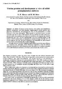
Effects on early pregnancy (see Text-fig. 1) a-Trichosanthin given intraperitoneally to mice on Day 4 of pregnancy resulted in an inhibition of pregnancy in 80% of the mice, and on Day 6, to a lesser extent of 35%. Severe bleeding from the vagina and abortion of conceptuses were observed in mice treated on Day 8. a-Momorcharin given to pregnant mice on Days 4 or 6 of pregnancy resulted in an inhibition of pregnancy in 57-64% of mice, but had no effect when given on Day 8 of pregnancy. This suggests that the abortifacient action of the proteins varies with the stages of implantation. The response of pregnant mice to either abortifacient followed an all-or-none pattern, i.e. the entire litter was aborted or survived the treatment. The average number of conceptuses per litter, which was about 12-9 (s.e.m. = 0-2, 63), was the same amongst the controls and the survivors in the treatment groups. The gross morphology of surviving embryos was normal and there were no signs of retarded development. =
*
Group
I
I II III IV 1511 9 9
II III IV
19 9 7 6
I II III IV 14 7 9 9
6
8
Day of pregnancy at treatment
Text-fig. 1. Effects of a-trichosanthin and a-momorcharin on early pregnancy (Days 1-4) of mice weighing 27-34 g. Data were pooled from 3 experiments. -^-Significantly different from the control at < 0-01 (Fisher's exact test applied to the raw data). I control (vehicle only), II a-trichosanthin (0-3 mg/25 g body wt), III a-momorcharin (0-1 mg/25 g body wt), IV a-momorcharin (0-2 mg/25 g body wt). no. of mice. =
=
=
=
=
Effects on hatching
in vitro
In our culture, 96-100% of the morulae developed to blastocysts after 18-24 h in the control groups and in those treated with a-trichosanthin or a-momorcharin. In the control group, about 50% of the embryos had hatched from the zona after 48 h in vitro and over 70% by 72 h in vitro. The
hatched embryos then attached to the culture plate, the trophoblasts formed outgrowths and the inner cell mass developed into an early egg cylinder (PI. 1, Figs 4 & 5). When 0-15 µg/ml or more of a-trichosanthin or a-momorcharin was present in the culture medium, significantly fewer embryos managed to hatch from the zona (Table 1). The unhatched embryos showed obvious signs of cell necrosis and the blastocoelic cavity rapidly collapsed. However, the embryos that did hatch successfully developed normally through the attachment and outgrowth stages in a fashion indistinguishable from untreated embryos (Table 1). Table 1. Effects of a-trichosanthin and a-momorcharin
on
in-vitro
No.
(%) of embryos hatched at :
Development subsequent 48 h
No. of Treatment
Control a-Trichosanthin
(Më/ml)
0015 015 0-75 1-50
Control o-Momorcharin
O^g/ml)
0015 015 0-75 1-50
embryos (2 exps)
24 h
48 h
37
2 (5)
19 (51)
22 30 19 19
0
49(2)
0
22(2) 22(2) 11(1) 23(2)
of zona-covered
development
embryos
2(7) 0 0
9(41) 7(23)* 3(16)* 0(0)
26(53)
72 h
hatching
to
72 h
Attached
Outgrowth
(70)
20(77)
6(23)
26(100) 24(92)
14(64) 12(40)* 4(21)* 0(0)
3(21) 10(83) 3(75)
0(0) 0(0) 2(50)
14(100) 14(100) 12(100) 10(83) 4(100) 4(100)
35(71)
20(57)
26
12(54) 16 (73) 5(23)* 12(54)* 0 1(9)* 0
0
0
8(50) 6(50)
0 0
0
Attached
0
Outgrowth
0
6(17)
35(100) 33(94)
0 0 0 0
16(100) 15(94) 12(100) 8(67) 1 (100) 1(100) 0
0
Values in *
parentheses are percentages. Significantly different from control values
at