polymers Article
Development of Poly(ε-Caprolactone) Scaffold Loaded with Simvastatin and Beta-Cyclodextrin Modified Hydroxyapatite Inclusion Complex for Bone Tissue Engineering Jung Bok Lee 1,2 , Ji Eun Kim 3 , Min Soo Bae 3 , Su A Park 4 , Daniel A. Balikov 1 , Hak-joon Sung 1,5,9 , Hoon Bong Jeon 6 , Hun Kuk Park 6 , Soong Ho Um 7 , Kook Sun Lee 8 and Il Keun Kwon 3, * 1
2 3 4 5 6 7 8 9
*
Department of Biomedical Engineering, Vanderbilt University, Nashville, TN 37212, USA;
[email protected] (J.B.L.);
[email protected] (D.A.B.);
[email protected] (H.S.) Department of Mechanical Engineering, Vanderbilt University, Nashville, TN 37235, USA Department of Maxillofacial Biomedical Engineering, Institute of Oral Biology, School of Dentistry, Kyung Hee University, Seoul 130-701, Korea;
[email protected] (J.E.K.);
[email protected] (M.S.B.) Department of Nature-Inspired Nanoconvergence Systems, Korea Institute of Machinery and Materials, 156 Gajeongbuk-ro, Yuseong-gu, Daejeon 304-343, Korea;
[email protected] Division of Cardiovascular Medicine, Vanderbilt University, Nashville, TN 37235, USA Department of Biomedical Engineering, College of Medicine, Kyung Hee University, 26, Kyungheedae-ro, Dongdaemun-gu, Seoul 130-701, Korea;
[email protected] (H.B.J.);
[email protected] (H.K.P.) School of Chemical Engineering & SKKU Advanced Institute of Nanotechnology, Sungkyunkwan University, Suwon, Gyeonggi-do 440-746, Korea;
[email protected] Department of Oral and Maxillofacial Radiology, College of Medicine, School of Dentistry, Kyung Hee University, Seoul 130-701, Korea;
[email protected] Severance Biomedical Science Institute, College of Medicine, Yonsei University, Seoul 120-752, Korea Correspondence:
[email protected]; Tel.: +82-10-7154-3000
Academic Editors: Esmaiel Jabbari and Frank Wiesbrock Received: 1 December 2015; Accepted: 5 February 2016; Published: 9 February 2016
Abstract: In this study, we developed poly(ε-caprolactone) (PCL) 3D scaffolds using a solid free form fabrication (SFF) technique. β-cyclodextrin (βCD) was grafted to hydroxyapatite (HAp) and this βCD grafted HAp was coated onto the PCL scaffold surface, followed by drug loading through an inclusion complex interaction between the βCD and adamantane (AD) or between βCD and simvastatin (SIM). The scaffold structure was characterized by scanning electron microscopy (SEM). The release profile of simvastatin in the β-CD grafted HAp was also evaluated. Osteogenic differentiation of adipose-derived stromal cells (ADSCs) was examined using an alkaline phosphatase activity (ALP) assay. The results suggest that drug loaded PCL-HAp 3-D scaffolds enhances osteogenic differentiation of ADSCs. Keywords: β-cyclodextrin; hydroxyapatite; poly(ε-caprolactone) 3-D scaffolds; simvastatin; bone regeneration
1. Introduction In the field of tissue engineering, three-dimensional (3D) porous scaffolds have been fabricated to mimic extra cellular matrix (ECM) found in native tissue for regeneration, restoration, and repair of damaged tissue and organs [1–3]. As their ECM mimetic structures are considered to be critical for successful clinical applications, several rapid prototyping (RP) techniques have been developed
Polymers 2016, 8, 49; doi:10.3390/polym8020049
www.mdpi.com/journal/polymers
Polymers 2016, 8, 49
2 of 10
including selective laser sintering (SLS) [4], stereolithography (SLA) [5], 3D printing [6] and solid free-form fabrication (SFF) [7–9]. In particular, the SFF technique can produce well-controlled, reproducible, scaffold structures such as pore size, porosity, and mechanical properties while varying their internal architecture [9–11]. Synthetic biodegradable polyesters such as poly(lactic-co-glycolic acid) (PLGA), poly(L-lactic acid) (PLLA), and poly(ε-caprolactone) (PCL) have been used as biomaterials in biomedical application due to their excellent biocompatibility and good mechanical properties. Among them, PCL is a thermosensitive polymer with low melting point of about 60 ˝ C and a glass transition temperature of about ´60 ˝ C, a highly desirable set of temperatures. Its low melting point (60 ˝ C) facilitates the SFF process compared to other polyesters. Furthermore, Several PCL-derived products have also been approved by the Food and Drug Administration (FDA) for clinical applications [12,13]. Although continuous progress has been made so far, PCL has not been applied extensively for bone regeneration, a niche field of tissue engineering that could benefit greatly from utilizing PCL [9,14]. In this study, hydroxyapatite (HAp) was used because it is an attractive material for bone tissue engineering due to its inorganic components boosting affinity for ECM proteins and induction of osteogenic differentiation and mineralization [15–17]. Since β-cyclodextrin (βCD) has been previously used for solubilizing and stabilizing drugs by host-guest type inclusion complexation [18], an inclusion complex system between βCD and adamantane (AD) was used to attach HAp onto the surface of the PCL scaffold. Inclusion complex systems have been widely used as a drug loading strategy that forms a bond between ferrocene and cyclodextrin [19]. In solution or in solid state, the cavity of the cyclodextrin generates a hydrophobic environment, thereby changing its physical and chemical interactions with a guest molecule (e.g., drug) [20,21]. For our study, the “driving force” of the complex formation is the fundamental interaction between the βCD as the host molecule and AD as the guest molecule. Using this inclusion complex, we produced an HAp-coated PCL scaffold as well as simvastin (SIM)-loaded on HAp that has been known to promote osteogenic differentiation of mesenchymal stem cell via Ras/Smad/Erk signaling derived from bone morphogenic protein (BMP)-2 treatment [22,23]. We used these scaffold templates to examine their osteoconductive/osteoinductive abilities in in vitro human adipose-derived stromal cell (hADSC) and in vivo animal models. 2. Experimental Section 2.1. Materials Poly(ε-caprolactone), hydroxyapatite (HAp) nanoparticles (NPs), 3-aminopropyltriethoxysilane (APTES), ascorbic acid, dexamethasone, β-glycerophosphate (glycerol 2-phosphate disodium salt hydrate), and adamantylamine were purchased from Sigma-Aldrich (St. Louis, MO, USA). β-cyclodextrin (β-CD) was purchased from TCI (TOKYO Chemical Industry Co. Ltd., Tokyo, Japan). Dulbecco’s modified Eagle’s medium (DMEM), fetal bovine serum (FBS), phosphate buffered saline (PBS), trypsin-EDTA and antibiotics (penicillin-streptomycin) were purchased from Gibco (Rockville, MD, USA). Simvastatin was purchase from Wako (Osaka, Japan). StemPro® Human Adipose-Derived Stem Cells (hADSCs) were obtained from Invitrogen (Carlsbad, CA, USA). 2.2. Poly(ε-caprolactone) (PCL) Scaffold Preparation PCL scaffolds were created using a 3D SFF plotting system consisting of a 3-axes machine with a 10 ˆ 10 ˆ 10 cm x–y–z stage, dispenser, nozzle, compression/heat controller, and software/hardware system. PCL pellets were melted in a cylinder at 100 ˝ C using the heating jacket of the plotting system. The hardware system controlled the scaffold structure by adjusting the pressure, feed rate, and nozzle size. The temperature, pressure, and feed rate were set at 85 ˝ C, 650 kPa, and 150 µm/s, respectively. For the reaction with adamantine, acrylic acid (AAc) was grafted onto the surface of the PCL scaffold. Briefly, the scaffolds were wetted with 70% ethanol and then immersed in an aqueous AAc solution (10% in distilled water (DW) with 0.01 M ammonium ferrous sulfate) and exposed to gamma ray
Polymers 2016, 8, 49
3 of 10
radiation (10 kGy) from a cobalt-60 source at ambient temperature. Scaffolds were washed with DW three times to remove any unreacted residual monomers or homoplymers. AAc-PCL scaffolds were immersed in adamantylamine (AD, 10 mM)/ethanol solution for 24 h with stirring to graft AD onto the PCL scaffold using 1-Ethyl-3-(3-dimethylaminopropyl)-carbodiimide (EDC)/N-hydroxysuccinimide (NHS). Finally, scaffolds were washed with DW three times and freeze dried. 2.3. Preparation of β-Cyclodextrin (βCD) Grafted Hydroxyapatite (HAp) βCD (10 mmol) was dissolved in anhydrous toluene. Then 15 mmol succinic anhydride was added to the solution and the polymers were reacted with stirring at 60 ˝ C for 12 h under N2 . After the reaction, the product was concentrated by rotary-evaporation. The polymer was then precipitated in diethyl ether. For amino-functionalization of HAp, 5 g of HAp NPs were immersed in 100 mL anhydrous toluene containing 10 mL APTES at 120 ˝ C under an N2 atmosphere with a reflux condenser for 24 h. The samples were then rinsed for 1 h in toluene to remove any unreacted silane, and βCD was grafted onto the surface of the HAp by a 1-ethyl-3-dimethylaminopropyl carbodiimide- (EDC) mediated reaction between the primary amine groups of the HAp NP surface and the carboxyl group of βCD. In order to quantify the amine concentration in the HAp surface, the HAp NPs were immersed in 500 µM acid orange 7 solution at pH 3 for 3 h at room temperature and then washed three times with pH 3 DI water. After reaction, the acid orange 7 dye was desorbed by placing the NPs in pH 12 DI water for 30 min. The absorbance was measured at a wavelength of 485 nm using a spectrophotometer (UV-1650PC, Shimadzu, Japan). 2.4. Surface Coating of PCL Scaffold with HAp and Simvastatin Loading SIM was dissolved in 50% ethanol at 5 wt % concentration and then βCD grafted HAp NPs (3 mg/mL) were suspended in the solution. AD-modified PCL scaffolds were immersed in the aforementioned solution and then subjected to ultra-sonication at room temperature for 1 h. SIM was loaded into the βCDs of the HAp NPs that were attached to ADs on the surface of the PCL scaffold. After the reaction, the surface of the PCL scaffolds was washed to remove any left over, unloaded HAp NPs and SIM. After 14 days of incubating SIM-loaded samples in a 15 mL conical tube containing 3 mL PBS (pH 7.4) at 37 ˝ C with orbital agitation at 100 rpm, the solution containing released SIM was freeze-dried and redissolved in 1 mL EtOH. The loading amount of SIM was analyzed by ultraviolet-visible (UV) spectroscopy (UV-1650PC, SHIMADZU, Kyoto, Japan) at a wavelength of 238 nm and compared to a calibration curve of standard solution at a certain concentration of SIM. 2.5. Characterization of βCD-Grafted HAp and PCL Scaffolds The surface modified HAp NPs were characterized by thermal gravimetric analysis (TGA, TGA Q5000 IR/SDT Q600, TA Instruments, New Castle, DE, USA) in a scanning range from 25–800 ˝ C at a constant heating rate of 5 ˝ C min´1 . The surface morphology of HAp-coated PCL scaffolds was analyzed with a scanning electron microscope (SEM, Hitachi S-2300, Tokyo, Japan). Images of samples were taken with 15 kV of an accelerating voltage after sputter-coating with gold. Surface chemical compositions of βCD-modified HAp and AAc-grafted PCL scaffolds (diameter: 8 mm and thickness: 3 mm) were determined using an X-ray photoelectron spectroscope (XPS, Thermo Fisher Scientific, MA, USA) at a grazing angle of 90˝ under high vacuum (600 µm, porosity of 91.15% and strands with a thickness of 380–400 µm [24]. After βCD-modified HAp coating, a homogeneous distribution of HAp NPs along the surface was observed on the AD-modified PCL scaffold surface whereas βCD-modified HAp NPs were not observed on non-coated PCL scaffolds.
Polymers 2016, 8, 49
5 of 10
Polymers 2016, 8, 49
5 of 9
Polymers 2016, 8, 49
5 of 9
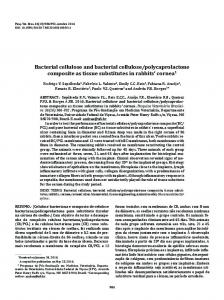
Figure 1. Schematic illustration of hydroxyapatite coated 3D poly(ɛ-caprolactone) (PCL) scaffolds.
Figure 1. Schematic illustration of hydroxyapatite coated 3D poly(ε-caprolactone) (PCL) scaffolds. Figure 1. Schematic illustration of(βCD)-grafted hydroxyapatite coated 3D poly(ɛ-caprolactone) (PCL) scaffolds. (1) Preparation of β-cyclodextrin hydroxyapatite (HAp) and (2) HAp coating and (1) Preparation of β-cyclodextrin (βCD)-grafted hydroxyapatite (HAp) and (2) HAp coating and (1) Preparation of β-cyclodextrin (βCD)-grafted (HAp) and (2) HAp coating and simvastatin loading on the surface of adamantane hydroxyapatite (AD)-modified PCL scaffold. simvastatin loading on on thethe surface (AD)-modifiedPCL PCL scaffold. simvastatin loading surfaceofofadamantane adamantane (AD)-modified scaffold.
Figure 2. Scanning electron microscopy (SEM) images of β-cyclodextrin (βCD)-grafted Figure 2. Scanning electronpoly(ɛ-caprolactone) microscopy (SEM)(PCL) images of β-cyclodextrin (βCD)-grafted hydroxyapatite (HAp)-coated scaffolds (A) without and (B) with Figure 2. Scanning electron microscopy (SEM) images of β-cyclodextrin (βCD)-grafted hydroxyapatite hydroxyapatite poly(ɛ-caprolactone) (PCL) scaffolds (A) without and (B) with adamantane (AD)(HAp)-coated grafting. (HAp)-coated poly(ε-caprolactone) (PCL) scaffolds (A) without and (B) with adamantane (AD) grafting. adamantane (AD) grafting.
Simulated body fluid (SBF) solution immersion has been commonly used to produce an Simulated body fluid (SBF)solution solution immersion has been usedused to produce an an Simulated body fluid immersion hasHowever, beencommonly commonly to produce osteoconductive surface for(SBF) bone tissue engineering [24,25]. its coating procedures, such osteoconductive surface for bone tissue engineering [24,25]. However, its coating procedures, such as using SBF solutions and direct coating of HAp powder, have some disadvantages because SFF osteoconductive surface for bone tissue engineering [24,25]. However, its coating procedures, such as asSBF using SBF solutions andofcoating direct of powder, HAp powder, have some disadvantages because extended periods time tocoating successfully work; and the surface structure of the scaffolds can usingrequires solutions and direct of HAp have some disadvantages because SFFSFF requires requires extended periods of time to successfully work; and the surface structure of the scaffolds be changed viaof indiscreet mineral formation, irreversible alteration scaffold properties. extended periods time to successfully work;resulting and the in surface structure of theofscaffolds can be can changed be changed viathese indiscreet mineral formation, resulting in irreversible scaffoldand properties. To overcome disadvantages, we used βCD-modified HAp as alteration a coating of applied via indiscreet mineral formation, resulting in irreversible alteration of scaffoldmaterial properties. To overcome To overcome these disadvantages, we used βCD-modified HAp as a coating material and this strategy to surface coating of PCL scaffold by an inclusion complex system between βCDapplied on the thesethis disadvantages, we used βCD-modified HAp as a coating material and applied this strategy to surface coating of PCL scaffold byproduced an inclusion complex system between βCDNPs on the HApstrategy and ADtoon the PCL scaffold. This reaction a homogeneous coating of HAp on surface coating of on PCL bythe anloading inclusion complex system between βCD on the HApNPs andon AD on HAp andscaffold AD thescaffold PCL scaffold. This reaction produced adrug homogeneous coating of HAp the PCL and enhanced efficiency of the [26]. the PCL scaffold. This reaction produced a homogeneous coating of HAp NPs on the PCL scaffold and the PCL scaffold and enhanced the loading efficiency of the drug [26]. enhanced the loading efficiency of theHAp drug 3.2. Characterization of βCD-Modified and[26]. AD-Modified PCL Scaffold 3.2. Characterization of βCD-Modified HAp and AD-Modified PCL Scaffold To analyze the surface characteristics of βCD-modified HAp and AD-modified PCL scaffolds, 3.2. Characterization of βCD-Modified HAp and AD-Modified PCL Scaffold analyze thespectroscopy surface characteristics of βCD-modified and After AD-modified PCL scaffolds, X-rayTo photoelectron (XPS) was conducted on theseHAp samples. amino-functionalization X-ray photoelectron (XPS) wasofconducted onand these samples. After amino-functionalization of and βCD, thespectroscopy binding energy peaks Ca2p, N1s of HApHAp NPs were (Figure 3) PCL ToHAp analyze the surface characteristics of P2p, βCD-modified andchanged AD-modified of HAp and βCD, thefacts binding peaks Ca2p, P2p,ofand N1s of HAp were changed (Figure 3) After as evidenced byphotoelectron the that energy thespectroscopy Ca2p andofP2p peaks pure HAp wereNPs reduced; and the N1s peak scaffolds, X-ray (XPS) was conducted on these samples. as evidenced by the facts that the Ca2p and P2p peaks of pure HAp were reduced; and the N1s peak was changed from 0.1 eV of HAp to 3.1 eV and 1.6 eV for HAp-NH 2, and HAp-βCD, respectively. amino-functionalization of HAp and βCD, the binding energy peaks of Ca2p, P2p, and N1s of was changed from 0.1 eV of HAp to 3.1 eV and 1.6 eV for HAp-NH2, and HAp-βCD, respectively.
HAp NPs were changed (Figure 3) as evidenced by the facts that the Ca2p and P2p peaks of pure HAp were reduced; and the N1s peak was changed from 0.1 eV of HAp to 3.1 eV and 1.6 eV for HAp-NH2 , and HAp-βCD, respectively. These results confirmed that the HAp surface was successfully
Polymers 2016, 8, 49
6 of 10
Polymers 2016, 8, 49 Polymers 2016, 8, 49
6 of 9
6 of 9
amino-functionalized by APTES, NH grafting ofamino-functionalized βCD onto the HAp NPs. Table 1 These results confirmed that leaving the HAp surface wasfor successfully by APTES, 2 groups These results confirmed that the HAp surface was successfully amino-functionalized by APTES, shows the quantified amine concentrations on the amino-functionalization of HAp. The amine leaving NH2 groups for grafting of βCD onto the HAp NPs. Table 1 shows the quantified amine leaving NH2 groups for grafting of βCD onto the HAp NPs. Table 1 shows the quantified amine concentration of HAp-βCD was 3.5 ˘ 0.4 µmol/mg and APTES-grafted HAp wasof21.9 2.9 µmol/mg. concentrations on the theamino-functionalization amino-functionalization HAp. The amine concentration of ˘ HAp-βCD concentrations on of of HAp. The amine concentration HAp-βCD was was 3.5 ± 0.4 µmol/mg and APTES-grafted HAp was 21.9 ± 2.9 µmol/mg. These results showed These results showed approximately 18.4 µmol/mg of NH groups were generated to graft βCD onto 3.5 ± 0.4 µmol/mg and APTES-grafted HAp was 21.9 2± 2.9 µmol/mg. These results showed approximately 18.4 µmol/mg of NH 2 groups were generated to graft βCD onto the surface of the HAp the surface of the HAp NPs. Unexpectedly, some remaining primary amines of the HAp NPs were approximately 18.4 µmol/mg of NH2 groups were generated to graft βCD onto the surface of the HAp NPs. Unexpectedly, some remaining primary amines of HAp were detected after βCD detected βCD grafting when fluorescent amine was of used (data not shown), indicating need to NPs.after Unexpectedly, some remaining primary amines thethe HAp NPsNPs were detected afterthe βCD grafting when fluorescent aminewas was used (data not shown), indicating the need to improve the βCD grafting when grafting fluorescent amine used (data not shown), the need to improve improve the βCD efficiency further. To determine theindicating amount of the grafted βCDthe onβCD the HAp efficiency further. ToHAp-βCD determinethe the amount grafted βCD on the HAp both grafting efficiency further. To determine amount of of thethe grafted βCD on the HAp NPs,NPs, both(TGA) non- nonNPs,grafting both non-treated HAp and were analyzed by thermal gravimetric analysis at a treated HAp and HAp-βCD were analyzed by thermal gravimetric analysis (TGA) at a temperature treated and HAp-βCD were analyzed by thermal gravimetric analysis (TGA) at a temperature ˝ temperature range of 0–800 C (Figure 4A). The TGA curve of both non-treated HAp and HAp-βCD range of 0–800 0–800 °C (Figure 4A). TGA of of both HAp andand HAp-βCD showed range 4A).The Therange TGAcurve curve both HAp showed ˝ Cnon-treated showed weight loss°C at (Figure a temperature of 20–100 asnon-treated expected given that HAp-βCD the scaffolds have weight loss at as as expected given thatthat the the scaffolds havehave somesome weight at aa temperature temperaturerange rangeofof20–100 20–100°C°C expected given scaffolds some moisture content that can evaporate within this temperature range. Between 300 and 600 ˝ C, moisture content content that this temperature range. Between 300 300 and and 600 °C, moisture thatcan canevaporate evaporatewithin within this temperature range. Between 600weight °C, weight weight loss of HAp-βCD was observed due to thermal degradation of grafted βCD,the confirming the loss of HAp-βCD was observed due to thermal degradation of grafted βCD, confirming loss of HAp-βCD was observed due to thermal degradation of grafted βCD, confirmingpresence the presence presence of grafted βCD. of grafted grafted βCD. of βCD.
Figure 3. X-ray photoelectron spectroscopy (XPS) analysis of β-cyclodextrin (βCD) grafted
Figure 3. X-ray photoelectron spectroscopy (XPS) analysis of β-cyclodextrin (βCD) grafted Hydroxyapatite (HAp), 3-aminopropyltriethoxysilane (APTES) grafted and pure HAp powders. Figure 3. X-ray spectroscopy (XPS) analysis ofHAp, β-cyclodextrin (βCD) grafted Hydroxyapatite (HAp),photoelectron 3-aminopropyltriethoxysilane (APTES) grafted HAp, and pure HAp powders. Hydroxyapatite (HAp), 3-aminopropyltriethoxysilane (APTES) grafted HAp, and pure HAp powders. Table 1. Amine contents of 3-aminopropyltriethoxysilane (APTES)-grafted hydroxyapatite (HAp)
Tableand 1. Amine contents of 3-aminopropyltriethoxysilane (APTES)-grafted hydroxyapatite (HAp) and β-cyclodextrin (βCD)-grafted HAp. Table 1. Amine contents of 3-aminopropyltriethoxysilane (APTES)-grafted hydroxyapatite (HAp) β-cyclodextrin (βCD)-grafted HAp. and β-cyclodextrin (βCD)-grafted HAp. Heading Amine contents (µmol/mg) Heading
Heading Amine(µmol/mg) contents (µmol/mg) Amine contents
Hap-NH2 21.9 ±Hap-NH 2.9 2
Hap-NH2 21.9 ˘ 2.9 21.9 ± 2.9
Hap-βCD
3.5 ± 0.4 Hap-βCD Hap-βCD 3.5 ˘ 0.4 3.5 ± 0.4
Figure 4. Thermal gravimetric analysis (TGA) of (A) surface-modified hydroxyapatite (HAp) composites and (B) HAp-coated poly(ɛ-caprolactone) (PCL) scaffolds.
Figure 4. Thermal gravimetricanalysis analysis(TGA) (TGA)ofof(A) (A)surface-modified surface-modified hydroxyapatite hydroxyapatite (HAp) Figure 4. Thermal gravimetric (HAp) composites and (B) HAp-coated poly(ɛ-caprolactone) (PCL) scaffolds. composites and (B) HAp-coated poly(ε-caprolactone) (PCL) scaffolds.
Polymers 2016, 8, 49 Polymers 2016, 8, 49
7 of 9 7 of 10
To determine the degree of AD grafting on to the PCL scaffold surface, acrylic acid (AAc) was grafted onto the surface as the functionalized carboxyl groups could then be stained by toluidine blue To determine the degree of AD grafting on to the PCL scaffold surface, acrylic acid (AAc) was (Figure S1). Using the same carboxyl groups, ADs were grafted onto the surface of the PCL scaffold. grafted onto the surface as the functionalized carboxyl groups could then be stained by toluidine blue Figure S2 shows the amount of grafted AD resulting from various AD concentrations. Nearly 100% (Figure S1). Using the same carboxyl groups, ADs were grafted onto the surface of the PCL scaffold. of AD in all treatment groups was grafted on the surface of the PCL scaffold regardless of the solution Figure S2 shows the amount of grafted AD resulting from various AD concentrations. Nearly 100% of concentration. To measure the amount of coated HAp on the PCL scaffolds, TGA was conducted at AD in all treatment groups was grafted on the surface of the PCL scaffold regardless of the solution a temperature range of 0–600 °C (Figure 4B). HAp was found to be 7 wt % whereas 0.7 wt % was concentration. To measure the amount of coated HAp on the PCL scaffolds, TGA was conducted at reported for non-AD grafted and 0.2 wt % for the PCL control. a temperature range of 0–600 ˝ C (Figure 4B). HAp was found to be 7 wt % whereas 0.7 wt % was reported for non-AD grafted and 0.2 wt % for the PCL control. 3.3. In vitro Cell Test of PCL Scaffolds 3.3. InThe Vitro Cell Test of PCL Scaffolds initial loading amount of SIM on the HAp-βCD-coated PCL scaffold was 9.56 µg/mg, and 100% of SIM was released after days not shown). Proliferation of hADSCs measured The initial loading amount of14SIM on(data the HAp-βCD-coated PCL scaffold was 9.56was µg/mg, and using the CCK-8 assay kit at 1, 4, and 7 days post culture. Figure 5A shows that there was no observed 100% of SIM was released after 14 days (data not shown). Proliferation of hADSCs was measured cytotoxicity among allkit groups and demonstrated similar Figure proliferation patterns. Figure ALP using the CCK-8 assay at 1, 4, and 7 days post culture. 5A shows that there was5Bnoshows observed activities of hADSCs on the test PCL scaffolds. Commonly, ALP activity is an early marker of cytotoxicity among all groups and demonstrated similar proliferation patterns. Figure 5B shows ALP immature osteoblast Moreover, has been ALP described organic activities of hADSCs on activity. the test PCL scaffolds.itCommonly, activitythat is anALP early cleavage marker ofof immature phosphate plays a role in the mineralization of the extracellular collagenous matrix by providing osteoblast activity. Moreover, it has been described that ALP cleavage of organic phosphate plays a calcium phosphate of ions generate new formation of the cell-mediated calcium phosphate role in theand mineralization theto extracellular collagenous matrix by providing calcium and phosphate mineralized matrix Elevated of ALP activity noticed on HAp-coated and ions to generate new[25,26]. formation of the levels cell-mediated calcium were phosphate mineralized matrixPCL [25,26]. SIM-loaded HAp-coated PCL scaffolds at day 14. The ALP activity was significantly higher on the Elevated levels of ALP activity were noticed on HAp-coated PCL and SIM-loaded HAp-coated PCL HAp-coated PCL PCL scaffolds PCL control at 14 days scaffolds at day 14.and TheSIM-loaded ALP activityHAp-coated was significantly higher onthan the the HAp-coated PCLscaffold and SIM-loaded of cell culture. HAp-coated PCL scaffolds than the PCL control scaffold at 14 days of cell culture.
Figure5.5.Human Humanadipose-derived adipose-derivedstem stemcell cell(hADSC) (hADSC)(A) (A)proliferation proliferationand and(B) (B)alkaline alkalinephosphatase phosphatase Figure (ALP)activity activityon onpoly(ε-caprolactone) poly(ɛ-caprolactone)(PCL) (PCL)scaffolds. scaffolds. (ALP)
3.4.In Invivo vivoAnimal AnimalStudy Study 3.4. Thebone bonedefect defectrepair repaireffect effectof ofscaffolds scaffoldswas wasinvestigated investigatedusing usingthe therabbit rabbitcalvarial calvarialdefect defectmodel model The by in vivo experiments. Figure 6 illustrates the raw µCT images and the corresponding analysis. by in vivo experiments. Figure 6 illustrates the raw µCT images and the corresponding analysis. HAp-coated and and SIM-loaded SIM-loaded HAp-coated HAp-coated PCL PCL scaffold scaffold groups groups had had noticeable noticeable increases increases in in bone bone HAp-coated formationafter after66weeks weeksof ofimplantation implantationas ascompared comparedto tothe thePCL PCLcontrol controlgroup. group.Using Usingimage imageanalysis analysis formation software,anan8 mm 8 mm circular was drawn µCTover images over site; the defect and the software, circular ROI ROI was drawn on the on µCTthe images the defect and thesite; regenerated regenerated total bone volume wasby obtained by measuring mean grayfor value this region. total bone volume (RBV) was (RBV) obtained measuring the meanthe gray value thisfor region. The The total RBV of all groups had increased over 6 weeks, however the RBV in the SIM-HAp loaded total RBV of all groups had increased over 6 weeks, however the RBV in the SIM-HAp loaded PCL PCL22.3 was˘22.3 1.9 3mm 6 weeks, which was significantly greater thanthat thatobserved observedininthe thePCL PCL was 1.9±mm at 36atweeks, which was significantly greater than scaffoldsgroups. groups. Bone Boneregeneration regeneration in inthe thedefects defectswas wasfound foundin inboth bothHAp-coated HAp-coatedand andSIM-loaded SIM-loaded scaffolds HAp-coated PCL scaffolds. However the SIM-loaded HAp-coated PCL scaffold group demonstrated HAp-coated PCL scaffolds. However the SIM-loaded HAp-coated PCL scaffold group demonstrated moreactive activeosteogenesis osteogenesisin inthe thedefect defectarea areacompared comparedwith withother othergroups groupsduring during inplantation inplantation periods. periods. more Theincorporation incorporationofofSIM SIMand andHAp HApwas wasshown shownto tosignificantly significantlyincrease increasethe thebone boneregeneration regenerationin invivo. vivo. The HAp NPs provide a suitable microenvironment for mimicking the inorganic phase of the native tissue HAp NPs provide a suitable microenvironment for mimicking the inorganic phase of the native tissue and SIM has a positive influence on accelerating mineral deposition in vivo. SIM is widely used in
Polymers 2016, 8, 49
8 of 10
Polymers 2016, 8, 49
8 of 9
and SIM has a positive influence on accelerating mineral deposition in vivo. SIM is widely used in clinical lipid-lowering drugs, and it it plays a role inin promoting new bone formation and regulation byby clinical lipid-lowering drugs, and plays a role promoting new bone formation and regulation BMP-2 and VEGF expression ofof osteoblasts [27,28]. BMP-2 and VEGF expression osteoblasts [27,28].
Figure Micro-computerized tomography (μCT) scan images rabbit calvarial defects after 6 weeks Figure 6. 6. Micro-computerized tomography (µCT) scan images of of rabbit calvarial defects after 6 weeks implantation various poly(ɛ-caprolactone) (PCL) scaffolds. < 0.05 and < 0.005 compared of of implantation of of various poly(ε-caprolactone) (PCL) scaffolds. * p*