CLINICAL AND VACCINE IMMUNOLOGY, Sept. 2007, p. 1182–1189 1556-6811/07/$08.00⫹0 doi:10.1128/CVI.00101-07 Copyright © 2007, American Society for Microbiology. All Rights Reserved.
Vol. 14, No. 9
Development of Recombinant Nucleoprotein-Based Diagnostic Systems for Lassa Fever䌤 Masayuki Saijo,1*† Marie-Claude Georges-Courbot,2 Philippe Marianneau,2 Victor Romanowski,3 Shuetsu Fukushi,1 Tetsuya Mizutani,1 Alain-Jean Georges,4 Takeshi Kurata,5 Ichiro Kurane,1 and Shigeru Morikawa1† Department of Virology 1, National Institute of Infectious Diseases, 4-7-1 Gakuen, Musashimurayama, Tokyo 208-0011, Japan1; Unit of Biology of Viral Emerging Infections, Institute Pasteur, IFR 128 Biosciences Lyon-Gerland, 21 Avenue Tony Garnier, 69365 Lyon Cedex 07, France2; Instituto de Bioquı´mica y Biologı´ca Molecular, Facultad de Ciencias Exactas, Universidad Nacional de a Plata, 1900 La Plata, Argentina3; Laboratory P4 Jean-Merieux-INSERM, 21 Avenue Tony Garnier 69365 Lyon Cedex 07, France4; and Department of Pathology, National Institute of Infectious Diseases, 1-23-1 Toyama, Shinjuku-ku, Tokyo 162-864, Japan5 Received 1 March 2007/Returned for modification 22 April 2007/Accepted 20 June 2007
Diagnostic systems for Lassa fever (LF), a viral hemorrhagic fever caused by Lassa virus (LASV), such as enzyme immunoassays for the detection of LASV antibodies and LASV antigens, were developed using the recombinant nucleoprotein (rNP) of LASV (LASV-rNP). The LASV-rNP was expressed in a recombinant baculovirus system. LASV-rNP was used as an antigen in the detection of LASV-antibodies and as an immunogen for the production of monoclonal antibodies. The LASV-rNP was also expressed in HeLa cells by transfection with the expression vector encoding cDNA of the LASV-NP gene. An immunoglobulin G enzymelinked immunosorbent assay (ELISA) using LASV-rNP and an indirect immunofluorescence assay using LASV-rNP-expressing HeLa cells were confirmed to have high sensitivity and specificity in the detection of LASV-antibodies. A novel monoclonal antibody to LASV-rNP, monoclonal antibody 4A5, was established. A sandwich antigen capture (Ag-capture) ELISA using the monoclonal antibody and an anti-LASV-rNP rabbit serum as capture and detection antibodies, respectively, was then developed. Authentic LASV nucleoprotein in serum samples collected from hamsters experimentally infected with LASV was detected by the Ag-capture ELISA. The Ag-capture ELISA specifically detected LASV-rNP but not the rNPs of lymphocytic choriomeningitis virus or Junin virus. The sensitivity of the Ag-capture ELISA in detecting LASV antigens was comparable to that of reverse transcription-PCR in detecting LASV RNA. These LASV rNP-based diagnostics were confirmed to be useful in the diagnosis of LF even in institutes without a high containment laboratory, since the antigens can be prepared without manipulation of the infectious viruses. Lassa fever (LF) is a viral hemorrhagic fever caused by Lassa virus (LASV), an Old World arenavirus. Many cases of LF occur in western Africa in countries such as Guinea, Sierra Leone, and Nigeria (7, 23, 27, 29–31). It is thought that LASV infects tens of thousands of humans annually and causes hundreds to thousands of deaths (34). Humans become infected through contact with infected excreta, tissue, or blood from the peridomestic rodent, Mastomys natalensis, the reservoir host of LASV (34). LASV can be transmitted to other humans via mucosal or cutaneous contact or through nosocomial contamination (27). More than 20 imported cases of LF have been reported outside the endemic region in areas such as the United States, Canada, Europe, and Japan (1, 2, 13, 15, 18, 24, 25). Recently, the potential for the use of hemorrhagic fever viruses, including LASV, as a biological weapon has been emphasized (5, 6). Therefore, the development of diagnostic systems for LF is important even in countries free from LF outbreaks to date.
Manipulation of infectious LASV is necessary for the detection of specific antibodies. However, a high-containment laboratory (biosafety level 4 [BSL-4]) is required for handling infectious LASV and, therefore, the preparation of LASV antigens cannot be implemented in institutes without a BSL-4 facility. Within this framework, it is important to develop sensitive and specific diagnostic systems for LF that eliminate the need for the manipulation of infectious LASV. In the present study, the recombinant nucleoprotein (rNP) of LASV (LASVrNP) was expressed and evaluated for its ability to detect LASV antibodies. LASV-rNP-based enzyme-linked immunosorbent and indirect immunofluorescence assays (ELISA and IIFA) were developed. Furthermore, novel monoclonal antibodies to LASV-rNP were generated and used in combination with the recombinant antigen to develop an LASV antigen (nucleoprotein) capture ELISA. The present study presents an alternative strategy to develop diagnostic systems without handling infectious LASV.
* Corresponding author. Mailing address: Special Pathogens Laboratory, Department of Virology 1, National Institute of Infectious Diseases, 4-7-1 Gakuen, Musashimurayama, Tokyo 208-0011, Japan. Phone: 81-42561-0771, ext. 320. Fax: 81-42-561-2039. E-mail:
[email protected]. † M.S. and S.M. contributed equally to this study. 䌤 Published ahead of print on 18 July 2007.
Cells. A HeLa cell line was cultured in the Eagle minimum essential medium supplemented with 10% fetal bovine serum and the antibiotics penicillin G and streptomycin (MEM-10FBS). Tn5 insect cells were used for the expression of the rNPs of arenaviruses (LASV, lymphocytic choriomeningitis virus [LCMV], and Junin virus [JUNV]) in a baculovirus system. The Tn5 insect cells were cultured as reported previously (38).
MATERIALS AND METHODS
1182
VOL. 14, 2007
RECOMBINANT NP-BASED DIAGNOSTICS FOR LASSA FEVER
Viruses. LASV (strain AV), which was isolated from an imported case of LF to Germany from West Africa, was used (13). The experimental process that required manipulation of infectious LASV was carried out in the BSL-4 laboratory in the P4 laboratory, INSERM, Lyon, France. Mopeia virus (MOPV), which belongs to the family Arenaviridae, genus Arenavirus, was also used. Recombinant NPs of LCMV (26) and JUNV (11), designated LCMV-rNP and JUNV-rNP, respectively, were also expressed in a baculovirus system and used in the study. A baculovirus (Ac-⌬P), which lacks polyhedrin expression, was used as a control virus (26). The virus titer of LASV in serum samples was determined by using a focus-forming unit (FFU) assay as described previously (3). Sera. Four human serum samples—three samples serially collected from one patient with LF and one additional sample from another patient with LF—and ninety-six human sera collected from Japanese subjects with no history of travel to areas where LF is endemic were used as positive and negative controls, respectively. The patient with LF, from whom three serial serum samples were collected, was the first case of LF to be imported in Japan in 1987 (15). The other human serum sample was provided from the Special Pathogens Branch, National Center for Infectious Diseases, Centers for Disease Control and Prevention, Atlanta, GA. Serum samples collected from five monkeys (Macaca fascicularis) subcutaneously infected with LASV strain AV at 103 FFU (two monkeys) or 107 FFU (three monkeys) and those collected from four monkeys with mock infection were also used. The serum samples used in the study were collected at 4 to 5 weeks postchallenge. Five hamsters were subcutaneously infected with 103 FFU of LASV, strain AV, and blood was drawn on days 0, 4, 11, and 16 postinfection, taking the day on which the virus was inoculated as day 0. Serum fractions of the collected blood specimens were separated and tested for LASV antigen by antigen capture (Ag-capture) ELISA and reverse transcription-PCR (RT-PCR). Rabbit sera (polyclonal antibodies) were raised against LASV-rNP, LCMVrNP, and JUNV-rNP by immunization of rabbits with the purified LASV-rNP, LCMV-rNP, and JUNV-rNP, respectively, in the form of a mixture with the adjuvant, Inject Alum (Pierce). Rabbits were immunized with sufficient amount of the purified nucleoproteins of each virus three times with an interval of 2 weeks. After confirmation of the increased titer, ⬎10,000 times as determined by indirect immunofluorescence assay, which was developed in the present study, blood was drawn from the rabbits, and the serum fraction was used in the present study. Recombinant baculovirus. In order to construct the transfer vector, a cDNA clone of NP from LASV strain Josiah was used. The cDNA was kindly provided by J. B. McCormick, former Director of the Special Pathogens Branch, National Centers for Infectious Diseases, Centers for Disease Control and Prevention. The complete nucleotide sequence of the NP gene is registered in GenBank under the accession number NC_004296. The DNA of the LASV-NP was amplified by PCR from the source using the primers LAS-NfB (5⬘-GTGGATCCA ACACAACAATCTGG-3⬘; the BamHI restriction site is underlined) and LASNrB (5⬘-CCGGATCCATTTACAGAACGACTC-3⬘). The PCR conditions were the same as previously reported (38). The 1,743-bp amplification product was digested with BamHI and subcloned into the BamHI site of pQE32 vector DNA (QIAGEN GmbH, Hilden, Germany) to construct pQE32-LASV-NP. The inserted LASV-NP DNA was sequenced by using appropriate primers with an ABI Prism 310 genetic analyzer (PE Applied Biosystems, Foster City, CA) and confirmed to be in proper orientation downstream the promoter and identical to the original sequence. The DNA fragment of LASV-NP with a histidine (His) tag was isolated from the plasmid, pQE32-LASV-NP, by digestion with EcoRI and HindIII. It was then blunt repaired with Klenow enzyme and ligated into the blunt-ended BamHI site of pAcYM1 (26). The resulting recombinant transfer vector with the correct orientation with respect to the polyhedrin promoter was constructed (pACYM1-His-LASV-NP). Tn5 insect cells were transfected with mixtures of purified Autographa californica nuclear polyhedrosis virus (AcMNPV) DNA and pAcYM1-His-LASV-NP according to the procedures described by Kitts et al. (20), with the modification of Matsuura et al. (26). Recombinant baculovirus was then isolated. The baculovirus, which expressed His-tagged LASV-rNP (His-LASV-rNP), was designated Ac-His-LASV-NP. The baculovirus, Ac-LCMV-NP, which expressed LCMV-rNP, was used in the study (26). The recombinant baculovirus that expressed JUNV-rNP, Ac-JUNV-NP, was generated as follows. The gene encoding the NP of JUNV (strain MC2) was reconstructed from cloned cDNA. The nucleotide sequence of the interest gene was deposited in GenBank under accession number D10072 (12). A complete NP gene with the initiation and stop codons amplified by PCR using appropriate primers, which possessed BamHI restriction sites. The entire DNA product of JUNV-NP was digested with BamHI and ligated into the transfer vector
1183
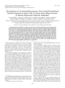
FIG. 1. Schematic representation of truncated LASV-rNP expressed as a form of GST fusion protein in E. coli transformed with the corresponding expression vector. The description “LASV-NP1-6” in the middle portion of the figure indicates full-length LASV-rNP.
pAcUW2B (28). Clones containing the insert in the correct orientation were selected, and the plasmid DNA was used for cotransfection in Sf21 cells with a polyhedrin-positive AcMNPV DNA, and the supernatant culture was screened for a polyhedrin-negative phenotype by plaque assay (19). Finally, recombinant baculovirus clones overexpressing JUNV-rNP were obtained after three successive plaque purifications. One of them, designated AcNMPV-Jun-N122, was used in the present study and is referred to hereafter as Ac-JUNV-NP. Expression and purification of His-LASV-rNP, LCMV-rNP, and JUNV-rNP. Tn5 cells infected with Ac-His-LASV-NP were incubated at 26°C for 72 h. The cells were then washed twice with cold phosphate-buffered saline (PBS) solution. A preliminary study demonstrated that most of the Tn5 cellular proteins were solubilized in PBS containing 2 M urea (PBS–2 M urea) but that the His-LASVrNP was insoluble and that the LASV-rNP could be solubilized in PBS containing 8 M urea (PBS–8 M urea). Therefore, the Tn5 cells infected with Ac-HisLASV-NP were first suspended in PBS–2 M urea. After the centrifugation of the cell suspensions at 15,000 ⫻ g for 10 min, the pellet fractions were collected and then were solubilized in PBS–8 M urea. After the centrifugation of the samples, the supernatant fractions were used as the purified antigens. LCMV-rNP and JUNV-rNP showed dissolution characteristics in urea similar to those of HisLASV-rNP; therefore, LCMV-rNP and JUNV-rNP were also fractioned in the same way as the His-LASV-rNP. The control antigen was produced from Tn5 cells infected with Ac-⌬P in the same manner as that for the positive antigens. The His-LASV-rNP was also purified by using the Ni2⫹ column purification method as reported previously (38). The source for His-LASV-rNP-purification was the supernatant fraction of the PBS–8 M urea-treated Tn5 cells infected with Ac-His-LASV-NP after sufficient dilution with PBS in order to reduce the urea concentration. SDS-PAGE. The expression and purification efficiency of His-LASV-rNP, LCMV-rNP, and JUNV-rNP were analyzed on sodium dodecyl sulfate-polyacrylamide gel electrophoresis (SDS-PAGE) gels (12% polyacrylamide) after staining with Coomassie blue. Establishment of MAbs. Monoclonal antibodies (MAbs) were generated as previously described (32, 41). BALB/c mice were immunized with purified HisLASV-rNP in the present study. Isotypes of the MAbs were determined by using a mouse MAb isotyping kit (Life Technologies). Expression of truncated NPs of LASV. In order to determine the epitope of the MAbs to the His-LASV-rNP, truncated LASV-rNPs were expressed as a form of fusion protein with glutathione S-transferase (GST) as shown in Fig. 1. The DNA corresponding to each of the truncated NP fragments was amplified with the designed primers. The amplified DNA was subcloned into the BamHI and EcoRI cloning sites of plasmid pGEX-2T (Amersham Pharmacia Biotech, Buckinghamshire, England). Each insert was sequenced and confirmed to be in the correct frame and identical to the original sequence. The GST-tagged nucleoprotein fragments were expressed in an Escherichia coli BL21 system. Western blotting. The MAbs were tested for reactivity to His-LASV-rNP and its fragments by Western blotting as reported previously (17, 32, 41). Pepscan analyses. ELISA was performed as reported previously with the purified rNP or partial nucleoprotein peptides as the antigen (33). The peptides
1184
SAIJO ET AL.
CLIN. VACCINE IMMUNOL.
FIG. 2. SDS-PAGE analyses of the purification of His-LASV-rNP using the Ni2⫹ column purification method (A) and of the semipurification strategy based on the hydrophobic property of arenavirus nucleoproteins (B). The supernatant fractions of the Ac-⌬P-, Ac-HisLASV-NP-, Ac-LCMV-NP-, or Ac-JUNV-NP-infected Tn5 cells treated with PBS–2 M urea (B, left part) are shown. The pellet fractions of these cells treated with PBS–2 M urea were further solubilized with PBS–8 M urea (B, right part).
were shifted by 1 amino acid (aa), with a consecutive overlap of 9 aa to cover the entire LASV-NP1 (aa 1 to 100) and LASV-NP5 (aa 361 to 460) fragments. Linear epitopes on the NP were determined by using Pepscan (Chiron Technologies, Clayton, Australia) according to the manufacturer’s instructions. Ninetysix peptides were prepared as 14-aa biotinylated peptides, including a 4-aa spacer sequence (SGSG) at the amino-terminal end, according to each of the amino acid sequences of the LASV-rNP1 and LASV-rNP5 of the LASV Josiah strain. The methods were previously described in detail (33). IgG-ELISA. Immunoglobulin G (IgG)-ELISA was performed as previously described except for the antigen preparation (38, 39). Briefly, ELISA plates (96-well type plate, Pro-Bind; Falcon; Becton Dickinson Labware, Franklin Lakes, NJ) were coated with the predetermined optimal quantity of purified His-LASV-rNP, LCMV-rNP, or JUNV-rNP (approximately 100 ng/well) at 4°C overnight. Then, each well of the plates was inoculated with 200 l of PBS containing 5% skim milk and 0.05% Tween 20 (M-T-PBS), followed by incubation for 1 h for blocking. The plates were washed three times with T-PBS and then inoculated with the test samples (100 l/well), which were diluted fourfold from 1:100 to 1:6,400 with M-T-PBS. After a 1-h incubation period, the plates were washed three times with T-PBS, and then the plates were inoculated with goat anti-human IgG antibody labeled with HRPO (1:1,000 dilution; Zymed Laboratory). After a further 1-h incubation period, the plates were washed and 100 l of ABTS [2,2⬘azinobis(3-ethylbenzthiazolinesulfonic acid)] solution (Roche Diagnostics, Mannheim, Germany) was added to each well. The plates were incubated for 30 min at room temperature, and optical density was at 405 nm (OD405) was measured against a reference of 490 nm. The adjusted OD405 was calculated by subtracting the OD of the negative antigen-coated wells from that of the corresponding wells. The means and standard deviations were calculated from the 96 control sera. The cutoff value for the assay was defined as the mean plus 3 standard deviations. Immunofluorescence. The pQE32-LASV-NP was digested with BamHI, and the insert was subcloned into the BamHI site of the pKS336 vector (40). The LASV-NP gene that was inserted into the pKS336 vector, pKS336-LASV-NP, was confirmed to be in the correct orientation to the promoter, tested for nucleotide sequencing as described above, and the nucleotide sequence of the gene was confirmed to be identical to the original sequence. HeLa cells were then transfected with pKS336-LASV-NP by using a FuGENE6 transfection reagent (Roche Diagnostics) according to the manufacturer’s instructions. The cells transfected with the plasmid were selected with 3 g of blasticidin Shydrochloride/ml in MEM-10FBS. The HeLa cell clones were analyzed for the expression of LASV-rNP by IIFA using the rabbit serum raised against HisLASV-rNP. The cells expressing LASV-rNP were subcloned and used as IIFA antigens. Ag-capture ELISA. Ag-capture ELISA was performed as previously described (32, 41). The purified MAb to His-LASV-rNP, MAb 4A5, produced in the present study was diluted in PBS solution, and 100 l was adsorbed overnight at 4°C onto the immunoplates (96-well type plate, Pro-Bind, Falcon; Becton Dickinson Labware). Purified MAb 4A5 was coated onto the immunoplates at a concentration of approximately 100 ng/well in 100 l of PBS. The difference in the procedures between the Ag-capture ELISA in the present study and those in
FIG. 3. Staining patterns of LASV-rNP-expressing HeLa cells by sera from an LF patient (A) and a healthy control (B) in an IIFA.
previous studies (32, 37, 41) is that the MAb, MAb 4A5, and rabbit serum raised to His-LASV-rNP were used as capture and detector antibodies, respectively. The procedure for the Ag-capture ELISA was performed as follows. The ELISA plate was coated with capture MAb, followed by blocking of the plate with M-T-PBS, addition of the samples to the ELISA plate, detection of the captured LASV-NP with rabbit serum raised to His-LASV-rNP, detection of rabbit IgG antibody that reacted with the captured antigen with goat anti-rabbit IgG antibodies conjugated with HRPO (Zymed Laboratories), and substrate reaction. In each run of the Ag-capture ELISA, the negative control antigen (M-T-PBS) was also tested. Serially diluted samples were added to the MAb-coated wells. The OD405 values of each well were adjusted by subtracting the OD405 value of the negative control antigen from the corresponding well. The adjusted OD405 was taken as a measure of the amount of antigen specifically bound. All samples were treated with 1% Nonidet-P40 (NP-40) in PBS to destroy the LASV virion and expose the nucleoprotein in the LASV virion. RT-PCR. RT-PCR was performed as previously described (10). The primers used in the RT-PCR were 36E2 (5⬘-ACCGGGGATCCTAGGCATT-3⬘) and 80F2 (5⬘-ATATAATGATGACTGTTGTTCTTTGTGCA-3⬘). The RT-PCR was carried out with a Ready-to-Go RT-PCR tube (Pharmacia). The amplified PCR products were visualized with ethidium bromide in 2% agarose gel after electrophoresis.
RESULTS Expression of His-LASV-rNP. Tn5 cells infected with each of the recombinant baculoviruses—Ac-His-LASV-NP, AcLCMV-rNP, and Ac-JUNV-rNP—were suspended in PBS–2 M urea. Most of the cell proteins were solubilized by this
VOL. 14, 2007
RECOMBINANT NP-BASED DIAGNOSTICS FOR LASSA FEVER
1185
FIG. 4. Reactivity of antibodies to arenaviruses (LASV, LCMV, and JUNV) to the rNPs of these viruses. The reactivities of rabbit sera raised to LASV-rNP (F), LCMV-rNP (f), or JUNV-rNP (Œ) with the antigens His-LASV-rNP (A), LCMV-rNP (B), and JUNV-rNP (C) in an IgG-ELISA are shown. The reactivities of the sera collected from patients with LF (D) and AHF (E) with the antigens LASV-rNP (F), LCMV-rNP (f), and JUNV-rNP (Œ) and negative control antigen (}) in an IgG-ELISA are also shown.
treatment, whereas the rNPs of these viruses remained insoluble. After centrifugation at 15,000 ⫻ g for 10 min, pellet fractions were collected. The rNPs, which were still present in the pellet fractions, were completely solubilized in PBS–8 M urea. The samples were then centrifuged at 15,000 ⫻ g for 10 min, and the supernatant fractions of the PBS–8 M urea were confirmed to contain highly purified recombinant rNPs of arenaviruses (Fig. 2). Development of indirect immunofluorescence. The LASVrNP was expressed in HeLa cells by transfection with the expression vector, pKS336-LASV-NP. The transfected cells were stained by anti-His-LASV-rNP rabbit serum and human serum samples from LF patients (Fig. 3). All 4 serum samples collected from two LF patients showed a positive staining, but 96 control serum samples did not. The LASV-rNP-based IIFA was also evaluated using serum samples collected from monkeys experimentally infected with LASV. All of the sera collected from five LASV-infected monkeys showed a positive staining, but those from four mock-infected monkeys did not. Development of His-LASV-rNP-based IgG-ELISA. Four serum samples collected from LF patients were determined to be positive by His-LASV-rNP-based IgG-ELISA, whereas 94 of the 96 control serum samples were determined to be negative. Thus, the sensitivity and specificity of the ELISA were 100 and 96%, respectively. All serum samples collected from five LASV-infected monkeys were determined to be positive,
whereas those from four mock-infected monkeys were negative. In order to examine cross-reactivity among arenaviruses in the LASV-rNP-based IgG-ELISA, antisera against LASVrNP, LCMV-rNP, or JUNV-rNP were examined (Fig. 4). The anti-LASV-rNP serum showed a strongly positive reaction, and anti-LCMV rNP and anti-JUNV-rNP sera showed strongly positive reactions in the IgG ELISA using the respective antigens (Fig. 4A, B, and C). Anti-LCMV-rNP and anti-JUNVrNP sera showed a less strongly positive reaction in the HisLASV-rNP-based IgG-ELISA than anti-LASV-rNP serum (Fig. 4A). Anti-LASV-rNP and anti-JUNV-rNP also showed a less strongly positive reaction in the His-LCMV-rNP-based IgG-ELISA than anti-LCMV-rNP serum (Fig. 4B). However, anti-LASV-rNP and anti-LCMV-rNP sera showed a negative reaction in the JUNV-rNP-based IgG-ELISA (Fig. 4C). Human sera from LF patients showed a highly positive reaction in the LASV-rNP-based IgG-ELISA, but sera from patients with Argentine hemorrhagic fever (AHF), which is caused by JUNV, did not (Fig. 4D). Serum from an AHF patient showed a highly positive reaction in the JUNV-rNP-based IgG-ELISA (Fig. 4E). These results suggest that cross-reactive antibody among arenaviruses may be detected by the newly developed LASV-rNP-based IgG-ELISA. Development of LASV Ag-capture ELISA. Three clones of a hybridoma that excreted an MAb to His-LASV-rNP were es-
1186
SAIJO ET AL.
CLIN. VACCINE IMMUNOL.
FIG. 5. Detection of the LASV genome by the RT-PCR (A), LASV-NP by the LASV-NP-Ag-capture ELISA (B), and the infectious dose of LASV (C) in serially collected sera of hamsters experimentally infected with LASV. The OD405 values in panel B were obtained at a dilution of 1:40.
tablished. The isotype of the three MAbs were identified as IgG1. These MAbs were designated MAb 4A5, MAb 6C11, and MAb 2-11. Of these MAbs, MAb 4A5 was the most efficient in capturing His-LASV-rNP in the Ag-capture ELISA format. The Ag-capture ELISA with MAb 4A5 detected HisLASV-rNP concentrations as low as 800 pg/ml (data not shown). Furthermore, the Ag-capture ELISA detected the MOPV-NP but not the rNPs of LCMV and JUNV (data not shown). All of the sera collected from five LASV-infected hamsters on days 11 and 16 postinfection were antigen positive in the Ag-capture ELISA using MAb 4A5 as a capture antibody, whereas the sera collected on days 0 and 4 were antigen negative. The OD405 values in the ELISA were highest on day 11. The reactivity patterns in each hamster in the ELISA were similar to the viremia levels (Fig. 5). The sera collected on days 11 and 16 were found to be LASV genome positive by RTPCR (10). Thus, the sensitivity of the Ag-capture ELISA was similar to that of the RT-PCR. Determination of the epitope recognized by the monoclonal antibodies. The epitope recognized by MAbs was determined. MAb-4A5 reacted in Western blots with GST-LASV-rNP1-6 (full-length LASV-rNP), GST-LASV-rNP1-5, and GSTLASV-rNP1-4 but not with the other truncated LASV-rNPs shown in Table 1, suggesting that MAb 4A5 reacted with a conformational epitope located on the amino-terminal portion of LASV-rNP. The epitope was maintained when the extreme amino-terminal portion, LASV-rNP1, was present but was lost
when LASV-rNP1 was removed. These results suggest that the extreme amino-terminal portion, LASV-rNP1, is essential for the maintenance of the conformational epitope. MAbs 6C11 and 2-11 reacted in Western blots with GST-LASV-rNP1 and GST-LASV-rNP5, respectively (Table 1). The Pepscan analyses indicated that MAbs 6C11 and 2-11
TABLE 1. Reactivities of the MAbs developed in the present study with the GST-tagged truncated LASV-rNP in Western blot analyses Reactivity with MAba: Truncated LASV-rNP
LASV-rNP1 LASV-rNP2 LASV-rNP3 LASV-rNP4 LASV-rNP5 LASV-rNP6 LASV-rNP1-2 LASV-rNP1-3 LASV-rNP1-4 LASV-rNP1-5 LASV-rNP1-6b LASV-rNP2-7 LASV-rNP3-6 LASV-rNP4-6 LASV-rNP5-6
6C11
4A5
2-11
⫹ – – – – – ND ND ND ND ND ND ND ND ND
– – – – – – – – ⫹ ⫹ ⫹ – – – –
– – – – ⫹ – ND ND ND ND ND ND ND ND ND
a “⫹” and “–” indicate positive and negative reactions, respectively. ND, not determined. b LASV-rNP1-6 indicates LASV-rNP.
VOL. 14, 2007
RECOMBINANT NP-BASED DIAGNOSTICS FOR LASSA FEVER
TABLE 2. Reactivities of the MAbs developed in the present study with the NPs of LASV, MOPV, LCMV, and JUNV in Western blot analyses Reactivity of MAba with NP of: MAb
4A5 6C11 2-11
LASV
MOPV
LCMV
JUNV
⫹ ⫹ ⫹
⫹ ND ND
– ⫹ –
– – –
a “⫹” and “–” indicate positive and negative reactions, respectively. ND, not determined. The reactivities of MAb 6C11 and MAb 2-11 were not evaluated with MOPV-NP. However, theoretically, MAb 6C11 should be reactive with MOPV-NP due to the presence of the amino acid residues that can react with MAb 6C11, but MAb 2-11 should not react with MOPV-NP due to the absence of the amino acid residues that can react with MAb 2-11.
recognized linear epitopes. MAbs 6C-11 and 2-11 recognized GLDFSEV (aa 41 to 47) within LASV-rNP1 and FATQP (aa 439 to 443) within LASV-rNP5, respectively (Fig. 6). The reactivity patterns of these MAbs with NPs of LASV, MOPV, LCMV, and JUNV are summarized in Table 2. DISCUSSION We report here the development of diagnostic systems (antibody and antigen detection systems) for LF using LASV-rNP.
1187
The LASV-rNP-based IgG-ELISA was sensitive and specific in detecting anti-LASV-IgG. Although the data were not shown, an IgM-capture ELISA using purified LASV-rNP as an antigen was developed in the same way as that shown in previous reports and detected LASV-IgM antibody (42, 43). All sera collected from LF patients and monkeys infected with LASV showed positive reactions in the LASV-rNP-based IIFA. The staining patterns of the rNP with these sera were granular in the IIFA (Fig. 3), making it easy to distinguish positives from negatives. IIFA using LASV-rNP-expressing HeLa cells was also highly sensitive and specific in detecting LASV-IgG. In the preliminary study, ca. 15% of the sera collected from 334 Ghanaians and only less than 1% of 280 Zambians showed positive reactions in the LASV-rNP-based IgG ELISA (our data). The results are considered to be compatible with the fact that LF is endemic to the western African region, including Ghana, but not to the eastern African region. The LASV-rNPbased antibody detection systems such as ELISA and IIFA were suggested to be useful not only in the diagnosis of but also in the seroepidemiological study of LF. The LASV-rNPs were expressed by a transformation system in E. coli or by recombinant baculovirus systems and have already been applied as antigens in ELISA, Western blotting, and IIFA for the detection of antibodies to LASV (4, 14, 16, 22, 23, 44). In the present study, an Ag-capture ELISA using
FIG. 6. Pepscan analyses to determine the epitopes of MAb 6C11 (A) and MAb 2-11 (B). The vertical bar indicates the amino acid residues with an amino acid position within the LASV-NP. MAb 6C11 was confirmed to react with 7 aa residues positioned from aa 42 to 48 (GLDFSEV) within LASV-NP1. MoAb-2-11 was confirmed to react with 5 aa residues positioned from aa 439 to 443 (FATQP) within LASV-NP5. (C) The corresponding amino acid residues to the epitope of the MAb 6C11 and MAb 2-11 among MOPV, LCMV, and JUNV are shown. The GenBank accession numbers for the S genes of LASV, MOPV, LCMV, and JUNV are NC_004296, AY772170, AY847350, and DQ272266, respectively. The epitope of the MAb 6C11 is present not only in the nucleoprotein of LASV but also in those of MOPV and LCMV—but not in that of JUNV.
1188
SAIJO ET AL.
CLIN. VACCINE IMMUNOL.
MAbs to LASV-rNP was also developed. Furthermore, detection of the cross-reactive antibody by LASV-rNP-based IgGELISA was examined. The results for cross-reactivity indicate that the LASV-rNP-based IgG-ELISA detects not only antibodies to LASV but also those to LCMV. The Ag-capture ELISA using MAb 4A5 was confirmed to be useful in the detection of authentic LASV antigen in sera serially collected from hamsters infected with LASV. The sensitivity of the MAb 4A5-based Ag-ELISA was similar to that of conventional RT-PCR, the efficiency of which in the diagnosis of LF was previously reported (10). Therefore, the MAb 4A5based Ag-capture ELISA is regarded as useful in the diagnosis of LF. Unfortunately, the efficacy of the MAb 4A5-based Agcapture ELISA in the diagnosis of LF was not evaluated using serum samples from patients. Thus, further study is still required. The three MAbs, including MAb 4A5, were characterized, and the corresponding amino acid residues within the nucleoproteins of LASV, MOPV, LCMV, and JUNV to the epitopes of MAb 6C11 and MAb 2-11 are summarized in Fig. 6C. It was of interest that LASV, MOPV, LCMV, and JUNV might be identified by analyses of the reactivity patterns of MAbs 4A5, 6C11, and 2-11 to the nucleoproteins of each virus. The nucleoproteins of all LASV strains circulating in the western and central parts of Africa would be detected by the MAb 4A5-based Ag-capture ELISA, since this ELISA was able to detect MOPV-NP that was different from LASV in terms of genetic and evolutional characteristics. We have thus far reported the development of antibody and antigen detection systems using the recombinant nucleoproteins of the viruses for Ebola hemorrhagic fever, Marburg hemorrhagic fever, and Crimean-Congo hemorrhagic fever (32, 33, 36–42). Recently, a number of highly pathogenic emerging virus infections in humans appeared, such as Nipah virus encephalitis (8), SARS-coronavirus infections (21, 35), and highly pathogenic avian influenza virus infections (9, 45, 46). The strategy shown here might be applicable to the development of diagnostic systems for severe viral infections whose etiologic agents are highly pathogenic to humans as an alternative method to methods using infectious viruses. ACKNOWLEDGMENTS We thank M. Ogata, Department of Virology 1, National Institute of Infectious Diseases, Tokyo, Japan, for great clerical assistance. This study was partly supported by a grant-in-aid for scientific research from the Ministry of Health, Labor, and Welfare of Japan; by the Japan Health Sciences Foundation (grant 16171301); and by the Japan Society for Promotion of Science (grant 18591210). REFERENCES 1. Anonymous. 2000. Lassa fever imported to England. Commun. Dis. Rep. CDR Wkly. 10:99. 2. Anonymous. 2000. Lassa fever, imported case, The Netherlands. Wkly. Epidemiol. Rec. 75:265. 3. Baize, S., D. Pannetier, C. Faure, P. Marianneau, I. Marendat, M. C. Georges-Courbot, and V. Deubel. 2006. Role of interferons in the control of Lassa virus replication in human dendritic cells and macrophages. Microbes Infect. 8:1194–1202. 4. Barber, G. N., J. C. Clegg, and G. Lloyd. 1990. Expression of the Lassa virus nucleocapsid protein in insect cells infected with a recombinant baculovirus: application to diagnostic assays for Lassa virus infection. J. Gen. Virol. 71(Pt. 1):19–28. 5. Borio, L., T. Inglesby, C. J. Peters, A. L. Schmaljohn, J. M. Hughes, P. B. Jahrling, T. Ksiazek, K. M. Johnson, A. Meyerhoff, T. O’Toole, M. S. Ascher, J. Bartlett, J. G. Breman, E. M. Eitzen, Jr., M. Hamburg, J. Hauer, D. A. Henderson, R. T. Johnson, G. Kwik, M. Layton, S. Lillibridge, G. J. Nabel,
6.
7.
8.
9.
10.
11.
12.
13.
14.
15.
16.
17.
18. 19. 20.
21.
22.
23.
24. 25.
26.
27.
28.
M. T. Osterholm, T. M. Perl, P. Russell, and K. Tonat. 2002. Hemorrhagic fever viruses as biological weapons: medical and public health management. JAMA 287:2391–2405. Bossi, P., A. Tegnell, A. Baka, F. Van Loock, J. Hendriks, A. Werner, H. Maidhof, and G. Gouvras. 2004. Bichat guidelines for the clinical management of haemorrhagic fever viruses and bioterrorism-related haemorrhagic fever viruses. Eur. Surveill. 9:E11–E12. Carey, D. E., G. E. Kemp, H. A. White, L. Pinneo, R. F. Addy, A. L. Fom, G. Stroh, J. Casals, and B. E. Henderson. 1972. Lassa fever. Epidemiological aspects of the 1970 epidemic, Jos, Nigeria. Trans. R. Soc. Trop. Med. Hyg. 66:402–408. Chua, K. B., K. J. Goh, K. T. Wong, A. Kamarulzaman, P. S. Tan, T. G. Ksiazek, S. R. Zaki, G. Paul, S. K. Lam, and C. T. Tan. 1999. Fatal encephalitis due to Nipah virus among pig-farmers in Malaysia. Lancet 354:1257– 1259. Claas, E. C., A. D. Osterhaus, R. van Beek, J. C. De Jong, G. F. Rimmelzwaan, D. A. Senne, S. Krauss, K. F. Shortridge, and R. G. Webster. 1998. Human influenza A H5N1 virus related to a highly pathogenic avian influenza virus. Lancet 351:472–477. Demby, A. H., J. Chamberlain, D. W. Brown, and C. S. Clegg. 1994. Early diagnosis of Lassa fever by reverse transcription-PCR. J. Clin. Microbiol. 32:2898–2903. Ghiringhelli, P. D., R. V. Rivera Pomar, N. I. Baro, M. F. Rosas, O. Grau, and V. Romanowski. 1989. Nucleocapsid protein gene of Junin arenavirus (cDNA sequence). Nucleic Acids Res. 17:8001. Ghiringhelli, P. D., R. V. Rivera-Pomar, M. E. Lozano, O. Grau, and V. Romanowski. 1991. Molecular organization of Junin virus S RNA: complete nucleotide sequence, relationship with other members of the Arenaviridae and unusual secondary structures. J. Gen. Virol. 72(Pt. 9):2129–2141. Gunther, S., P. Emmerich, T. Laue, O. Kuhle, M. Asper, A. Jung, T. Grewing, J. ter Meulen, and H. Schmitz. 2000. Imported Lassa fever in Germany: molecular characterization of a new Lassa virus strain. Emerg. Infect. Dis. 6:466–476. Gunther, S., O. Kuhle, D. Rehder, G. N. Odaibo, D. O. Olaleye, P. Emmerich, J. ter Meulen, and H. Schmitz. 2001. Antibodies to Lassa virus Z protein and nucleoprotein co-occur in human sera from Lassa fever endemic regions. Med. Microbiol. Immunol. 189:225–229. Hirabayashi, Y., S. Oka, H. Goto, K. Shimada, T. Kurata, S. P. Fisher-Hoch, and J. B. McCormick. 1988. An imported case of Lassa fever with late appearance of polyserositis. J. Infect. Dis. 158:872–875. Hummel, K. B., M. L. Martin, and D. D. Auperin. 1992. Baculovirus expression of the glycoprotein gene of Lassa virus and characterization of the recombinant protein. Virus Res. 25:79–90. Ikegami, T., M. Niikura, M. Saijo, M. E. Miranda, A. B. Calaor, M. Hernandez, L. P. Acosta, D. L. Manalo, I. Kurane, Y. Yoshikawa, and S. Morikawa. 2003. Antigen capture enzyme-linked immunosorbent assay for specific detection of Reston Ebola virus nucleoprotein. Clin. Diagn. Lab. Immunol. 10:552–557. Jeffs, B. 2006. A clinical guide to viral haemorrhagic fevers: Ebola, Marburg, and Lassa. Trop. Doct. 36:1–4. King, L., and R. Possee. 1992. The baculovirus expression system: a laboratory guide. Chapman and Hall, New York, NY. Kitts, P. A., M. D. Ayres, and R. D. Possee. 1990. Linearization of baculovirus DNA enhances the recovery of recombinant virus expression vectors. Nucleic Acids Res. 18:5667–5672. Ksiazek, T. G., D. Erdman, C. S. Goldsmith, S. R. Zaki, T. Peret, S. Emery, S. Tong, C. Urbani, J. A. Comer, W. Lim, P. E. Rollin, S. F. Dowell, A. E. Ling, C. D. Humphrey, W. J. Shieh, J. Guarner, C. D. Paddock, P. Rota, B. Fields, J. DeRisi, J. Y. Yang, N. Cox, J. M. Hughes, J. W. LeDuc, W. J. Bellini, and L. J. Anderson. 2003. A novel coronavirus associated with severe acute respiratory syndrome. N. Engl. J. Med. 348:1953–1966. Lloyd, G., G. N. Barber, J. C. Clegg, and P. Kelly. 1989. Identification of Lassa fever virus infection with recombinant nucleocapsid protein antigen. Lancet ii:1222. Lukashevich, L. S., J. C. Clegg, and K. Sidibe. 1993. Lassa virus activity in Guinea: distribution of human antiviral antibody defined using enzymelinked immunosorbent assay with recombinant antigen. J. Med. Virol. 40: 210–217. Macher, A. M., and M. S. Wolfe. 2006. Historical Lassa fever reports and 30-year clinical update. Emerg. Infect. Dis. 12:835–837. Mahdy, M. S., W. Chiang, B. McLaughlin, K. Derksen, B. H. Truxton, and K. Neg. 1989. Lassa fever: the first confirmed case imported into Canada. Can. Dis. Wkly. Rep. 15:193–198. Matsuura, Y., R. D. Possee, and D. H. Bishop. 1986. Expression of the S-coded genes of lymphocytic choriomeningitis arenavirus using a baculovirus vector. J. Gen. Virol. 67(Pt. 8):1515–1529. McCormick, J. B., P. A. Webb, J. W. Krebs, K. M. Johnson, and E. S. Smith. 1987. A prospective study of the epidemiology and ecology of Lassa fever. J. Infect. Dis. 155:437–444. Merryweather, A. T., U. Weyer, M. P. Harris, M. Hirst, T. Booth, and R. D. Possee. 1990. Construction of genetically engineered baculovirus insecticides
VOL. 14, 2007
29. 30. 31.
32.
33. 34. 35.
36.
37.
38.
RECOMBINANT NP-BASED DIAGNOSTICS FOR LASSA FEVER
containing the Bacillus thuringiensis subsp. kurstaki HD-73 delta endotoxin. J. Gen. Virol. 71(Pt. 7):1535–1544. Monath, T. P. 1975. Lassa fever: review of epidemiology and epizootiology. Bull. W. H. O. 52:577–592. Monath, T. P., P. E. Mertens, R. Patton, C. R. Moser, J. J. Baum, L. Pinneo, G. W. Gary, and R. E. Kissling. 1973. A hospital epidemic of Lassa fever in Zorzor, Liberia, March-April 1972. Am. J. Trop. Med. Hyg. 22:773–779. Monson, M. H., J. D. Frame, P. B. Jahrling, and K. Alexander. 1984. Endemic Lassa fever in Liberia. I. Clinical and epidemiological aspects at Curran Lutheran Hospital, Zorzor, Liberia. Trans. R. Soc. Trop. Med. Hyg. 78:549–553. Niikura, M., T. Ikegami, M. Saijo, I. Kurane, M. E. Miranda, and S. Morikawa. 2001. Detection of Ebola viral antigen by enzyme-linked immunosorbent assay using a novel monoclonal antibody to nucleoprotein. J. Clin. Microbiol. 39:3267–3271. Niikura, M., T. Ikegami, M. Saijo, T. Kurata, I. Kurane, and S. Morikawa. 2003. Analysis of linear B-cell epitopes of the nucleoprotein of Ebola virus that distinguish Ebola virus subtypes. Clin. Diagn. Lab. Immunol. 10:83–87. Peters, C. J. 2002. Clinical virology, p. 949–969. In D. D. Richman, R. J. Whitley, and F. G. Hayden (ed.), Arenaviruses, 2nd ed. ASM Press, Washington, DC. Rota, P. A., M. S. Oberste, S. S. Monroe, W. A. Nix, R. Campagnoli, J. P. Icenogle, S. Penaranda, B. Bankamp, K. Maher, M. H. Chen, S. Tong, A. Tamin, L. Lowe, M. Frace, J. L. DeRisi, Q. Chen, D. Wang, D. D. Erdman, T. C. Peret, C. Burns, T. G. Ksiazek, P. E. Rollin, A. Sanchez, S. Liffick, B. Holloway, J. Limor, K. McCaustland, M. Olsen-Rasmussen, R. Fouchier, S. Gunther, A. D. Osterhaus, C. Drosten, M. A. Pallansch, L. J. Anderson, and W. J. Bellini. 2003. Characterization of a novel coronavirus associated with severe acute respiratory syndrome. Science 300:1394–1399. Saijo, M., M. Niikura, T. Ikegami, I. Kurane, T. Kurata, and S. Morikawa. 2006. Laboratory diagnostic systems for Ebola and Marburg hemorrhagic fevers developed with recombinant proteins. Clin. Vaccine Immunol. 13: 444–451. Saijo, M., M. Niikura, A. Maeda, T. Sata, T. Kurata, I. Kurane, and S. Morikawa. 2005. Characterization of monoclonal antibodies to Marburg virus nucleoprotein (NP) that can be used for NP-capture enzyme-linked immunosorbent assay. J. Med. Virol. 76:111–118. Saijo, M., M. Niikura, S. Morikawa, T. G. Ksiazek, R. F. Meyer, C. J. Peters, and I. Kurane. 2001. Enzyme-linked immunosorbent assays for detection of
39.
40.
41.
42.
43.
44.
45.
46.
1189
antibodies to Ebola and Marburg viruses using recombinant nucleoproteins. J. Clin. Microbiol. 39:1–7. Saijo, M., T. Qing, M. Niikura, A. Maeda, T. Ikegami, C. Prehaud, I. Kurane, and S. Morikawa. 2002. Recombinant nucleoprotein-based enzymelinked immunosorbent assay for detection of immunoglobulin G antibodies to Crimean-Congo hemorrhagic fever virus. J. Clin. Microbiol. 40:1587– 1591. Saijo, M., T. Qing, M. Niikura, A. Maeda, T. Ikegami, K. Sakai, C. Prehaud, I. Kurane, and S. Morikawa. 2002. Immunofluorescence technique using HeLa cells expressing recombinant nucleoprotein for detection of immunoglobulin G antibodies to Crimean-Congo hemorrhagic fever virus. J. Clin. Microbiol. 40:372–375. Saijo, M., Q. Tang, B. Shimayi, L. Han, Y. Zhang, M. Asiguma, D. Tianshu, A. Maeda, I. Kurane, and S. Morikawa. 2005. Antigen-capture enzymelinked immunosorbent assay for the diagnosis of Crimean-Congo hemorrhagic fever using a novel monoclonal antibody. J. Med. Virol. 77:83–88. Saijo, M., Q. Tang, B. Shimayi, L. Han, Y. Zhang, M. Asiguma, D. Tianshu, A. Maeda, I. Kurane, and S. Morikawa. 2005. Recombinant nucleoproteinbased serological diagnosis of Crimean-Congo hemorrhagic fever virus infections. J. Med. Virol. 75:295–299. Tang, Q., M. Saijo, Y. Zhang, M. Asiguma, D. Tianshu, L. Han, B. Shimayi, A. Maeda, I. Kurane, and S. Morikawa. 2003. A patient with Crimean-Congo hemorrhagic fever serologically diagnosed by recombinant nucleoproteinbased antibody detection systems. Clin. Diagn. Lab. Immunol. 10:489–491. Ter Meulen, J., K. Koulemou, T. Wittekindt, K. Windisch, S. Strigl, S. Conde, and H. Schmitz. 1998. Detection of Lassa virus antinucleoprotein immunoglobulin G (IgG) and IgM antibodies by a simple recombinant immunoblot assay for field use. J. Clin. Microbiol. 36:3143–3148. Tran, T. H., T. L. Nguyen, T. D. Nguyen, T. S. Luong, P. M. Pham, V. C. Nguyen, T. S. Pham, C. D. Vo, T. Q. Le, T. T. Ngo, B. K. Dao, P. P. Le, T. T. Nguyen, T. L. Hoang, V. T. Cao, T. G. Le, D. T. Nguyen, H. N. Le, K. T. Nguyen, H. S. Le, V. T. Le, D. Christiane, T. T. Tran, J. Menno de, C. Schultsz, P. Cheng, W. Lim, P. Horby, and J. Farrar. 2004. Avian influenza A (H5N1) in 10 patients in Vietnam. N. Engl. J. Med. 350:1179–1188. Yuen, K. Y., P. K. Chan, M. Peiris, D. N. Tsang, T. L. Que, K. F. Shortridge, P. T. Cheung, W. K. To, E. T. Ho, R. Sung, and A. F. Cheng. 1998. Clinical features and rapid viral diagnosis of human disease associated with avian influenza A H5N1 virus. Lancet 351:467–471.