CANCER GENOMICS & PROTEOMICS 4: 157-164 (2007)
Review
Development of Reverse Phase Protein Microarrays for Clinical Applications and Patient-tailored Therapy RUNA SPEER1, JULIA WULFKUHLE2, VIRGINIA ESPINA2, ROBYN AURAJO2, KIRSTEN H. EDMISTON3, LANCE A. LIOTTA2 and EMANUEL F. PETRICOIN III2* 1University
of Tübingen, Faculty of Medicine, Department of Obstetrics and Gynecology, Calwer Str. 7, 72076 Tübingen, Germany; 2Center for Applied Proteomics and Molecular Medicine, George Mason University, Manassas, VA; 3Department of Surgery, Inova Fairfax Hospital Cancer Center, 3300, Gallows Road, Falls Church, VA, U.S.A.
Abstract. While genomics provide important information about the somatic genetic changes, and RNA transcript profiling can reveal important expression changes that correlate with outcome and response to therapy, it is the proteins that do the work in the cell. At a functional level, derangements within the proteome, driven by post-translational and epigenetic modifications, such as phosphorylation, is the cause of a vast majority of human diseases. Cancer, for instance, is a manifestation of deranged cellular protein molecular networks and cell signaling pathways that are based on genetic changes at the DNA level. Importantly, the protein pathways contain the drug targets in signaling networks that govern overall cellular survival, proliferation, invasion and cell death. Consequently, the promise of proteomics resides in the ability to extend analysis beyond correlation to causality. A critical gap in the information knowledge base of molecular profiling is an understanding of the ongoing activity of protein signaling in human tissue: what is activated and "in use" within the human body at any given point in time. To address this gap, we have invented a new technology, called reverse phase protein microarrays, that can generate a functional read-out of cell signaling networks or pathways for an individual patient obtained directly from a biopsy specimen. This "wiring diagram" can serve as the basis for both, selection of a therapy and patient stratification.
Correspondence to: Emanuel F. Petricoin, George Mason University, Center for Applied Proteomics and Molecular Medicine, 10900 University Blvd. MS 4E3, Discovery Hall Room 181A, Manassas, VA 20110, U.S.A. Tel: +1 703 993 8646, Fax: +1 703 993 4288, e-mail:
[email protected] Key Words: Cancer, combinatorial therapy, laser capture microdissection, microarray, molecular profiling, protein, proteomics, tissue heterogeneity, review.
1109-6535/2007 $2.00+.40
While medicine practice has always aimed at individualizing treatment of disease, biomedical research has only recently begun to realize this goal and empower the physician with tools and information to truly implement personalized medicine. The deciphering of the human genome is accompanied by growing knowledge in bioinformatics, the tool that helps us to understand the huge amount of generated data and therewith the functionality of cells, tissues, and organs, in both diseased and healthy states. Together with high throughput techniques these elements pave the way to the revolution in biology and medicine that we are experiencing today. In a highly dynamic research landscape we are enabled to analyze complex biological systems and to continuously refine ‘omics’-techniques and methods that promise to elicit major advances in personalized medicine in the near future. What is proteomics’ contribution to this new scientific era in terms of cancer research and treatment? Cancer affects a significant share of the population and the challenge to treat cancer patients lies in the complexity of the disease. Most cancers are driven by and dependent upon multiple genetic and genomic changes, as well as by a variety of aberrant signalling pathways, because genetic alterations such as amplification of oncogenes or loss of tumor suppressor genes cause malignant transformation, as well as changes and interferences in the functional executive level of a cell’s proteomic equilibrium. The heterogeneous nature of cancer helps to explain unpredictable responses to existing drug therapies observed to date. These characteristics make a malignant disease a prime target for new proteomic molecular technologies, expediting the applications of basic research findings into daily clinical practice through translational research. The unique signature of each individual patient’s malignant disease lies in the molecular level of the cancer
157
CANCER GENOMICS & PROTEOMICS 4: 157-164 (2007) cell’s genome, which is considered a rather static entity, and its proteome, which is more complex and dynamic than the genome, which includes splice variants, post-translational modifications and cleavage products and which is characterized by a wide dynamic range of protein expression expanding over several orders of magnitude. The dynamic nature of the proteome allows us to monitor closely changes in the state of a cell, tissue or organism over time and therewith to follow the course of a disease and track its pathogenetic mechanisms, as well as its response to therapy. The platform for this complex approach is offered by proteomics, because it provides powerful (high throughput) tools for profiling and comparing protein compositions within defined cell populations as well as for the identification of novel diagnostic and prognostic biomarkers for cancer. Proteomic offers powerful technologies for protein separation and identification, for characterising their biomolecular interactions, function and regulation, as well as for storing and distributing protein information (1-3). In the context of translational research, proteomics emphasizes on the analysis of clinically relevant biological samples like biopsy specimens and body fluids in order to find key defects in a diseased tissue and approach the ultimate goal, patient tailored therapies (4, 5). Until now, a missing key analytical component for the effective molecular analysis of human tissue, and ultimately the personalization of therapy, was the ability to generate a portrait of the activity of the cellular "circuitry" – the actual drug targets that are the signalling pathways and molecular networks within a cell from tissue from clinical biopsy material. The development of certain classes of protein microarrays is looking to overcome this hurdle.
identification and molecular profiling of cellular and biological material. Reverse phase protein microarrays (RPA) are a subcategory of protein arrays, and unlike forward phase arrays (e.g. antibody array), are comprised of an immobilized cellular lysate that is probed with a primary antibody (Figure 1). Signal amplification is independent of the immobilized protein, permitting the coupling of detection strategies with highly sensitive tyramide amplification chemistries (18-21). RPA technology is well-suited and in fact, specifically developed for clinical proteomics and clinical applications. Currently, one of the most important attributes of the platform lies in the ability to provide a "map" of the state of multiple in vivo kinase driven signal pathways, and to provide critical information about protein post-translational modifications, such as the phosphorylation states of these proteins (4, 6, 22, 23). The post-translational modifications reflect the activity state of signaling pathways and networks, and is information that cannot be generated from gene transcript profiling or DNA analysis. Identification of disease-related alterations with the kinase-driven cellular networks is a key starting point for both drug development as well as design of individual therapy regimens. Importantly, proteomic analysis of cell signaling and signaling networks, unlike genomic endpoints, represent the actual drug targets. RPA may be used to monitor changes in protein phosphorylation over time, before and after treatment, between disease and non-disease states and responders versus non-responders, allowing one to infer the activity levels of the proteins in a particular pathway in real time to tailor treatment to each patient’s cellular "circuitry".
Protein Microarrays
The Growing Role of Protein Arrays in Molecular Diagnostics
In their basic form, protein microarrays are simply proteins immobilized within distinct regions on a substratum as a solid support (6-10). The immobilized material may be heterogeneous or homogeneous in nature, and may consist of cell or phage lysates, body fluids, an antibody, body fluid, or recombinant/expressed proteins (6-17). The immobilized molecules are then queried by probing with an analytespecific molecule (e.g. antibody) that is coupled to a second signal-generating molecule such as a tagged antibody, ligand, serum or cell lysate. The signal can be from a chemiluminescent, colorimetric, fluorescent, radiometric or electrochemical read-out. The resulting signal intensity of each spot is, in theory, proportional to the quantity of applied tagged molecules bound to the bait molecule. The spot pattern image is then captured, analyzed and correlated with biological information. Protein microarrays have broad applications for both discovery and quantitative analysis. Currently, these formats have applications in drug discovery, drug screening, biomarker
New classes of molecular diagnostics and molecular classification techniques are still largely building upon traditional pathological techniques for tissue analysis over the past decade. In oncology, overall histopathological measurements, such as tumor size, degree of differentiation, degree of metastases and cytogenetic analysis, are being supplemented with new clinically important analytes such as HER-2/neu. However, while these newer analytes are playing an expanding role in therapeutic decision making, they do not begin to address or solve the complexity and heterogeneity in individual tumors that can lead to success or failure of a targeted therapeutic agent. Molecular classification using gene expression microarray profiling has demonstrated considerable potential for molecular classification (24). However transcript profiling provides an incomplete picture of the ongoing molecular network for a number of clinically important reasons. As previously stated, while it is certain that genetic mutations cause human diseases such as cancer, the analysis of the genome or transcriptome (i.e. gene transcript profiling) has
158
Speer et al: Reverse Phase Protein Microarrays (Review)
Figure 1. Example format for forward (top) and reverse (bottom) phase protein microarrays. In contrast to forward phase arrays, the reverse phase array is constructed by directly printing the material that is being analyzed, usually in the form of a whole cell lysate or body fluid. The reverse phase array requires only one primary antibody, and can employ third generation immunoassay amplification chemistries such as tyramide precipitation. These features can greatly expand the sensitivity of the assay, as well as the number of analytes that can be measured in a given sample.
never been shown to be able to predict or measure the protein signaling networks that ultimately produce the cancer. The idea of a gene network itself is in many ways a misnomer, as genes do not really form interacting networks- it is the protein gene products that form the linked and interacting enzymatic networks. Indeed, gene transcript levels have not been found to significantly correlate with total protein expression for many analytes and, much less with the phosphorylated or otherwise functional forms of the encoded proteins (25). RNA transcripts also provide little information about protein–protein interactions and the state of the cellular signaling pathways (26, 27). Finally, most new molecularly targeted therapeutics are directed at protein targets (e.g. HERCEPTIN and GLEVEC target c-erbB2 and c-kit/abl/PDGF proteins, respectively), and these targets in the cellular proteome are constantly fluctuating depending on the cellular microenvironment, tissue microecology and patient-specific alterations.
Reverse Phase Arrays: Enabling Technology for Patient-tailored Therapeutics The effective combination of diagnostic platforms with targeted therapeutics is optimal when the diagnostic platform provides information about the drug target itself and thus identifies patients who will likely respond to a given therapy.
Likely, this analysis would come from the profiling of target organ/cells taken from the patient through biopsy procurement. Most proteomic technologies have significant technological limitations due to analytical sensitivity, and these limitations are dramatically revealed when the analysis is of the very small tissue samples. Most tissue biopsy specimens contain only a few thousand cells. Widely used proteomic platforms such as two-dimensional polyacrylamide gel electrophoresis, isotope-coded affinity tagging (ICAT) and multidimensional LC-MS platforms, and antibody arrays, require relatively large numbers of cells for any significant results – many orders of magnitude greater than the quantity procured during a clinical biopsy (28-32). Currently, there is no direct PCR-like technology for amplifying proteins. New technologies are needed that can utilize microscopic amounts of cellular material. A new type of protein array, the RPA (46, 22-24, 33-39) has demonstrated the critical ability to measure, in multiplex, the level of phosphorylation of signaling pathways using small numbers of microdissected human tissue cells from biopsy specimens. Because human tissues are composed of hundreds of interacting cell populations, they provide both a unique opportunity, as well as a significant barrier in the quest for discovery disease-related information that reflect the cellular microenvironment. However, established technologies such as
159
CANCER GENOMICS & PROTEOMICS 4: 157-164 (2007)
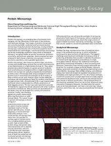
Figure 2. Workflow for reverse phase array construction. Patient tissue was obtained from tissue biopsies using laser capture microdissection, and cellular lysate generated. Experimental test samples were arrayed in a series of 1:2 dilutions (from left to right). Each sample is printed in duplicate. On the bottom of every slide, a positive and negative control is printed, as well as a calibration standard. These standards serve to normalize the results across slides and different experiments.
laser capture microdissection (LCM) now provide for routine procurement of desired cell populations directly from tissue sections, while maintaining the fidelity of the cellular DNA, RNA and protein molecular content (40). Combining LCM with RPA technology provides a facile means of isolating pure cell populations from human biopsy specimens, generating a protein lysate, and spotting this lysate onto nitrocellulosecoated slides using a robotic arrayer. The resulting RPA can thus be composed of hundreds of patient samples or cellular lysates from cell lines. In the RPA, each array is incubated with a single primary antibody (e.g. phospho-ERK kinase) and a single analyte endpoint is measured and directly compared across multiple samples (Figure 2). During printing, each patient sample is arrayed as a series of dilutions, usually at a 1:2, providing an internal standard curve. This printing strategy provides for direct quantitative measurement once the linear range of detection is established. Positive and negative controls, along with calibrators are also printed concurrently, providing facile quantitative analysis and inter/intra assay comparisons (Figure 2). In addition, the RPA is a flexible insofar as non-denatured lysates can also be directly printed, so that protein–protein, protein–DNA and/or protein–RNA complexes can be detected and characterized (33-40).
160
Several critical technological components of the RPA offer unique advantages over other array based platforms such as tissue arrays (41) or antibody (forward phase) arrays (7, 10, 42, 43). The RPA can employ denatured lysates, so that antigen retrieval, a significant limitation for tissue arrays, antibody arrays, and immunohistochemistry technologies, is not problematic. RPAs only require a single class of antibody per analyte protein and do not require direct tagging of the protein as readout for the assay. Other technologies, such as suspension bead array platforms, have significant limitations in the portfolio of analytes that can be measured, even in multiplex, because of the requirement of a two-site assay. The ability to generate quantitative data from minute quantities of cellular input without a two-site assay also enables a marked improvement in reproducibility, sensitivity and robustness of the assay over other techniques (6).
Use of Reverse Phase Arrays for Signal Pathway Profiling of Human Cancer Recent case studies demonstrate the ability of RPA for the analysis of surgically obtained tissues and demonstrate the potential for aiding in therapeutic decision-making by providing information about the activity of signaling proteins.
Speer et al: Reverse Phase Protein Microarrays (Review)
Figure 3. Proteomics-guided treatment of metastatic disease using targeted inhibitors. Metastasis-specific cell signaling defects are used to select the appropriate therapy.
Figure 4. Individualized cancer therapy and how it could work. Following tissue procurement, cell signaling analysis was performed using reverse phase protein microarrays. The specific "circuit map" was used to select and guide therapy. If the patient relapsed, a new fingerprint was obtained by rebiopsy and the cellular "circuitry" was measured to select the next round of therapy.
161
CANCER GENOMICS & PROTEOMICS 4: 157-164 (2007) The first published demonstration of RPA signal pathway profiling revealed that members of the PI3 kinase/pro-survival protein pathways are activated at the invasion front during prostate cancer progression (33). In another study, Zha et al. examined the differences in pro-survival signaling between Bcl-2+/– lymphomas (38). Comparison of various pro-survival proteins in Bcl-2+ and Bcl-2– follicular lymphoma subtypes by reverse phase protein microarrays suggested that there are pro-survival signals independent of Bcl-2 (38). Reverse phase protein microarrays have also been used to compare cell signaling portraits in patient-matched primary and metastatic cancer lesions (4, 37). Because the tissue microecology of the metastatic lesion is inherently different from the environment within the primary tumor, cell signaling events may be significantly altered depending on the site of metastasis. Since the signaling changes in the metastasis would be the most appropriate for the selection of targeted therapy due to the fact that metastasis most often determines mortality, it might be critical to develop a profile of metastatic cells themselves. In a view of the future, a patient that presents with advanced stage disease and multiple metastatic sites could be treated with a selected combination of different targeted therapies, tailored to the different signaling changes (Figure 3). Preliminary published data supports this concept. A small case study set of three laser capture microdissected, patient-matched primary colorectal tumor cells and the corresponding cells from the hepatic metastasis (obtained simultaneously at surgery) were analyzed for the status of multiple phosphoprotein endpoints involved in mitogenesis and survival including growth factor receptors, signal transducing proteins, and nuclear transcription factors (4). Unsupervised hierarchical clustering of the data suggested that cell signaling in metastatic hepatic lesions differed significantly from the matched primary lesions, yet, appeared very similar to each other (4). Significant changes in cell signaling pathways depending on the underpinning microenvironment were also observed in a similarly constructed study of six primary ovarian tumors and patient-matched metastases taken simultaneously at surgery (37). In this study, signaling within the metastatic lesion was dramatically changed compared with their matched primary counterparts, with phosphorylation of c-Kit dramatically elevated in five of the six metastatic tumors compared to the primary lesions. The clinical implications that the metastatic cell signaling is so dissimilar to the primary tumor are important, if validated in further studies. Patient-tailored therapy that is designed to mitigate the metastatic process could have significant implications in the clinic. Reverse phase protein microarrays are also well-suited to the analysis of clinical trial material in that they can provide signaling network information that complements standard histological analysis of patient specimens collected before, during and after treatment.
162
Use of Reverse Phase Arrays: A View to the Future Molecular profiling of the human "kinome", and ongoing signaling cascades produced within and as a consequence of the tumor microenvironment, host and peripheral circulation holds great promise in effective selection of therapeutic targets as well as patient stratification (Figure 4). As our understanding of human diseases such as cancer expands, we are presented with information that points to the individuality of cancer at the molecular level. This is manifest, of course, in the frustratingly unpredictable nature of response to cancer to currently available therapies. Protein-based analysis where phosphorylation-driven information can be gleaned is particularly useful in this area since these endpoints are the direct drug targets themselves. The generation of a portrait of the state of these networks will provide the data necessary for a rationally based formulation of targeted therapies, perhaps in combination with each other. Monitoring different phosphoprotein levels will also help to identify treatmentacquired resistance to chemotherapy. Probably the greatest promise of this technology is the opportunity to identify early disease pathway changes and provide information that could lead to prevention. For example, molecular network analysis of the ductal carcinoma in situ from women who progress to develop invasive breast cancer may have a distinct signaling portrait that discriminates them from patients who have indolent disease. The promise of proteomic based profiling, that is critically distinct from gene transcript profiling, is that the resulting prognostic signatures are derived from drug targets (e.g. activated kinases) not genes, so the pathway analysis provides a direct translation to potenital therapies. In effect, the phosphoproteomic pathway analysis becomes both a diagnostic/prognostic signature, as well as a guide to therapeutic intervention. Before the widespread adoption of RPA and the resulting cell signaling results in clinical practice, several challenges will need to be addressed. The technology must generate reproducible results, with standardized and highly validated controls, and be cost effective. Focused and standardized specimen acquisition is critical to ensure that low abundance proteins and post-translationally modified isoforms are kept intact and can be correctly identified. The RPA tests themselves will have to be performed in a CAP/CLIA laboratory setting, a standardization that is gradually implemented. Under CAP/CLIA regulations, the RPA will require the development of reference standards, controls and calibrators, and measures of proficiency. Changes in tissue fixation and pathological workflow will also have to occur for the facile phosphoprotein and protein-based analysis. Currently, formaldehyde fixation and paraffin embedding is the standard method for tissue preservation. Current proteomic analysis requires snap freezing of tissues to avoid cross linking of proteins by
Speer et al: Reverse Phase Protein Microarrays (Review)
formalin and the resulting difficulties of extraction from fixed material. The requirement of frozen material for molecular analysis can be exceedingly challenging from a logistical perspective in clinical offices and practices where blood is drawn and biopsies are performed. Many clinical practices do not have infrastructure for immediate freezing. However, new classes of embedding material and the growing use of alcohol fixation as a replacement to formalin (precipitation methods such as alcohol fixation do not adversely effect final protein yield) illustrate progress that could accelerate the use of molecular information for clinical decision making. The co-evolution of proteomic technologies such as the RPA, the growing cadre of molecular information produced by molecular profiling techniques, and changes in pathology practices and tissue fixation/storage/handling, are synergistically producing a paradigm shift towards personalized medicine.
References 1 Aebersold R and Goodlett DR: Mass spectrometry in proteomics. Chem Rev 101(2): 269-295, 2001. 2 Chakravarti DN, Chakravarti B and Moutsatsos I: Informatic tools for proteome profiling.Biotechniques Suppl 4-10: 12-15, 2002. 3 Panisko EA, Conrads TP, Goshe MB and Veenstra TD: The postgenomic age: characterization of proteomes. Exp Hematol 30(2): 97-107, 2002. 4 Petricoin III EF et al: Mapping molecular networks using proteomics: a vision for patient-tailored combination therapy. J Clin Oncol 23: 3614-3621, 2005. 5 Wulfkuhle JD, Edmiston KH, Liotta LA and Petricoin EF: Technology Insight: pharmacoproteomics for cancer-promises of patient-tailored medicine using protein microarrays. Nat Clin Pract Oncol 3(5): 256-268, 2006. 6 Liotta LA, Espina V, Mehta AI et al: Protein microarrays: Meeting analytical challenges for clinical applications. Cancer Cell 3(4): 317-325, 2003. 7 Haab BB, Dunham MJ and Brown PO: Protein microarrays for highly parallel detection and quantitation of specific proteins and antibodies in complex solutions. Genome Biol 2(2): RESEARCH0004, 2001. 8 Macbeath G and Schreiber SL: Printing proteins as microarrays for high-throughput function determination. Science 289(5485): 1760-1763, 2000. 9 Macbeath G: Protein microarrays and proteomics. Nat Genet 32 Suppl: 526-532, 2002. 10 Zhu H and Snyder M: Protein chip technology. Curr Opin Chem Biol 7(1): 55-63, 2003. 11 Wilson DS and Nock S: Recent developments in protein microarray technology. Angew Chem Int Ed Engl 42(5): 494500, 2003. 12 Templin MF, Stoll D, Schrenk M et al: Protein microarray technology. Trends Biotechnol 20(4): 160-166, 2002. 13 Schaeferling M, Schiller S, Paul H et al: Application of selfassembly techniques in the design of biocompatible protein microarray surfaces. Electrophoresis 23(18): 3097-3105, 2002.
14 Weng S, Gu K, Hammond PW et al: Generating addressable protein microarrays with PROfusion covalent mRNA-protein fusion technology. Proteomics 2(1): 48-57, 2002. 15 Petach H and Gold L: Dimensionality is the issue: use of photoaptamers in protein microarrays. Current Opinion in Biotechnology 13: 309-314, 2002. 16 Lal SP, Christopherson RI and Dos Remedios CG: Antibody arrays: an embryonic but rapidly growing technology. Drug Discov Today 7(18 Suppl): S143-149, 2002. 17 Humphery-Smith I, Wischerhoff E and Hashimoto R: Protein arrays for assessment of target selectivity. Drug Discovery World 4(1): 17-27, 2002. 18 Bobrow MN, Harris TD, Shaughnessy KJ and Litt GJ: Catalyzed reporter deposition, a novel method of signal amplification. Application to immunoassays. J Immunol Methods 125(1-2): 279-285, 1989. 19 Bobrow MN, Shaughnessy KJ and Litt GJ: Catalyzed reporter deposition, a novel method of signal amplification. II. Application to membrane immunoassays. J Immunol Methods 137(1): 103-112, 1991. 20 Hunyady B, Krempels K, Harta G and Mezey E: Immunohistochemical signal amplification by catalyzed reporter deposition and its application in double immunostaining. J Histochem Cytochem 44(12): 1353-1362, 1996. 21 King G, Payne S, Walker F and Murray GI: A highly sensitive detection method for immunohistochemistry using biotinylated tyramine. J Pathol 183(2): 237-241, 1997. 22 Grubb RL, Calvert VS, Wulkuhle JD et al: Signal pathway profiling of prostate cancer using reverse phase protein arrays. Proteomics 3(11): 2142-2146, 2003. 23 Wulfkuhle JD, Aquino JA, Calvert VS et al: Signal pathway profiling of ovarian cancer from human tissue specimens using reverse-phase protein microarrays. Proteomics 3(11): 20852090, 2003. 24 Brennan DJ et al: Application of DNA microarray technology in determining breast cancer prognosis and therapeutic response. Expert Opin Biol Ther 5: 1069-1083, 2005. 25 Nishizuka S et al: Proteomic profiling of the NCI-60 cancer cell lines using new high-density reverse-phase lysate microarrays. Proc Natl Acad Sci USA 100: 14229-14234, 2003. 26 Celis JE and Gromov P: Proteomics in translational cancer research: toward an integrated approach. Cancer Cell 3: 9-15, 2003. 27 Hunter T: Signaling-2000 and beyond. Cell 100: 113-127, 2000. 28 Gorg A, Weiss W and Dunn MJ: Current two-dimensional electrophoresis technology for proteomics. Proteomics 4: 36653685, 2004. 29 Gygi SP et al: Quantitative analysis of complex protein mixtures using isotope-coded affinity tags. Nat Biotechnology 17: 994999, 1999. 30 Krutchinsky AN, Kalkum M and Chait BT: Automatic identification of proteins with a MALDI-quadrupole ion trap mass spectrometer. Analytical Chemistry 73: 5066-5077, 2001. 31 Washburn MP, Wolters D and Yates JR: Large scale analysis of the yeast proteome by multidimensional protein identification technology. Nat Biotechnology 19: 242-247, 2001. 32 Zhou G et al: 2D differential in-gel electrophoresis for the identification of esophageal scans cell cancer-specific protein markers. Molecular and Cellular Proteomics 1: 117-124, 2002.
163
CANCER GENOMICS & PROTEOMICS 4: 157-164 (2007) 33 Paweletz CP, Charboneau L, Bichsel VE et al: Reverse phase protein microarrays which capture disease progression show activation of pro-survival pathways at the cancer invasion front. Oncogene 20(16): 1981-1989, 2001. 34 Petricoin E, Wulfkuhle J, Espina V and Liotta LA: Clinical proteomics: revolutionizing disease detection and patient tailoring therapy. J Proteome Res 3(2): 209-217, 2004. 35 Wulfkuhle JD et al: Signal pathway profiling of ovarian cancer from human tissue specimens using reverse-phase protein microarrays. Proteomics 3: 2085-2090, 2003. 36 Gulmann C et al: Proteomic analysis of apoptotic pathways reveals prognostic factors in follicular lymphoma. Clin Cancer Res 11: 5847-5855, 2005. 37 Sheehan KM et al: Use of reverse-phase protein microarrays and reference standard development for molecular network analysis of metastatic ovarian carcinoma. Mol Cell Proteomics 4: 346-355, 2005. 38 Zha H et al: Similarities of prosurvival signals in Bcl-2-positive and Bcl-2-negative follicular lymhomas identified by reverse phase protein microarray. Lab Invest 84: 235-244, 2004.
164
39 Espina V et al: Protein microarrays: Molecular profiling technologies for clinical specimens. Proteomics 3: 2091-2100, 2003. 40 Emmert-Buck MR et al: Laser capture microdissection. Science 274: 998-1001, 1996. 41 Giltrane JM and Rimm DL: Technology insight: identification of biomarkers with tissue microarray technology. Nature Clin Pract Oncol 1: 104-111, 2005. 42 Eckel-Passow JE, Hoering A, Therneau TM and Ghobrial I: Experimental design and analysis of antibody microarrays: applying methods from cDNA arrays. Cancer Res 65: 29852989, 2005. 43 Haab BB: Antibody arrays in cancer research. Mol Cell Proteomics 4: 377-383, 2005.
Received March 26, 2007 Accepted April 17, 2007