255
SHORT REPORT
Development of selective verbal memory impairment secondary to a left thalamic infarct: a longitudinal case study J M Schott, S J Crutch, N C Fox, E K Warrington .............................................................................................................................
J Neurol Neurosurg Psychiatry 2003;74:255–257
A 68 year old man suffered an acute dysphasic episode with persistent memory disturbance while taking part as a control in a longitudinal magnetic resonance imaging (MRI) study. A small new left thalamic infarct involving the mamillo-thalamic tract could be demonstrated on volumetric MRI, coinciding with the development of a selective verbal memory impairment. This suggests that lateralisation of cognitive processing of visual and verbal material exists at the thalamic as well as the cortical level. High resolution volumetric MRI may be helpful in demonstrating small subcortical infarcts that may not be seen using computed tomography or conventional MRI.
C
erebrovascular disease may lead to memory impairment because of involvement of cortical or subcortical structures. It has long been recognised that discrete focal thalamic lesions may be associated with acute and persistent memory dysfunction.1 We report the case of a control subject taking part in a prospective longitudinal magnetic resonance imaging (MRI) and neuropsychology study who suffered a left thalamic stroke during the course of follow up. We were able to demonstrate the appearance of this lesion on MRI, and the coincident onset of new focal memory impairment.
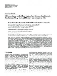
CASE REPORT ANC, a 68 year old right handed man, volunteered and consented to take part as a control subject in a longitudinal MRI study at the Institute of Neurology, London. He was seen for his first assessment in February 2001 when he was well, with no cognitive symptoms. He was a retired management consultant, whose past medical history included a hip replacement, mild renal impairment, and benign prostatic hypertrophy, for which he was taking finasteride. He did not smoke and he drank less than 21 units of alcohol a week. Examination revealed hypertension (blood pressure 170/ 95) and a body mass index of 28 kg/m2, but was otherwise unremarkable. He scored 29/30 on a mini-mental state examination (MMSE).2 Baseline neuropsychology was undertaken (see below) and a volumetric MRI brain scan acquired. Over the following nine months he remained well and had five further volumetric MRI scans as part of the study. There was no evidence of sustained hypertension. He was started on orlistat during this time by his general practitioner to help weight control. He scored 30/30 on the MMSE done six months into the study. Ten months into the study he suffered an acute dysphasic episode when he was suddenly unable to get the correct words out or to complete sentences. There was no associated limb or facial weakness. He retired to bed and woke two hours later, by which time his speech had virtually returned to normal. Over
the next few days, his wife reported that he was slightly confused and had continued difficulties in word finding, as well as new difficulties in remembering names. He saw his general practitioner who diagnosed a probable stroke, began treatment with aspirin, and referred him to the local neurology service. One month later he attended for his final study visit. He and his wife reported that his cognition had improved, but that although his ability to recognise faces was unimpaired, he had persistent difficulties recalling people’s names. He had also become more reliant on his diary. On examination, the MMSE was 26/30, the blood pressure was 130/85, and a neurological examination was normal. Imaging All volumetric imaging was done on the same 1.5 Tesla Signa unit, using a spoiled gradient echo technique with the same imaging parameters (TR/TE/TI/Theta 17/4.2/450/20, field of view 24 × 18 cm, 256 × 192 image matrix). All scans were reported by an expert neuroradiologist. The first scan revealed evidence of mild small vessel disease especially affecting the pallidum, with normal hippocampi and no evidence of global or regional cerebral atrophy. The next five scans (over the following nine months) revealed no significant atrophy and no new ischaemic lesions. The final scan, one month after the acute event, revealed a discrete new infarct in the left thalamus, involving the medial thalamic nuclei and interrupting the mamillo-thalamic tract (fig 1). Neuropsychology ANC was assessed at the start and end of the study using a standard battery of neuropsychological tests. A summary of these results is shown in table 1. At the start of the study, he performed in the superior range on tests of verbal and non-verbal intelligence, recognition memory, reading, and picture naming. Calculation was in the average range, and visuoperceptual and visuospatial skills were satisfactory. Reassessment was carried out at one year, a month after the acute event. The only change noted was a subtle decline in naming skills on the graded naming test7; nonetheless, his score still fell within the superior range (90th centile). In the light of his persistent memory complaint, additional tests of word retrieval skills and episodic memory were administered.
Language skills Two further stringent graded difficulty naming tests were attempted. ANC was able to name 22 of 30 objects and 28 of 30 animals (> 50th centile and > 90th centile, respectively) from the McKenna category specific names test.9 On a comparable proper noun retrieval test (historical figures, countries, buildings),10 his performance was extremely competent (25/30). His good verbal comprehension was demonstrated on a test of knowledge of synonyms11 (concrete words: 22/25, > 50th
www.jnnp.com
256
Schott, Crutch, Fox, et al
Figure 1 Coronal volumetric magnetic resonance brain scan at the start of the study (A) and one year later (B). In (B), there is an infarct in the left thalamus (arrowed) involving the medial thalamic nuclei and interrupting the mamillo-thalamic tract.
centile; abstract words: 25/25, > 90th centile). He also expressed himself fluently using a wide vocabulary. Thus at this stage there was no evidence of a dysphasic syndrome.
Episodic memory The routine neuropsychological battery contained only the easy recognition memory test, on which ANC scored at ceiling. He was also tested on the standard recognition memory test,12 which allows visual and verbal memory to be assessed independently with an identical test design for each component. He scored 41/50 on the visual section of the test (faces: 50th centile). By contrast, on the verbal section of the test he scored only 36/50 (words: 5th centile). This discrepancy of five points represents a selective verbal memory deficit (p < 0.02).12 On a demanding test of visual recognition memory—the topographical recognition memory test13—he scored 26/30 (75th centile). His performance was also impaired on a measure of verbal recall: he scored at the 5th centile on the Camden paired associate learning test (CPALT).13 In an attempt to document this recall deficit in more detail, ANC was assessed with part of a longer famous faces test previously used by Cipolotti et al 14 to provide evidence of retrograde amnesia in their patient vc. This test consists of 39 monochrome photographs of people from the 1990s famous in the fields of politics, entertainment, sport, and because of particular newsworthy events. The subject was first requested to recall the name of each individual orally. Subsequently, each photograph was re-presented with a choice of three names in a forced choice recognition paradigm—the target name alongside two equally famous distractor personalities. His scores were compared with those of 20 age matched control subjects tested by Cipolotti et al.14 He was able to recall only 21% of names correctly, significantly fewer than the control
Table 1
subjects (50%; z = 2.0, p < 0.02, two tailed test). However, no such difference was found using the forced choice measure (ANC 92%, controls 85%). These findings corroborate the evidence from the anterograde memory tests, providing further evidence for a selective verbal memory deficit.
DISCUSSION Using longitudinal imaging in this individual, we have been able to demonstrate the appearance of a new discrete left sided thalamic infarct. At follow up, both ANC and his wife reported memory deficits occurring during the period over which the lesion appeared. Although he scored at ceiling on the easy recognition memory test at both the start and end of the study, his new reported memory impairment prompted us to undertake more detailed testing at follow up. Clear focal cognitive deficits were determined at this assessment, which we conclude are highly likely to be the result of the localised thalamic infarct. Thalamic lesions may produce a wide range of neuropsychological deficits. The memory dysfunction associated with thalamic lesions appears to be best correlated with disruption of the mamillo-thalamic tract, as seen in this case. The role of the medial thalamic nuclei in memory dysfunction is less clear (for a review, see Van Der Werf et al 15). We have shown that ANC developed a selective episodic memory impairment for verbal material with preservation of visual memory—as demonstrated by his performances on the recognition memory test, the Camden paired associate learning test, and the topographical recognition memory test—and his poor recall of names of famous faces despite good recognition. There was no evidence of semantic memory impairment, as evidenced by the high score on the graded naming test. Thalamic lateralisation (where dominant lesions result in verbal memory impairment, and non-dominant lesions in visual
Neuropsychological raw scores and centile rankings at the initial and one year assessments
Neuropsychological tests
Assessment 1 (Feb 2001)
Assessment 2 (Feb 2002)
77 28 134 (99%ile)
76 29 135 (99%ile)
5 (124)
4
3
The Wechsler abbreviated scale of intelligence : Vocabulary (raw score) Matrix reasoning (raw score) Predicted full scale IQ National adult reading test4: Number of errors (Predicted IQ) Easy recognition memory test5 Words Faces Graded difficulty arithmetic test6 Graded naming test7 Visual object and space perception battery8 Silhouettes Number location %ile, centile.
www.jnnp.com
25/25 25/25 14/24 29/30
(>50%ile) (>50%ile) (>50%ile) (>99%ile)
21/30 (>25%ile) 8/10 (>5%ile)
25/25 25/25 14/24 25/30
(>50%ile) (>50%ile) (>50%ile) (>90%ile)
21/30 (>25%ile) 10/10 (>50%ile)
Memory impairment with left thalamic infarct
memory problems) has been proposed by several investigators,16–18 although others have failed to demonstrate a consistent effect.19 20 Our findings support the hypothesis that lateralisation of cognitive processing of visual and verbal material exists at the thalamic as well as at the cortical level. While lesions throughout the dominant hemisphere can cause a selective impairment of verbal memory,12 it is rare for such lesions to be demonstrated longitudinally. Small thalamic or other subcortical lesions may produce defects of memory or other cognitive functions. It is likely that many of these lesions will not be visualised using computed tomography, and may even be missed by conventional MRI. The possibility of a thalamic lesion should be considered in patients presenting with new persistent memory dysfunction in the absence of cortical signs or abnormalities on conventional imaging. In these cases, small lesions may be demonstrated using the higher resolution afforded by volumetric MRI.
ACKNOWLEDGEMENTS This study was funded by GlaxoSmithKline. We would like to thank ANC for his help with the study, and to acknowledge the assistance of David MacManus (scan acquisition), Jennifer Whitwell (image analysis), and Dr John Stevens (neuroradiology). Dr Lisa Cipolotti and Professor Tim Shallice kindly provided test material and Professor Martin Rossor offered useful comments on the manuscript. NCF is a Medical Research Council senior clinical scientist. .....................
Authors’ affiliations J M Schott, S J Crutch, N C Fox, E K Warrington, Dementia Research Group, Institute of Neurology, Queen Square, London, UK Competing interests: none declared Correspondence to: Dr J M Schott, Dementia Research Group, Institute of Neurology, Queen Square, London WC1N 3BG, UK;
[email protected] Received 25 June 2002 In revised form 18 September 2002 Accepted 26 September 2002
257
REFERENCES 1 Bogousslavsky J, Regli F, Uske A. Thalamic infarcts: clinical syndromes, etiology, and prognosis. Neurology 1988;38:837–48. 2 Folstein M, Folstein S, McHughs P. The “mini mental state”: a practical method for grading the cognitive state of patients for the clinician. J Psychiatr Res 1975;12:189–98. 3 Wechsler D. The Wechsler abbreviated scale of intelligence. San Antonio: The Psychological Corporation, 1999. 4 Nelson H. The national adult reading test (NART): test manual. Windsor: NFER-Nelson, 1982. 5 Clegg F, Warrington EK. Four easy memory tests for older adults. Memory 1994;2:167–82. 6 Jackson M, Warrington EK. Arithmetic skills in patients with unilateral cerebral lesions. Cortex 1986;22:611–20. 7 McKenna P, Warrington EK. The graded naming test. Windsor, UK: NFER-Nelson, 1983. 8 Warrington EK, James M. The visual object and space perception battery. Bury St Edmunds: Thames Valley Test Co, 1991. 9 McKenna P. Category specific names test. Hove, UK: Psychology Press Ltd, 1997. 10 McKenna P, Warrington EK. Testing for nominal dysphasia. J Neurol Neurosurg Psychiatry 1980;43:781–8. 11 Warrington EK, McKenna P, Orpwood L. Single word comprehension: a concrete and abstract word synonym test. Neuropsychol Rehab 1998;8:143–54. 12 Warrington EK. Recognition memory test. Windsor, UK: NFER-Nelson, 1984. 13 Warrington EK. The Camden memory tests manual. Hove: Psychology Press, 1996. 14 Cipolotti L, Shallice T, Chan D, et al. Long term retrograde amnesia. The crucial role of the hippocampus. Neuropsychologia 2001;39:151–72. 15 Van der Werf YD, Witter MP, Uylings HB, Jolles J. Neuropsychology of infarctions in the thalamus: a review. Neuropsychologia 2000;38:613–27. 16 Clarke S, Assal G, Bogousslavsky J, et al. Pure amnesia after unilateral left polar thalamic infarct – topographic and sequential neuropsychological and metabolic (PET) correlations. J Neurol Neurosurg Psychiatry 1994;57:27–34. 17 Parkin AJ, Rees JE, Hunkin NM, et al. Impairment of memory following discrete thalamic infarction. Neuropsychologia 1994;32:39–51. 18 Mori E, Yamadori A, Mitani Y. Left thalamic infarction and disturbance of verbal memory: a clinicoanatomical study with a new method of computed tomographic stereotaxic lesion localization. Ann Neurol 1986;20:671–6. 19 Exner C, Weniger G, Irle E. Implicit and explicit memory after focal thalamic lesions. Neurology 2001;57:2054–63. 20 Wallesch CW, Kornhuber HH, Kunz T, et al. Neuropsychological deficits associated with small unilateral thalamic lesions. Brain 1983;106:141–52.
www.jnnp.com