Jan 15, 1996 - protein was detected first at postnatal day (P) 8. The Kv3.1 .... properties of these neurons has been investigated (Buhl et al., ... and initial .... ered to pH 7.4 and -275 mOsm. .... considered it possible that the amount of KvS.lb protein extracted ... mine whether Kv3.lb was expressed in any cell type in younger.
The Journal
of Neuroscience,
January
Developmental Expression and Functional Characterization Potassium-Channel Subunit Kv3.1 b in Parvalbumin-Containing Interneurons of the Rat Hippocampus Jing
Du,l Lei Zhang,l
Michael
Weiser,*
Bernard0
Rudy,*
and Chris
15, 1996,
76(2):506-518
of the
J. McBainl
‘Unit on Cellular and Synaptic Physiology, Laboratory of Cellular and Molecular Neurophysiology, National Institute of Child Health and Human Development, Bethesda, Maryland 20892-4495, and *Department of Physiology and Neuroscience, New York University Medical Center, New York, New York 10016
The expression of the voltage-gated K+-channel subunit Kv3.1 b in the developing hippocampus was determined by immunoblot and immunohistochemical techniques. Kv3.1 b protein was detected first at postnatal day (P) 8. The Kv3.1 bimmunopositive cell number per tissue section reached a maximum at PI4 and was maintained through P40. In contrast, the Kv3.1 b protein content of isolated membrane vesicles in immunoblots progressively increased through P40, suggesting an increase in Kv3.1 b content per cell throughout this time period. Kv3.1 b protein was expressed selectively in the somata, proximal dendrites, and axons of cells lying within or near the pyramidal cell layer, consistent with their being GABAergic inhibitory interneurons. Kv3.lb was present in -80% of parvalbumin-positive interneurons. The developmental onset of Kv3.1 b and parvalbumin immunoreactivity was identical. In contrast, Kv3.1 b was mostly absent from the subset of somatostatin-positive inhibitory interneurons. Electrophysiological recordings were made from stratum pyramidale inter-
neurons in which morphology and Kv3.1 b-positive immunoreactivity were confirmed post hoc. Outward currents had voltage-dependent and biophysical properties resembling those of channels formed by Kv3.1 b. The current blocked by low concentrations of 4-aminopyridine (4-AP) showed marked inactivation, suggesting that Kv3.1 b may coassemble with other members of the Kv3 subfamily. In current-clamp recordings, concentrations of 4-AP that blocked the current through Kv3.1 b channels allowed us tentatively to assign a role to Kv3.1 b-containing channels in action-potential repolarization. These data demonstrate that Kv3.1 b is regulated developmentally in a specific subpopulation of hippocampal interneurons and that channels containing this subunit may be a major determinant in imparting “fast-spiking” characteristics to these and other cells throughout the central nervous system containing the Kv3.1 b subunit. Key words: hippocampal interneurons; K+-channel subunits; Kv3.1; GABA; parvalbumin; immunohistochemistry
Despite the wealth of data concerning the structure and function of voltage-gated K+-channel subunits, little is known concerning their developmental regulation, the molecular composition of native channels in the mammalian CNS, or the actual physiological roles they play (Jan and Jan, 1990; Pongs, 1992; Vega-Saenz de Miera et al., 1994; Gutman and Chandy, 1995). In situ hybridization and immunohistochemical techniques have demonstrated that many voltage-dependent Kt channel subunits show regionspecific expression, which occurs in a cell-specific or subcellular manner, consistent with a highly specialized role for each subunit in distinct physiological processes. The mRNAs of members of the Kv3 subfamily of voltage-gated K+ channels are distributed selectively throughout the mammalian CNS (Vega-Saenz de Miera et al., 1994). High levels of Kv3.lb mRNA (designated Kv3.la in Perney et al., 1992) are expressed in the reticular thalamus and cerebellum and are under developmental regulation (Perney et al., 1992; Rudy et al., 1992;
Weiser et al., 1994). In the hippocampus, Kv3.lb mRNA expression occurs at low levels and has been shown to be restricted to cells tentatively identified as GABAergic inhibitory interneurons (Perney et al., 1992; Weiser et al., 1994). Recently, Weiser et al. (1995), using immunohistochemical techniques, have confirmed that Kv3.lb protein is expressed in a selective population of parvalbumin-containing cells, tentatively identified as hippocampal interneurons. Hippocampal inhibitory interneurons are GABA-containing local-circuit cells that mediate both feedforward and feedback inhibition of pyramidal neurons (Lacaille et al., 1989; Traub and Miles, 1991). Parvalbumin-containing interneurons belong to the class of “fast-spiking” interneurons in which cell bodies are located within or near stratum (St.) pyramidale (Kawaguchi et al., 1987; Kosaka et al., 1987). It is of interest that some parvalbuminpositive interneurons have been shown to be resistant to both ischemic damage (Schlander et al., 1988) after bilateral carotid occlusion (Tortosa and Ferrer, 1993) and electrographic seizureinduced neuronal damage (Sloviter, 1989). The actual mechanisms responsible for the selective resistance of these cells are unclear at present because little more than the basic physiological properties of these neurons has been investigated (Buhl et al., 1994a,b). With the exception of interneurons of the st. oriensalveus (Zhang and McBain, 1995a,b), no data exist concerning the developmental expression and the biophysical properties of voltage-gated Kt channels on any of the other hippocampal
Received Aug. 10, 1995; revised Oct. 10, 1995; accepted Oct. 12, 1995. This work was supported by National Institutes of Health Grant NS30989 to B.R. We thank Dr. Vittorio Gallo for help with immunohistochemistry, Dr. Joanna Hill for help with the whole-animal perfusion, and Dr. Mark Maye; and Dr. Vittorio Gallo for critically reading this manuscript. CorresDondence should be addressed to Chris J. McBain. Unit on Cellular and Synaptic ‘Physio!ogy, Laboratory of Cellular and Molecular Neurophysiology, Room 5A72, Building 49, NICHD-LCMN, 49 Convent Drive, MSC 4495, Bethesda, MD 20892.4495. Copyright 0 1996 Society for Neuroscience 0270-6474/96/160506-13$05.00/O
Du et al. . Kv3.1 b in Hippocampal
interneurons
interneuron populations. It is a reasonable assumption, however, that the action-potential properties and the specific firing patterns of these cells are determined, at least in part, by the complement of K+ channels expressed by these cells. In this study, we have investigated the developmental expression and the functional role of Kv3.lb subunits in an immunologically distinct subpopulation of hippocampal interneurons. Western blot analysis and immunocytochemistry were used to determine the developmental expression of Kv3.lb. Doublestaining techniques demonstrated that Kv3.lb is expressed predominantly in parvalbumin-containing cells, but not somatostatinpositive interneurons, commencing at approximately postnatal day (P) 8. Finally, we have identified currents through native Kt channels that likely contain the Kv3.lb subunit and have identified a role for such currents in the action-potential waveform of morphologically identified and Kv3.lb-positive interneurons. The nomenclature of Shaker genes and products proposed by Chandy et al. (1991) was used throughout this article.
MATERIALS AND METHODS Kv3.Ib antibody production. Antibody production, purification, and initial characterization have been described previously (Weiser et al., 1995). Briefly, a peptide corresponding to the C-terminal sequence of the rat Kv3.lb protein (residues 567-585) (L uneau et al., 1991) was synthesized, and antiKvS.lb antibodies were raised in rabbits and were affinity-purified by coupling to a Sulfolink Sepharose resin (Pierce, Rockford, IL). Western blot analysis of hippocampal membrane vesicles. SpragueDawley rats aged P6-P40 were anesthetized deeply with Forane (Ohmeda, Liberty Corner, NJ). The crude membrane fractions were prepared as described previously by Sheng et al. (1992) with slight modifications. Briefly, the hippocampus was dissected and homogenized in homogenization buffer [20 IIIM Tris-HCI, pH 7.4, 0.3 M sucrose, 1 mM phenylmethanesulfonyl fluoride (PMSF), 1 FM pepstatin A, and 2 Fgiml leupeptin] using a Dounce tissue grinder with a pestle at 4°C. The homogenates were centrifuged at 1000 X g for 10 min at 4°C. The supernatants then were centrifuged at 100,000 X g for 1 hr at 4°C and the pellets were dissolved in suspension buffer (20 mM Tris-HCI, pH 7.4, 1 mM EDTA, 1 ITIM PMSF, 1 pM pepstatin A, and 2 p&ml leupeptin). The resulting solution was centrifuged at 40,000 X g for 20 min at 4°C. The pellets were resuspended in suspension buffer, and aliquots were used immediately or stored at -80°C for later use. Protein concentrations were determined by Bio-Rad protein assay (Hercules, CA) according to the manufacturer’s instructions with bovine serum albumin (BSA) as a control. Fifty micrograms of membrane protein were resuspended in equal amounts of sample buffer (100 mM Tris, pH 7.6, 1% SDS, 10% glycerol, 0.05% bromphenol blue, 5% P-mercaptoethanol), denatured at 37°C for 1 hr, and separated by 8% SDS-PAGE. Proteins then were transferred to nitrocellulose membranes in a transfer tank containing 25 mivt Tris, 0.2 M glycine, and 20% methanol at 150 mA for 17 hr at room temperature. Blocking of nonspecific binding was achieved by incubating the nitrocellulose -membrane with Trisy buffered saline/Tween-20 (TBST: 20 miw Tris. oH 7.6.137 mM NaCl. 0.2% Tween-20) with 5% nonfat dry ‘milk. The blots were probed with antiKv3.lb antibody (1:3000) in TBST with 1% nonfat dry milk for 1 hr at room temperature. The secondary antibody was peroxidase-conjugated goat anti-rabbit antibody (1:lOOO). Enhanced chemiluminescence (ECL) was used as a secondary detection system according to the manufacturer’s instructions. K+-channel Kv3.Ib immunostaining. For the developmental study of the K’ -channel subunit Kv3.1 b or parvalbumin immunoreactivity, P6-P40 animals were anesthetized deeply using Forane. The brain tissue was dissected free, and fresh-frozen tissue sections (12 pm) were processed commercially by American Histolab (Gaithersburg, MD). The freshfrozen sections were allowed to come to room temperature before fixation in 4% p-formaldehyde in PBS for 5 min on ice. The sections were then washed with PBS once and taken through an alcohol series (50, 70, and 95%) before being stored in 95% alcohol at 4°C until needed. The tissue sections were rehydrated with an alcohol series (90, 70, and 50%) and PBS for 5 min each. They then were incubated in 0.3% H,O, in methanol for 3 min to remove endogenous peroxidase activity. After washing three times in PBS, a 30 min immersion in 0.2% Triton X-100 and 1% BSA in PBS was followed by overnight incubation with rabbit
J. Neurosci.,
January
15, 1996,
16(2):506-518
507
anti-Kv3.lb (1:3000) in 0.5% BSA in PBS. All incubations and wash steps were performed in 0.5% BSA in PBS. For the secondary antibody, biotinylated goat anti-rabbit antibody (1:200) was added for 1 hr, followed by an incubation with horseradish peroxidase (HRP)-avidinbiotin complex (l:SO, Vector Elite ABC kit, Burlingame, CA) for an additional 1 hr. The color was developed by treatment with 0.05% diaminobenzidinc (DAB) and 0.003% H,02 in PBS. The sections then were dehydrated, cleared, and mounted before photomicroscopy. The Kv3.lb-positive cell number per section was counted under microscope for later analysis. A cell was considered Kv3.lb-positive if the soma and proximal dendrites were appreciably darker than the surrounding tissue when observed under high-power magnification. Double staining of parvalbumin, somatostatin, and Kv3.Ib. For the experiments involving double immunostaining with parvalbumin or somatostatin and KvS.lb, P6-P20 animals were anesthetized with Forane and perfused transcardially with ice-cold PBS followed by ice-cold 4% p-formaldehyde in PBS for 10 min. The animals were decapitated and post-fixed overnight in 4% p-formaldehyde in PBS at 4°C. The brains were dissected from the skull and stored in 5% sucrose in PBS at 4°C until needed. Serial sagittal sections (20 Fm) of the middle portion of the hippocampus were cut on a freezing microtome (Zeiss, Jena, Germany). Sections were mounted onto gelatin coated-slides and air-dried. Sections were incubated in a blocking solution of 10% normal goat serum and 0.2% Triton X-100 in PBS for 30 min. The monoclonal antibody [mouse anti-parvalbumin (1:200) or mouse anti-somatostatin (l:lO)] and rabbit polyclonal anti-Kv3.lb antibody (1:400) were diluted in 5% normal goat serum in PBS and incubated with the sections overnight at 4°C. The sections were rinsed and incubated with fluorescein isothiocyanate (FITC)-conjugated anti-mouse antibody (1:50, green stain) and Cy3conjugated goat anti-rabbit antibody (1:300, red stain) in 5% normal goat serum in PBS for 1.5 hr at room temperature. The sections then were rinsed, mounted, and observed by using fluorescence microscopy with the appropriate filters. Whole-cell recordings from visually identified interneurons in hippocampal slices. Hippocampal slices were prepared as described previously (McBain, 1994). Briefly, Sprague-Dawley P14-P18 rats were killed by decapitation after deep anesthesia using Forane. The brain was removed rapidly and placed in ice-cold artificial CSF (ACSF) (in mM): 130 NaCI, 24 NaHCO,, 3.5 KCI, 1.25 NaH,PO,, 1.5 CaCl,, 1.5 MgSO,, and 10 glucose saturated with 95% 0,/5% CO,, pH 7.4, 307 mOsm. Sagittal slices (250- to 350-pm-thick) were cut from the middle third of the hippocampus using a Vibratome (Oxford series 1000, Polysciences, Warrington, PA). Slices were allowed a recovery period of 45 min before use, during which they were held in oxygenated media at 27°C. During recording, slices were bathed in a tissue chamber of 1 ml capacity and perfused at a rate of 2-4 mlimin with ACSF at room temperature (24-26°C). For electrophysiological recordings, Ca’+ was omitted and Co’+ (1.5 mM) was added to the recording solution to remove any Ca’+-dependent components of the outward current. Tetrodotoxin (0.51.0 pM) and the AMPA-receptor antagonist 6,7-dinitroquinoxaline-2,3dione (20 FM) were added to the perfusate to block voltage-dependent sodium channels and excitatory synaptic activity, respectively. All drugs were applied in known concentrations by direct addition to the perfusate via a three-way tap. Tight-seal (>l Gn), whole-cell recordings (Hamill et al., 1981; Edwards et al., 1989) were obtained from the cell bodies of interneurons located at the border between st. pyramidale and st. oriens or radiatum. The cell bodies of these cells were resolved easily and were identified tentatively as interneurons before confirmation by post hoc biocytin processing. Electrodes were fabricated from borosilicate glass and were not fire-polished. Patch electrodes had resistances of 3-6 Ma when filled with (in mM): 130 K-eluconate. 10 NaCl. 2 Na,-ATP. 0.3 Na-GTP. 10 HEPES, 0.6 ‘EGTA, 1”tetraethylammonium (TEA), 5 ‘glutathione, buffered to pH 7.4 and -275 mOsm. Biocytin (0.4%) was added to the internal solution for post hoc histochemical study. All data are represented as mean -C SEM. Seals were attained by the method originally described by Blanton et al. (1989). The electrode was positioned under visual control within the CA1 subfield; individual cells were visualized by using a Zeiss water-immersion objective modified with Hoffman (Nutley, NJ) optics (overall magnification 500X). Cell sealing and breakthrough to whole-cell mode were performed under current-clamp conditions to allow an initial evaluation of cell viability. The series resistance usually ranged from 20 to 33 Ma; membrane potentials were not corrected for these errors. Linear leak current and the capacitive artifacts were subtracted digitally by using Pi4 subtraction before data acquisition and
508
J. Neuroscl.,
January
15, 1996,
76(2):506-518
Du et al. . Kv3.1 b in Hippocampal
Interneurons
analysis. All recordings were obtained with an Axopatch 1D amplifier (Axon Instruments, Foster City, CA). Records were filtered at l-5 kHz and digitized at 3-10 kHz on a 486 PC. Data were acquired and analyzed using the pClamp suite of programs (Axon) and Microcal Origin (Boston, MA). Biocytin detection and immunostaining after electrophysiological recording. After electrophysiological recording, the slice was fixed in 4%
p-formaldehyde in PBS for 1 hr on ice and stored in 1% p-formaldehyde in PBS overnight at 4°C. The slice was incubated in 30% sucrose in PBS for 30 min at room temperature and resectioned on a freezing microtome at -70°C in 40-70 Km sections. The sections were mounted and dried on gelatin-coated slides. Before biocytin detection, the sections were rehydrated in PBS for 10 min and preincubated in HEPES buffer (10 mM HEPES, pH 8.5, 0.15 M NaCl, and 0.08% sodium azide) for 10 min. The sections then were transferred to a solution containing fluorescenceconjugated Avidin-D (1:200) and 0.2% Triton X-100 in HEPES buffer for 20 min in the dark at room temperature. The sections were washed in PBS (3 times). A 30 min immersion in 0.2% Triton X-100 and 10% normal goat serum in PBS was followed by overnight incubation with rabbit anti-Kv3.lb (1:400) in 5% normal goat serum in PBS. After rinsing in 5% normal goat serum in PBS (3 times), the sections were placed in CyS-conjugated goat anti-rabbit antibody’ (1:300) in 5% normal goat serum in PBS for 1.5 hr. Thev then were rinsed three times in PBS. mounted, and observed with fluorescence microscopy with appropriate filters. Cells that were filled with biocytin and were Kv3.lb-positive were photographed and drawn using camera lucida techniques to allow the complete reconstruction of the cell morphology. Materials. The parvalbumin monoclonal antibody was obtained from Sigma (St. Louis, MO). The somatostatin monoclonal antibody was from Biomeda (Foster City, CA). The fluorescence-conjugated Avidin D, biotinylated anti-rabbit IgG, normal goat serum, and Vectastain ABC kits were from Vector. The Cy3-conjugated goat anti-rabbit antibody was from Jackson Immunoresearch (West Grove, PA). The fluorescenceconjugated anti-mouse antibody was from Cappel (Durham, NC). The peroxidase-conjugated goat anti-rabbit antibody and ECL were purchased from Amersham (Arlington Heights, IL). The molecular weight markers were purchased from Bio-Rad (Melville, NY).
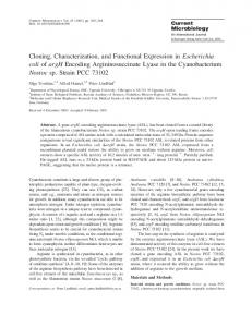
RESULTS Expression of Kv3.1 b in the developing hippocampus In situ hybridization techniques have shown previously that Kv3.lb mRNA is expressed as early as embryonic day (E) 19 in rat brain; however, a significant increase in mRNA was detected between P7 and P14 (Perney et al., 1992). In the hippocampus, only a small number of cells had grain densities greater than background levels at early stages, and only by P7 were detectable levels of Kv3.lb mRNA observed. In the first series of experiments, we performed Western blot analysis of membrane preparations purified from the developing postnatal hippocampus. Kv3.lb protein was detected first in membrane preparations derived from P8 animals (Fig. 1). In the hippocampus, anti-Kv3.lb recognized a single, prominent band with an M, of -96 kDa at P8, consistent with the data obtained using the identical antibody by Weiser et al. (1995). In the present study, the mean M, of the band recognized by the Kv3.lb antibody is -10 kDa larger than that described for total brain by Weiser et al. (1995). The reasons for this discrepancy are unclear at present; however, this may reflect a more heavily glycosylated form of the Kv3.lb protein in the hippocampus than found in the total brain, which is dominated by the higher levels of Kv3.lb protein derived from the cerebellum compared with the small amounts of hippocampal protein. As the hippocampal developmental age progressed, the anti-Kv3.lb band on the immunoblot broadened and had an M, ranging from 90 to 110 kDa (Fig. 1). These data were confirmed in three independent experiments. In two experiments, densitometric measurement revealed 7.1- and 6.0-fold increases in the protein content from immunoblots obtained at P20 compared with that observed at P14.
v3. lb
1. The developmental expression of KvS.lb protein as detected by immunoblot analysis. Equal amounts (50 pg) of protein isolated from hippocampal membrane vesicles from P6-P40 animals were fractionated on SDS-polyacrylamide gels, transferred to nitrocellulose membrane, and incubated with anti-Kv3.lb antibody (1:3000). ECL was used as a secondary-detection system. Detection of Kv3.lb protein commenced in membrane preparations isolated from P8 animals and was detected as a prominent band ranging from 90 to 110 kDa. The Kv3.1 protein content was observed to increase continuously through P40, the oldest age tested. These data were confirmed in three independent experiments. Figure
Despite the previous observation that Kv3.lb mRNA was detected first at El9 in total rat brain, we were unable to detect KvS.lb protein in hippocampal membrane tissue derived before P8. Because Kv3.lb mRNA has been shown to be expressed only in a selective subpopulation of neurons of the hippocampus, we considered it possible that the amount of KvS.lb protein extracted from younger tissue was low compared with the total amount of protein derived from the principal pyramidal neurons, which outnumber these interneurons by at least lo-fold. Next, to determine whether Kv3.lb was expressed in any cell type in younger hippocampus, we performed immunohistochemical detection on tissue sections derived from PO-P40 rat brains. Kv3.lb protein expression was not detected in hippocampal tissue derived at PO, P6, and P7 (n = 4; data not shown). At P8, a few nonpyramidal cells located at the borders between strata radiatum and pyramidale and strata oriens and pyramidale were Kv3.lb-immunopositive (Fig. 2A). At P14 and P20, there was a marked increase in the number of cells that were Kv3.lbimmunopositive (1 tissue section from each animal; II = 4 animals). In general, these cells were found throughout all hippocampal subfields from the hilus to the subiculum. Figure 2B shows high-power images of Kv3.lb-positive cells in tissue derived from P14 animals. In the CA1 and CA3 subfields, Kv3.lb-positive cells usually lay at the border between st. pyramidale or st. oriens. Occasionally, cells were found deep in the st. pyramidale proper. Kv3.lb-positive cells rarely were observed in the st. lucidum and lacunosum-moleculaie. The anatomical localization of these Kv3.lb-positive cells is consistent with their being GABAergic inhibitory interneurons (Buhl et al., 1994a,b). In the dentate gyrus, on the basis of their positioning at the border of the granule cell layer, the Kv3.lb-positive cells likely were inhibitory basket cells. The granule cells of the dentate gurus did not appear to express Kv3.lb at high levels. Kv3.lb protein was restricted primarily to the somata and the proximal dendrites of positive cells, and only extremely rarely were we able to observe Kv3.lb immunostaining on the distal dendrites of these cells (see Fig. 2Z?). Of interest, we were always able to distinguish Kv3.lb staining above background in the pyramidal cell layer (see P20 in Fig. 2 and fluorescent images in Fig. 6). This staining clearly was not in
Du et al.
l
Kv3.1 b in Hippocampal
Interneurons
J. Neurosci.,
January
15, 1996,
76(2):506-518
509
Figure 2. The developmental distribution of Kv3.lb in tissue sections of the hippocampus. Low-power (34X) bright-field photomicroscopy of hippocampal sections (12 pm) from P8, P14, and P20 animals (A) that were immunostained with anti-Kv3.lb antibody (1:3000). The secondary antibody was biotinylated goat anti-rabbit antibody (1:200). The sections were incubated with HRP-avidin-biotin complex to amplify the signal using DAB (0.05%) as the chromogen. Consistent with data obtained from immunoblot experiments, Kv3.lb immunoreactivity was detected first in tissue derived from P8 animals. In this section, two cells located at the border of the st. pyramidale and st. radiatum were Kv3.lb-immunoreactive (filled arrowhen&). By P14, numerous cells located throughotrt the entire hippocampus and dentate gyms are Kv3.lb-positive. In the hippocampus proper, cells were restricted primarily to the borders between st. pyramidale and strata oriens and radiatum. A large fraction of KvS.lb-positive cells also was identified in the subicular and hilar regions. Note the distinct absence of Kv3.lb-positive cells in the principal cell layers of st. pyramidale and the dentate gyrus. However, a diffuse band of KvS.lb immunoreactivity could be detected in the st. pyramidale of CA3-CAl, which is consistent with the presence of Kv3.lb immunoreactivity in the axons of these cells. This pattern of Kv3.lb immunoreactivity was identical in tissue derived from P20 animals. B, Higher-power (333X) photomicrographs from boxes indicated in A of the st. pyramidale of CA1 and CA3 and the dentate gyrus (DG) show that the KvS.lb immunostaining is restricted primarily to the somata and proximal dendrites of these cells. The control photomicrograph (COW depicts an identical experiment performed in the absence of primary antibody to demonstrate the specificity of the Kv3.lb antibody. Scale bars: A, 200 pm; B, 20 pm. the pyramidal neuron cell bodies per se; rather, it was restricted to the extracellular matrix of the pyramidal cell layer. This is evident particularly in the photomicrograph depicted in Figure 6. Because the axonal projections of this subtype of inhibitory neuron are known to be restricted to the pyramidal cell layer and the regions closely adjacent to it, it is possible that the axon projections of these cells also contain Kv3.lb immunoreactivity (Weiser et al., 1995). At this time, however, we are unable to resolve the finer axonal projections of these cells by using standard light microscopy. The use of confocal microscopic techniques will be required to resolve the immunoreactivity of the interneuron axons. The somatodendritic immunostaining pattern of the Kv3.lb antibody was consistent even when concentrations of antibody of up to
1:lOO were used, suggesting that poor penetration of the antibody is not responsible for the observation that Kv3.lb is restricted to the soma and proximal dendrites of these cells. At this higher concentration of antibody, we were unable to resolve immunostaining of the distal portions of the dendritic tree further. It was observed often at higher concentrations of antibody, however, that the staining within the pyramidal cell layer was more intense, which is consistent with the heavier staining of the axonal projections of the Kv3.lb-positive interneurons. On no occasion were glial cells found to be Kv3.lb-positive. These data support and extend the recent findings of Weiser et al. (1995). In an attempt to quantify the developmental expression of Kv3.lb in cells throughout the hippocampus, we counted the
510
B
J. Neurosci.,
January
15, 1996,
76(2):506-518
Du et al. . Kv3.1 b in Hippocampal
60
0-l
-*-
Sub
8
10
I
-
Postnatal Days
12
14
Postnatal Days
Figure 3. A schematic representation of the developmental time course of Kv3.lb-immunoreactive cell number across various regions in rat hippocampus. To characterize the region-specific localization of hippocampal Kv3.lb-positive cells, we divided the hippocampus into four principal regions as depicted inA: st. oriens-alveus (Sf.otiens-alveus), st. pyramidale (St.pyramidale), Subiculum, and Dentate gyms-hilus. Kv3.lb immunostaining was performed on hippocampal sections derived from four animals from two litters at each age (P6, P7, P8, P9, PlO, Pll, P14, P20, and P40). The cell number per section was counted and is represented graphically in 8. Data are presented as mean k SE (n = 4). The developmental onset of Kv3.lb immunoreactivity progressed in parallel throughout each region of the hippocampus. Of interest, KvS.lb was maintained in all regions between P14 and P40 except the st. oriens-alveus, in which a significant drop in the expression was observed over this time period. By P40, the highest numbers of KvS.lb-positive cells were found as follows: subiculum (34%) > st. pyramidale (28%) > dentate gyrus (22%) > st. oriens-alveus (9%). C, To determine whether the onset of Kv3.lb immunoreactivity was identical in each region, we repeated the experiment using tissue sections derived from animals of the same litter at 24 hr time points between P7 and P14 (n = 4 experiments). Kv3.lb expression started at approximately P8 in st. pyramidale and was followed closely in all subfields by P9. The Kv3.lb immunoreactivity progressively increased through P14. Sub, Subiculum; 4/r, st. pyramidale; DG, dentate gyrus; OA, st. oriens-alveus; Tot, total.
number of cells observed per section in the subfields of the dentate gyrus proper, the st. oriens-alveus region of CA3-CAl, st. pyramidale, and its borders with the strata radiatum and oriens and the subiculum. Figure 3A shows the divisions of the hippocampal subfields used. In data obtained from four experiments, the highest number of Kv3.lb-positive cells was observed in the subiculum (33.8 ? 2.0%), followed by cells at the borders of CA1 and CA3 st. pyramidale (28.2 i- 0.8%) and the dentate gyrus (22.4 ? 1.8%; Fig. 3B). In contrast, only a small number of cells was detected in the subfields of st. oriens-alveus (9.4 ? 2.1%) and st. lacunosum-moleculare (6.5 -C 1.6%). Our data show a close correlation with the distribution of 3.lb mRNA and protein described by Weiser et al. (1994, 1995).
Interneurons
It should be noted that despite the different absolute numbers of cells per subfield, the onset of Kv3.lb expression occurred primarily around P8 in all subfields (Fig. 3). Because there appeared to be an abrupt change in the number of cells expressing Kv3.lb between P8 and P14, we repeated this experiment using daily time points on tissue derived from P7-P14 littermates (n = 4) to determine a finer pattern of developmental expression. This experiment revealed that the expression of Kv3.lb increased up to P14 (Fig. 3B). After P14, the number of KvS.lb-positive cells remained constant up to P40 (the oldest age tested) in all subfields with the exception of the st. oriens-alveus, in which there was a small but significant decrease in the number of Kv3.lb-positive cells (Fig. 3B). Interestingly, despite the observation that the number of cells expressing Kv3.lb remains constant after P14, the data obtained from Western blot analysis (Fig. 1) clearly show that the Kv3.lb protein content of the hippocampus continues to increase over this period (an approximately sixfold increase between P14 and P20 tissue). The most parsimonious explanation for this observation is that the membrane surface area of the axon and dendritic processes of these Kv3.lb-containing interneurons continue to develop over this period, allowing the insertion of greater amounts of Kv3.lb protein per cell or, alternatively, that for a constant membrane surface area the channel density is greater. It is unlikely that the discrepancy between constant cell number and the continued increase in Kv3.lb protein levels is attributable to the developmental changes in the hippocampal volume “diluting” the cell number observed per tissue section. Although the increase in Kv3.lb-positive cells reaches a maximum by P14, the volume of the hippocampus develops linearly between P7 and P21 (-10%/d) (Bayer, 1980b), suggesting that changes in hippocampal volume and positive cell number per section are unrelated. Colocalization of Kv3.1 b with parvalbumin-containing, but not somatostatin-containing, interneurons The distribution of Kv3.lb-containing neurons suggests that these cells correspond to a particular subset of hippocampal inhibitory interneurons. Differentiation and identification of particular subsets of GABAergic inhibitory neurons have been demonstrated using the cell-specific detection of a variety of neuropeptides or calcium-binding proteins in these cells (Morrison et al., 1983; Nunzi et al., 1985; Katsumaru et al., 1988; Gulyas et al., 1992). In both the hippocampus and the striatum, Kv3.lb has been shown to colocalize in cells predominantly expressing the calcium-binding protein parvalbumin (Lenz et al., 1994; Weiser et al., 1995). These experiments, however, did not address whether Kv3.lb was colocalized with other markers for other classes of inhibitory interneurons. In the hippocampus, parvalbumin-containing interneurons have a roughly similar distribution to that of those cells shown to be Kv3.lb-positive (Sloviter, 1989). In contrast, somatostatin is known to label a differ&t subset of interneurons from those known to express parvalbumin (Sloviter, 1989; Nitsch et al., 1990a,b; Bergmann et al., 1991). Therefore, we chose to perform double immunohistochemistry using antibodies against either somatostatin or parvalbumin with anti-Kv3.lb (2-3 tissue sections/ experiment; y1 = 2). Figure 4A demonstrates that in P20 hippocampus Kv3.lb is coexpressed with parvalbumin. At P20, 82% of all cells containing parvalbumin were Kv3.lb-positive (Fig. 4Aii). Furthermore, ~90% of Kv3.lb-positive cells were parvalbumin-positive. In contrast, somatostatin preferentially labeled a distinct subset of interneurons from those that were parvalbumin-positive. As demonstrated previously, somatostatincontaining interneurons were restricted primarily to the st. oriens-
Du et al.
l
Kv3.1 b in Hippocampal
Interneurons
J. Neuroscl.,
January
15, 1996,
16(2):506-518
511
Figure 4. Kv3.lb colocalizes with parvalbumin, but not somatostatin, in distinct populations of hippocampal interneurons. Double immunostaining was performed on tissue sections derived from P20 animals to determine whether Kv3.lb colocalized with the calcium-binding protein parvalbumin or with somatostatin, markers for distinct subsets of hippocampal interneurons. The secondary antibodies were fluorescence-conjugated anti-mouse antibodies for parvalbumin and somatostatin (1:50, green) and Cy3-conjugated goat anti-rabbit antibody for Kv3.lb (1:300, red). Sections were observed by using fluorescence microscopy with the appropriate filters. A, Kv3.lb is expressed in pan/albumin-containing cells. In this double cxposurc, cells that were immunopositive for both Kv3.lb (red) and parvalbumin (green) appear yellow (magnification 250X). Note the absence of immunoreactivity for either marker in the st. pyramidale. Aii, Schematic representation of double immunostaining with parvalbumin and Kv3.lb antibodies in hippocampal sections. Cells that were immunopositive for both parvalbumin and Kv3.lb are depicted by solid squares. Approximately 80% of all cells containing parvalbumin also were Kv3.lb-positive. The remaining cells were parvalbumin-positive only (stars); 100% of the Kv3.1-positive cells also were parvalbumin-positive. Cells surrounded by the rectangle in Aii arc the cells depicted in Ai. B, Kv3.lb is expressed in only a very small percentage of somatostatin-positive cells. Bi, Double immunostaining with somatostatin (green) and Kv3.lb (red) reveals little overlap in the cells containing either Kv3.lb or somatostatin (magnification 200X). In this panel and in the schematic representation of this experiment (Bii), Kv3.lb was observed to colocalize with somatostatin in -5% of somatostatin-positive cells (yellow cells marked by white arrowheads in Bi). Bii, Schematic representation of the distributions of Kv3.lb-positive cells (triangles), somatostatin-positive cells (solid circles), and cells containing positive signals for both markers (hatched circles). Note the nearly distinct pattern of distribution of Kv3.lb cells compared with somatostatin-positive cells. Scale bars: Ai, 30 pm; Bi, 40 pm.
alveus and the hilar subfield (Bakst et al., 1985; Kohler and Chan-Palay, 1982). A small number of somatostatin cells (40 recordings). Sustained outward currents were evoked from a holding potential of - 40 to - 60 mV (Fig. 7), which was chosen to prevent contamination by any transient current components (Zhang and McBain, 1995a,b). Sustained outward currents were not activated until test potentials were positive to -20 mV. Plots of the voltage dependence of the sustained current activation
Du et al.
l
Kv3.1 b in Hippocampal
Interneurons
J. Neuroscl.,
January
15, 1996,
16(2):506-518
513
Eiguve 6. The morphological identification of Kv3.lb-positive cells is revealed by filling with biocytin during whole-cell recording. Biocytin was introduced into inhibitory neurons by its inclusion in the internal solution used during whole-cell patch-clamp recordings. After electrophysiological characterization of individual intcrncurons, slices ‘wcrc fixed, resectioned, and processed for biocytin (L3) followed by the detection of Kv3.lb immunoreactivity (A). This figure depicts one such successful experiment in -which electrophysiology, morphology, and Kv3.lb imrnunoreactivity could be confirmed on a single cell. In A the cell from which recordings were made is indicated by an arrowhead. It can be seen clearly using a fluorescently labeled Q.3 secondary antibody (positive cells appear red) that the cell from which the recording was made is positive for Kv3.lb. By using fluorescein-conjugated avidin-I) (positive cells appear green) as a secondary marker for biocytin, the morphology of the interneuron of interest can be recovered (b), rhe photomicrograph in L) is a double exposure to show both Kv3,lb immunostaining (red) and the cell from which the recording was made. The overlap of the Cy3 signal and the avidin-D signal combines to give a yellow fluorescence. C, Camera lucida drawing of the cell depicted in A and B allowed the exact reconstruction of the morphological identification of Kv3.lb-containing cells. In the illustrated cell, the soma of the interneulon lies close to the border between strata pyramidale (St. Pyramid&) and oriens (St. &ens). The dendrites of this cell extended into both st. radiaturn (St. Kadiatum) and st. orien-alveus. The fine processes represent the axonal arborization of this cell that are observed to ramify in the subfields straddling the st. pyramidale, an area containing the proximal dendrites and axons of pyramidai neurons. Scale bar, SO ptt,
were constructed by dividing the peak current by the current driving force (Vrcbt-- V,), where vt:,,t 1sthe step depolarization potential and U; is the reversalpotential. The activation profiles -werefitted with a Boltzmann equation of the form:
where GIG,,, is the conductance normalized to its maximum value, I/is the membranepotential, L’,,,, isthe membranevoltage at which the current amplitudeis half-maximum,and V, is a slope factor. A fit of the data with the Boltzmann isotherm revealed a Vhaifof +14.5 t 1.0 mV (TZ= 8; Fig. 7C). A number of K.‘- channelsdemonstratecumulativeinactivatiou during repetitive depolarizingpulses(Aldrich et al., 19’79),including severalheterologouslyexpressedfrom cloned subunits(Grissmer et aLj 1992).This use-dependentinactivation is attributable to a test pulseinterval of insufficient duration to permit complete recovery from time-dependentinactivation. Homomericchannels formed by Kv3.1. however, do not demonstrateuse-dependent cumulative inactivation (Grissmer et al., 1992, 1994). To determine whether any cumulative inactivation occurred in inhibitory
neuron-sustainedcurrents, we delivered repetitive test pulsesto I 40 n1V at 1 HL (I/hold = 40 mV). Figure 7L, showsthat repetitive stepsLOthis test potential did not causethe progressive decreaseof the outward current amplitude, demonstratinga lack of cumulative inactivatron of the whole-cell current (n s=3). Currents through Lhannelsformed by heterologousexpression of membersof the Kv3 subfamilyare blocked by low concentra tions of both external IbA (Kv3.la and KvLlb, IC,,, = lOU-2UO PM)
(Yukoyarna
et
al.,
et al,, 1992, 1994; Vega
1989;
Luneau
et al., 1991; Grissnler
Saenz de Miena et al., 1994) and 4-aminopyridine (4-AP: IC,, = 3U-200 p~hl~) (Grissmer et al., 1992, 1994; Kirsch and &ewe, 1993).1 his low IC,,, of 4-AP for Kv3.lb makesit one of the most scnsltive of all of the cloned Kt -channel subunits, a property that can be used to identify currents through these charmelsbecause“native” hippocdmpal interneuron-delayedrectifier currents generallyare insensitiveto 4-AP up to high tnillimolar concentrations(Lhang aud McBain, 1995a,b). In the next series of expelinrents, we determined whether auy componentof the sustainedoutward current at a test potential of t-40 niV (I/;lc,,d -= ~40 mV, 4 trials at 0.1 Hr) was
514
J. Neurosci.,
January
15, 1996,
76(2):506-518
7. Whole-cell recordings of outward currents from cells shown by post hoc immunohistochemistry to contain the Kv3.lb subunit. Whole-cell recordings made from Kv3.lbpositive cells (A) possess outward currents that activate at potentials positive to -20 mV. Designations as in Figure 6C. Interneurons were voltage-clamped at -60 mV, and families of outward currents were elicited by steps from -20 to 60 mV (in 10 mV increments, 200 msec duration). Outward currents were not observed until voltages were positive to -20 mV. C, Voltage-dependent activation curves were constructed for all cells subsequently shown to be Kv3.lb-positive. The outward currents in these cells possessed a half-activation at +14.5 2 1.0 mV (n = 8). D, Repetitive voltage steps (1 Hz) to +40 mV reveal a lack of cumulative inactivation in cells possessing the Kv3.lb subunit. The traces depicted in D show five overlapping current traces.
Du et al.
l
Kv3.1 b in Hippocampal
interneurons
InA
Figure
C.
sensitive to low concentrations of 4-AP (100 PM). Hippocampal slices were exposed to 4-AP for a period of 5-10 min to permit the drug concentration to reach equilibrium within the slice. In all Kv3.lb-positive cells, the sustained current possessed a component that was blocked by 100 pM 4-AP. 4-AP removed a considerable fraction of the total outward current (mean 38.1 +- 8.0%, y1 = 8; Fig. SA). Subtraction of the current remaining in 4-AP from the total current allowed the isolation of the 4-AP-sensitive current (Fig. 8B). In all cells, the current blocked by 4-AP at +40 mV was rapidly activating and had a time-to-peak of 3.9 + 0.4 msec (n = 8). The 4-AP-sensitive current contained a partially inactivating component for which decay was best-fit by a single exponential with a time constant of 6.9 -t 1.5 msec (n = 7). The 4-AP-sensitive current never demonstrated complete time-dependent inactivation, even when pulses of 2 set duration were given (data not shown). Although the fraction of current blocked by 4-AP in any given Kv3.lb-positive cell showed a considerable range (20-90%) the isolated 4-AP-sensitive current always had an inactivating component that was independent of the isolated current amplitude (n = 7). In only one cell were we able to determine the conductance-voltage (g-v) relationship of the 4-AP-sensitive component. The Vhalf of the g-I/ relationships was + 12 mV (data not shown), a value close to the Vhalf for the total whole-cell current (+14 mV; Fig. 7C). In cells subsequently shown to lack the Kv3.lb protein, the voltage dependence of sustained current activation also was in the positive range (mean I/half = 7.7 t 1.3 mV, n = 8; Fig. 9A); however, 4-AP had no effect on this current component (mean reduction at +40 mV was 4.9 t 2.6%, n = 14; Fig. 9B). In three cells, however, which were shown subsequently to be Kv3.lbnegative, 100 F.M 4-AP attenuated the sustained current component by 30-50%. These cells, however, possessed morphologies consistent with their belonging to a distinct set of interneurons located high in st. oriens-alveus (McBain et al., 1994) suggesting that other functional subunits of the Kv3 subfamily are expressed in different hippocampal interneuron subpopulations. The kinetics of closure of recombinant Kv3.lb channels is extremely rapid and has been shown to deactivate with a time constant of -2 msec at -60 mV (Grissmer et al., 1994). This rate of channel deactivation was found to be almost lo-fold faster than
-20
0
20
40
60
Vtest any other K+-channel subunit current characterized in the study of Grissmer et al. (1994). In addition, in T lymphocytes, current through Kv3.1 channels has been suggested to underlie the l-type Kt channel (Grissmer et al., 1992). In these cells, the tail currents of the l-type channel also have a very brief deactivation time constant of -1-3 msec at -60 mV. In the present experiments, deactivation rates of the total sustained current were determined by first opening the channels with a 200 msec depolarizing pulse to +40 mV and then forcing the channels to close by repolarization with a step to -40 mV. The resulting tail currents obtained under control conditions were best-fit by the sum of two exponentials (Fig. 8C). The mean 7 values observed were 4.2 + 0.7 and 26.5 t3.8 msec, which comprised 38.3 and 61.7% of the total current, respectively (n = 5). Analysis of the tail currents of the isolated 4-AP-sensitive component (100 FM 4-AP) of the outward current revealed currents that were best-fit by a single exponential with a time constant of 2.7 + 0.7 msec (n = 5), which accounted for 100% of the 4-AP-sensitive current. These data suggest that the total sustained outward current in these interneurons is comprised of two distinct current components under the present recording conditions. At a concentration of 100 PM, 4-AP removed a single current component possessing deactivation kinetics identical to Kv3.lb. In cells that were shown post hoc to be Kv3.lb-negative, no fast-deactivating tail current component was observed (n = 9; data not shown). In current-clamp recordings, all cells subsequently shown to be Kv3.lb-positive possessed short-duration, fast-spiking action potentials and sustained repetitive firing, which is consistent with the known properties of interneurons of st. pyramidale (data not shown) (Buhl et al., 1994). To determine the physiological role of Kv3.lb channels in these inhibitory neurons, we next made current-clamp recordings and determined the effect of low concentrations of 4-AP on the action-potential waveform. Although the lack of selective antagonists precludes making a definitive conclusion of the role of Kv3.lb in any physiological process, we chose extremely low concentrations of 4-AP (lo-50 PM) that would have no effect on the transient current of interneurons of the st. oriens-alveus (A-current IC,, = 1.8 mM) (Zhang and McBain, 1995). In two cells, application of 4-AP at concentrations of 10, 30, and 50 PM caused a progressive broadening of the
Du et al.
l
Kv3.1 b in Hippocampal
Interneurons
Control Wash
J. Neurosci.,
B. Subtraction
1OOuM 4AP
1
SOOpA 20ms
C.Control I 1 tau l= 5.5 msec, 48%
4AP-sensitive e2.9
msec,
100>tiOPA 2ms
Figure 8. Low concentrations of 4-AP block an inactivating current component in Kv3.lb-positive cells. A, Repetitive test pulses to +20 mV (0.1 Hz, 4 pulses in each condition) were used to evoke outward currents in Kv3.lb-positive interneurons in control conditions (Control), during 4-AP (100 uM). and after 5 min of wash in drug-free solution (Wash). 4-AP blocked a &&cant component of the outwaid current that‘was partially reversible after return to drug-free saline. B, Subtraction of the current remaining in 4-AP from the control outward current revealed a rapidly activating current that partially inactivates. B, Four overlapping, isolated current traces obtained from the data depicted in A. C, Tail currents caused by channel deactivation were obtained by stepping back to -40 mV after a depolarizing test pulse to +40 mV for 200 msec. Tail currents obtained under control conditions were best-fit by the sum of two exponentials with time constants of 5.5 and 46.0 msec that contributed 48 and 52%, respectively, to the total current (top). Isolation of the 4-AP (100 PM)-sensitive current component revealed tail currents that were best-fit by a single exponential with a time constant of 2.9 msec (bottom). D, Current-clamp experiments show that the repolarization phase of the action potential in Kv3.lb-positive cells is sensitive to low concentrations of 4-AP (lo-50 PM). At a concentration of 50 pM, the action-potential duration was increased by 228% of the control value. Note the augmentation of the action potential amplitude in the presence of 4-AP, which suggests that the conductance blocked by 4-AP is involved in the repolarization at the peak of the action potential. repolarization phase of the action potential (Fig. 80). In both cells, at a concentration of 50 ,CLM, the spike width was 237 and 275% of control. Interestingly, these low concentrations of 4-AP also caused an augmentation of the peak amplitude of the action potential, which is consistent with a role for this current in the initiation of the spike repolarization. These concentrations of 4-AP had no effect on the resting membrane properties of the cell, and they did not alter the spike-firing pattern or frequency. In a single cell subsequently shown to lack the Kv3.lb protein, 4-AP (lo-50 pM) had only a modest effect (, although the sustained current component in these cells was blocked by