RESEARCH ARTICLE
Developmental Expression of the Actin Depolymerizing Factor ADF in the Mouse Inner Ear and Spiral Ganglia Michel K. Herde,1 Eckhard Friauf,2 and Marco B. Rust1* 1 2
Neurobiology/Neurophysiology Group, Department of Biology, University of Kaiserslautern, 67663 Kaiserslautern, Germany Animal Physiology Group, Department of Biology, University of Kaiserslautern, 67663 Kaiserslautern, Germany
ABSTRACT Hair cells, the inner ear’s sensory cells, are characterized by tens to hundreds of actin-rich stereocilia that form the hair bundle apparatus necessary for mechanoelectrical transduction. Both the number and length of actin filaments are precisely regulated in stereocilia. Proper cochlear and vestibular function also depends on actin filaments in nonsensory supporting cells. The formation of actin filaments is a dynamic, treadmill-like process in which actin-binding proteins play crucial roles. However, little is known about the presence and function of actin binding molecules in the inner ear, which set up, and maintain, actin-rich structures and regulate actin turnover. Here we examined the expression and subcellular location of the actin filament depolymerizing factor (ADF) in the cochlea and vestibular organs. By means of immunocytochemistry and confocal microscopy, we analyzed whole-
mount preparations and cross-sections in fetal and postnatal mice (E15–P26). We found a transient ADF expression in immature hair cells of the organ of Corti, the utricle, and the saccule. Interestingly, the stereocilia were not labeled. By P26, ADF expression was restricted to supporting cells. In addition, we localized ADF in presynaptic terminals of medio-olivocochlear projections after hearing onset. A small population of spiral ganglion neurons strongly expressed ADF. Based on their relative number, peripheral location within the ganglion, smaller soma size, and coexpression of neurofilament 200, we identified these cells as Type II spiral ganglion neurons. The developmentally regulated ADF expression suggests a temporally restricted function in the stereocilia and, thus, a hitherto undescribed role of ADF. J. Comp. Neurol. 518: 1724 –1741, 2010. © 2009 Wiley-Liss, Inc.
INDEXING TERMS: cochlea; planar cell polarity; actin treadmilling; stereocilia; efferent innervations
Actin depolymerizing factor (ADF) is a member of the ADF/cofilin family of actin-binding proteins. ADF/cofilin bind cooperatively to filamentous actin (F-actin) (Hawkins et al., 1993; Hayden et al., 1993), resulting in filament severing and dissociation of ADP-bound actin subunits at the filament’s minus end (Carlier et al., 1997; McGough et al., 1997). ADF/cofilin thus speeds up actin dynamics as they increase the number of actin filaments that can either elongate or shorten, depending on the local set of actin binding proteins (Condeelis, 2001). Furthermore, ADF/ cofilin accelerate actin treadmilling, a permanent process of ATP-actin incorporation at the plus end as well as hydrolysis and ADP-actin dissociation at the minus end (Bamburg and Wiggan, 2002). ADF and its homolog n-cofilin display a broad tissue distribution with substantial overlap (Gurniak et al., 2005; Bellenchi et al., 2007). Whereas inactivation of n-cofilin results in severe phenotypes, such as defective neural tube closure and a lissencephaly-like disorder (Gurniak et
© 2009 Wiley-Liss, Inc.
1724
al., 2005; Bellenchi et al., 2007), deletion of ADF induces a rather mild phenotype of corneal hypertrophy, combined with stromal neovascularization (Bellenchi et al., 2007). These defects have also been described for a spontaneous mouse mutant (Ikeda et al., 2003). Functional inactivation of twinstar, the Drosophila homolog of ADF/cofilin, produces defective planar cell polarity in several epithelia, including wing and eye (Blair et al., 2006). Planar cell polarization, a process of tissue organization in which cells adopt a uniform orientation within the horizontal plane of an epithelium, occurs in a great number of epithelia throughout the metazoans and is regulated by Additional Supporting Information may be found in the online version of this article. Grant sponsor: German National Academic Foundation (to M.K.H.). *CORRESPONDENCE TO: Marco B. Rust, PhD, Neurobiology/ Neurophysiology Group, Department of Biology, University of Kaiserslautern, Erwin-Schro ¨dinger-Strae 13, D-67663 Kaiserslautern, Germany. E-mail:
[email protected] Received 28 May 2009; Revised 14 August 2009; Accepted 21 November 2009 DOI 10.1002/cne.22298 Published online December 17, 2009 in Wiley InterScience (www.interscience. wiley.com).
The Journal of Comparative Neurology 円 Research in Systems Neuroscience
518:1724 –1741 (2010)
-------------------------------------------------------------------------------------------------------------------------------------------------- ADF development in the inner ear
a conserved signaling cascade that also includes the rearrangement and polarization of cytoskeletal structures (Lewis and Davies, 2002; Kelly and Chen, 2007; Zallen, 2007). The organ of Corti, the mammalian auditory sensory epithelium, features a remarkable form of planar cell polarity as various cell types are precisely arranged in lines along the length of the cochlear duct. Moreover, cochlear hair cells possess several rows of highly organized stereocilia that form a “V”-shaped hair bundle in a staircase pattern with the vertices pointing away from the center of the cochlea (review: Kelly and Chen, 2007). The structural backbone of stereocilia is formed by actin filaments (Drenckhahn et al., 1991), and actin undergoes permanent remodeling in the stereocilia of cochlear hair cells (Schneider et al., 2002). Interestingly, the velocity of actin treadmilling is tightly coupled to the stereocilia length, which implies a concerted activity of various actin-regulating proteins, including actin depolymerizing activity (Manor and Kachar, 2008). Thus, several findings, like defective planar cell polarity in twinstar mutants and the importance of planar cell polarity signaling cascades during inner ear development, as well as the persistent precise regulation of actin turnover in stereocilia, imply an important role of actin depolymerization for inner ear development and maintenance. ADF expression was recently shown in the avian auditory sensory epithelium (Oh et al., 2002). ADF and its interaction partner WDR1 (WD40 repeat protein 1), the vertebrate homologue of yeast actin-interacting protein 1 AIP1, are upregulated in cochlear structures subsequent to acoustic overstimulation (Oh et al., 2002), suggesting that ADF is important for cochlear function and regenerative processes taking place in the avian inner ear. However, the expression of ADF has not been investigated as yet in the mammalian inner ear. Choosing the mouse as a model system, we performed an in-depth characterization of the developmental and spatial expression pattern of ADF in the mammalian cochlea and in the vestibular sensory epithelia. By exploiting an ADF-specific antibody, we show that ADF is transiently expressed in developing hair cells of the organ of Corti and vestibular sensory epithelia, namely, the utricular macula and crista ampullaris. Whereas ADF expression was broad in mature vestibular organs, it became restricted to supporting cells in the mature cochlea. Moreover, after hearing onset, ADF was localized to presynaptic structures of medio-olivocochlear (MOC) projections innervating outer hair cells (OHCs). This synaptic location was timeparalleled by a strong ADF expression in the somata of a subset of spiral ganglion neurons (SGNs), which we identified as Type II SGNs. We therefore conclude that ADF is a
useful molecular tool for marking the small population of Type II SGNs.
MATERIALS AND METHODS Animals C57BL/6 mice of both genders were kept with access to food and water ad libitum according to local and National Institutes of Health (NIH) guidelines. At least two animals were investigated at each age. If not indicated specifically, chemicals and antibodies were purchased from Sigma Aldrich (Munich, Germany). Primary antibodies used in the present study are listed in Table 1. The ADF antibody (GV13, #D8815, Sigma Aldrich) recognized a single band of 19 kD on Western blots of mouse brain, cochlear, and modiolar lysates. No band was detectable in brain lysates of ADF-deficient mice (Bellenchi et al., 2007) (see also Fig. 2). Specificity of the calbindin antibody (clone CL-300, #C8666, Sigma Aldrich) was controlled by 2D immunoblotting in which the antibody specifically stained a 28 kD spot that was identified as calbindin (manufacturer’s technical information). In Western blots the antibody recognizes a single 28 kD band in rabbit cerebellum (Celio et al., 1990). Its specificity was further confirmed by failure to detect immunoreactivity in various tissues of calbindin knockout mice (Airaksinen et al., 1997; Wassle et al., 1998). Within the cochlea the antibody yielded hair cellspecific staining identical to that reported before (Rabie et al., 1983; Dechesne and Thomasset, 1988). The neurofilament 200 antibody (clone NE14, #N5389, Sigma Aldrich) specifically recognizes phosphorylated neurofilaments of 200 kD in immunohistochemistry. The antibody shows no immunoreactivity with dephosphorylated neurofilaments or with other intermediate filament proteins (manufacturer’s technical information). In immunohistochemistry the antibody stained neurofilament-rich structures with an identical pattern of immunoreactivity when compared to other antibodies directed against neurofilament 200 (Mansour et al., 1989). The antibody is frequently used in immunohistochemical analyses (Peichl and Gonzalez-Soriano, 1993; Paysan et al., 2000; Leclere et al., 2007). In our study the antibody was used as a marker for Type II SGNs and, within the inner ear, yielded a pattern of immunoreactivity similar to what was shown before with different neurofilament 200 antibodies (Berglund and Ryugo, 1991; Hafidi, 1998). The parvalbumin antibody (clone PA-235, #P3171, Sigma Aldrich) was raised in mouse using purified carp muscle parvalbumin as the immunogen. Specificity of the antibody was controlled by immunoblotting in which a single 12 kD band identified as parvalbumin was detected (manufacturer’s technical information). The antibody does
The Journal of Comparative Neurology 円 Research in Systems Neuroscience
1725
Herde et al. ----------------------------------------------------------------------------------------------------------------------------------------------------------------------------------------
TABLE 1. Primary Antibodies Used in this Study Antibody ADF Calbindin-D Neurofilament-200 Parvalbumin Phalloidin-TRITC SK2 Sox2
Synaptophysin Synaptophysin1
Immunogen
Species
Catalog No. Lot No.
Supplier
Dilution
hADF, aa 153–165; Hm. Calbindin-D (28 kD) purified from chicken gut; Ch. Neurofilament purified from pig spinal cord; Pg. Parvalbumin purified from carp muscle; Cp. Isolated from Amanita phalloides, fluorescent conjugate Synthetic peptide corresponding to aa 542–559 of rtSK2; Rt. Synthetic peptide corresponding to aa 277–293 of hmSOX2; Hm. Synthetic peptide corresponding to aa 270–291 of hmSYP; Hm. Synthetic peptide corresponding to aa 301–313 of hmSYP; Hm.
Rb. Ms.
D8815 032K4885 C8666 074H4816
Sigma-Aldrich Sigma-Aldrich
1:400 (IHC) 1:1,500 (IB) 1:1,000
Ms.
N5389 056K4810
Sigma-Aldrich
1:80
Ms.
P3171 017H4821
Sigma-Aldrich
1:5,000
x
P1951
Sigma-Aldrich
1:500
Rb.
P0483 066K1451
Sigma-Aldrich
1:100
Gt.
sc-17320 I1708
Santa Cruz
1:200
Gt.
sc-7568 E2308
Santa Cruz
1:50
Rb.
101002 101002/14
Synaptic Systems
1:100
aa, amino acids; IB, immunoblot; IHC, immunohistochemistry; Ms., mouse; Rb., rabbit; Gt., goat; Hm., human; Rt., rat; Cp., carp; Ch., chicken; Pg., pig.
not react with other members of the EF-hand family of calcium-binding proteins (Rymar and Sadikot, 2007). Preadsorption experiments using purified carp muscle parvalbumin eliminated parvalbumin staining in rat brain tissue (Heizmann and Celio, 1987). Within the cochlea the antibody yielded hair cell-specific staining identical to that reported before (Pack and Slepecky, 1995; Eybalin et al., 2002). Specificity of the SK2 antibody (#P0483, Sigma Aldrich) was verified by loss of immunoreactivity in myocytes isolated from SK2-deficient mice and by loss of immunoreactivity in myocytes of wildtype mice after preincubation with the respective antigenic peptide (Li et al., 2009). Within the organ of Corti the antibody yielded a specific staining at postsynaptic structures of medio-olivocochlear/outer hair cell synapses as reported before (Oliver et al., 2000; Ruttiger et al., 2004; Winter et al., 2007). In Western blots using human or mouse protein lysates the Sox2 antibody (Y-17, #sc-17320, Santa Cruz Biotechnology, Santa Cruz, CA) recognizes a single band at 34 kD (manufacturer’s technical information). Specificity was verified by using a Sox2 blocking peptide that abolished immunoreactivity of the antibody in the mouse cochlea (Hume et al., 2007). Within the postnatal mouse cochlea the antibody yielded supporting cell nuclei-specific staining identical to that reported before (Hume et al., 2007; Oesterle et al., 2008). In immunoblotting the synaptophysin antibody (#sc7568, Santa Cruz) recognizes a single band at 38 kD in mouse and rat brain lysates (manufacturer’s technical information). Based on mass spectrometry we revealed the sequence of the synaptophysin blocking peptide (#sc-
1726
7568P, Santa Cruz), which is identical to the antigen used for immunization, as PQGGYQPDYGQPAGSGGSGYGP (corresponding to aa 270 –291 of human synaptophysin). The antibody is frequently used to label synaptophysin-rich presynaptic structures in immunohistochemical analyses (Leshchyns’ka et al., 2006; Maas et al., 2006; Sytnyk et al., 2006). The synaptophysin1 antiserum (#101002, Synaptic Systems, Goettingen, Germany) stains a single band of ⬇40 kD in synaptosomal fraction of rat brain (manufacturer’s technical information). The specificity of the antibody has been confirmed using immunoblot and immunocytochemistry analysis of hippocampal neurons (Synaptic Systems technical information). Specificity was confirmed in various immunohistochemical studies using mammalian retina or mouse brain (Von Kriegstein et al., 1999; Leitch et al., 2009). Our staining results in mouse cochlea produced a similar pattern of immunoreactivity for both antibodies. Moreover, these patterns are in accordance with previous studies in which different synaptophysin antibodies were used as markers for presynaptic structures of outer hair cell-innervating medio-olivocochlear projections (Liberman et al., 1990; Engel et al., 2006).
Western blot analysis Protein lysates were prepared from tissue of at least three animals per age. In case of the embryonic day (E) 15 sample, the cochlear cartilage was removed and the entire tissue content was collected. At older ages the stria vascularis was removed and the organ of Corti and the modiolus were collected and processed separately. Tissue was homogenized in 20 mM TRIS-HCl (pH 8.0), 100 mM NaCl, 5 mM EGTA, 2 mM EDTA, and 0.5% Triton X-100 containing the protease inhibitor
The Journal of Comparative Neurology 円 Research in Systems Neuroscience
-------------------------------------------------------------------------------------------------------------------------------------------------- ADF development in the inner ear
complete mini (Roche Diagnostics, Mannheim, Germany). Cell debris was removed by centrifuging at 20,800g for 20 minutes at 4°C. Sodium dodecyl sulfate-polyacrylamide gel electrophoresis (SDS-PAGE) was performed and stained with Coomassie to adjust for equal protein amounts of the samples. Incubation of the ADF antibody was performed in 5% milk powder in TRIS-buffered saline (20 mM TRIS-HCl pH 7.5, 150 mM NaCl, and 0.1% Tween-20) at 4°C overnight. After rinsing with TRIS-buffered saline, the secondary antibody goat antirabbit/HRP (Pierce, Bonn, Germany #31460; 1:5,000) was incubated at room temperature for 1 hour. Finally, the blot was developed using chemiluminescence (PerkinElmer, Waltham, MA).
Tissue preparation For cryostat sections, animals were sacrificed by cervical dislocation and the inner ears were dissected and fixed in 2% paraformaldehyde (PFA) in phosphate-buffered saline (PBS, pH 7.0) for 2 hours at room temperature. Cochleae of animals older than postnatal day (P)8 were decalcified using RDO Rapid Bone Decalcifier (Apex Engineering, Aurora, IL), and duration of decalcification was calculated according to the following equation: age (in days)/2 ⫽ duration (in minutes). Subsequently, cochleae were cryoprotected at 4°C overnight in 25% sucrose in a 1 mM HEPES-buffered solution containing (in mM) 5.36 KCl, 0.5 MgCl2, 0.4 MgSO4, 141.7 NaCl, 1.56 CaCl2, 3.42 L-glutamine, and 6.3 D-glucose. Inner ears of mice younger than P8 were cryoprotected without decalcification at 4°C overnight in 25% sucrose in PBS. Midmodiolar sections of 10 m thickness were prepared using a cryostat (Leica CM3000, Leica Microsystems, Wetzlar Germany). A midmodiolar cochlear cross-section stained with propidium iodide is shown in Figure 1A and various regions of interest are marked. The organization of hair cells and supporting cells is illustrated in Figure 1B,E as seen in cochlear and vestibular cross-sections. For whole-mount preparations, the inner ear was removed as described above. The oval window and the helicotrema were opened and the tissue was fixed in 4% PFA/ PBS for 2 hours at room temperature. The organ of Corti was dissected by removing the cochlear bone, stria vascularis, Reissner membrane, and tectorial membrane and by pulling off the basilar membrane from the modiolus. Figure 1C illustrates schematically the arrangement of various cell types of the organ of Corti in a top view. Vestibular epithelia were carefully dissected by removing overlying membranes.
Immunohistochemistry Immunohistochemistry with antibodies against ADF, calbindin, neurofilament-200, and parvalbumin was performed after permeabilizing in 0.5% Triton X-100 in PBS for
10 minutes. After rinsing with PBS, nonspecific binding was blocked in 4% goat serum (Invitrogen, Karlsruhe, Germany) in PBS for 30 minutes. Primary antibodies were incubated at 4°C overnight in PBS containing 1% goat serum, 2% NaCl, and 0.1% Triton X-100. After rinsing with PBS, secondary antibodies (goat antirabbit/Alexa-488 #A11008, 1:1,000; goat antimouse/Alexa-546 #A-11003, 1:500; both obtained from Invitrogen, Karlsruhe, Germany) were incubated for 1 hour at room temperature. In some experiments a nuclear counterstaining with propidium iodide (Sigma Aldrich #P4170; 1:1,000) was performed for 10 minutes in PBS. Whole-mounts were counterstained with the F-actin marker Phalloidin-TRITC (Sigma Aldrich #P1951, 1:500) for 20 minutes in PBS. Specimens were then mounted using a homemade mounting medium based on glycerol, polyvinylalcohol, and DABCO. Immunohistochemical labeling with antibodies against SK2 and synaptophysin was performed by permeabilizing in 0.1% Triton X-100 in PBS for 3 minutes. After rinsing with PBS, nonspecific binding was blocked in 1% bovine serum albumin (BSA) in PBS for 30 minutes. Primary antibodies were incubated at 4°C overnight in PBS containing 0.5% BSA. After rinsing with PBS, secondary antibodies (donkey antirabbit/Cy3 1:1,500, Jackson Laboratories, West Grove, PA; #711-166-152; donkey antigoat/Alexa-488 1:500, Invitrogen #A-11055; goat antimouse/Alexa-546, 1:500, Invitrogen #A-11003) were incubated for 1 hour at room temperature. Specimens were mounted as described above. Sox2 staining was performed by permeabilizing in 0.1% saponin and 0.1% Tween-20 in PBS for 30 minutes. After rinsing with PBS, nonspecific binding was blocked in 4% BSA, 0.03% saponin, and 0.1% Triton X-100 in PBS for 1 hour. Primary antibodies were incubated at 4°C overnight in PBS containing 0.2% BSA, 0.03% saponin and 0.1% Triton X-100. After rinsing with PBS, secondary antibodies (donkey antigoat/Alexa-546 1:500, Invitrogen #A-11056; goat antirabbit/Alexa-488 1:1,000, Invitrogen #A-11008) and Hoechst 33342 (Invitrogen #H1399; 1:1,000) were applied for 1 hour at room temperature. Specimens were mounted as described above.
Microscopy and image analysis Images were acquired on a Zeiss LSM 510 confocal microscope (Carl Zeiss, Go ¨ttingen, Germany) using 10⫻/ 0.3, 40⫻/1.3, and 63⫻/1.4 objectives. Image stacks were projected with the Zeiss LSM Image Browser 4.2 in the maximum intensity mode. Images were then exported to the tiff-format and processed for brightness and contrast with the Adobe Photoshop CS4 software (San Jose, CA). Figure panels were generated using Adobe Illustrator CS4 software.
The Journal of Comparative Neurology 円 Research in Systems Neuroscience
1727
Herde et al. ----------------------------------------------------------------------------------------------------------------------------------------------------------------------------------------
Figure 1. Cell architecture in the mouse inner ear. A: Midmodiolar cochlear cross-section stained with propidium iodide. Various regions of the organ of Corti analyzed in this study are marked. B: Illustration of a cross-section through a mature organ of Corti. Inset shows the actin-rich structures (red) at the apical pole of a hair cell. C: Top view illustration of a cochlear whole-mount preparation. D: Spiral ganglion illustration demonstrating how the relative border distance was determined (see Materials and Methods). E: Schematic 3D illustration of the hair cell and supporting cell organization in the vestibular sensory epithelium. Notice that the color of individual cells in B,C,E is consistent with the color code of arrows and arrowheads used in Figures 3, 4, 8, 9. SG, spiral ganglion; bm, basomedial turn; m, medial turn; ma, medioapical turn; a, apical turn; Type I, neurites of Type I spiral ganglia neurons (SGNs); Type II, neurites of Type II SGNs; MOC, medio-olivocochlear projections; IPhC, inner phalangeal cell; IPC, inner pillar cell; OPC, outer pillar cell; DC, Deiters cells; HeC, Hensen cell; GER, greater epithelial ridge; BC, border cell; IHC, inner hair cell; OHC, outer hair cells. cb: circumferential belt; cp, cuticular plate; st, stereocilia. SC: supporting cell; HC, hair cell; st, stereocilia; kin, kinocilium. Scale bar ⫽ 100 m in A.
For the quantitative analysis, only SGNs with a mean gray value ⬎25 in ADF signal intensity were referred to as strongly ADF-expressing SGNs (ADFstrong). Morphometric analyses were performed using Zeiss LSM Image Browser 4.2 and ImageJ software (NIH, Bethesda, MD).
1728
The relative distance of SGNs from the border of the spiral ganglion was calculated by dividing the distance “individual SGN-spiral ganglion border” through the distance “spiral ganglion center-spiral ganglion border” (cf. Fig. 1D).
The Journal of Comparative Neurology 円 Research in Systems Neuroscience
-------------------------------------------------------------------------------------------------------------------------------------------------- ADF development in the inner ear
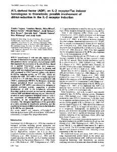
Figure 2. Developmental expression of ADF in the mouse inner ear. A: ADF presence in brain lysates of wildtype mice (WT) and absence in brain lysates of ADF-deficient mice (ADF⫺/⫺), demonstrating the specificity of the antibody. At E15, ADF was found in total cochlea protein lysates. Analysis of modiolus protein lysates revealed stronger ADF expression after hearing onset (P18, P26) compared to the younger stages (P0, P9). B: During postnatal development, ADF expression gradually decreased in the organ of Corti until P26.
Statistical analysis Statistical significance was calculated using Student’s t-test. Values P ⬍ 0.05 were referred to as statistically significant and marked by an asterisk (*P ⬍ 0.05). Data in Figure 7A–C are presented as mean ⫾ standard error of the mean.
RESULTS ADF expression in the inner ear during embryonic and postnatal development In a first step, the expression of ADF in the fetal and postnatal murine inner ear was investigated by immunoblot analysis, using total cochlear protein lysates of embryos at day 15 and protein lysates of the modiolus as well as the organ of Corti at various postnatal stages (P0 –P26). The specificity of the antibody was confirmed by using total brain lysates of wildtype (WT) and ADF-deficient (ADF⫺/⫺) mice (Fig. 2A, left lanes). In the cochlea, ADF was present throughout fetal and postnatal development. Whereas the expression became upregulated in the modiolus after hearing onset (Fig. 2A) which, in mice, occurs at about P10 –12 (Mikaelian et al., 1965; Ehret, 1976), it gradually decreased in the organ of Corti (Fig. 2B). We therefore decided to investigate the expression and location of ADF in the modiolus and various turns of the organ of Corti in more detail. To do so, immunohistochemistry was performed on both cochlear whole-mount preparations and cross-sections.
ADF expression in hair cells and supporting cells during organ of Corti development In whole-mount preparations of the organ of Corti at E15, weak ADF expression occurred in inner hair cells
(IHCs) of basomedial turns (Fig. 3A,B) and, to some extent, in medial turns (Fig. 3C). Furthermore, ADF was observed in epithelial structures strial to IHCs. No ADF expression was observed in apical turns of the cochlea (Fig. 3D), where cochlear development is delayed compared to the basal parts (Bruce et al., 1997). At P0, both IHCs and outer hair cells (OHCs) showed ADF expression with a gradual decrease in signal intensity from basomedial to apical turns (Fig. 3E–H). Within hair cells, ADF was absent from the base of stereociliary bundles (Fig. 3F). Around birth, ADF expression was not restricted to sensory hair cells. Hensen cells, which constitute the outer border of the organ of Corti, showed a substantial amount of ADF expression in all turns of the cochlea (Fig. 3E–H). Aside from Hensen cells, inner pillar cells (IPCs), inner phalangeal cells (IPhCs), and border cells also expressed ADF. No ADF was found in Deiters cells and outer pillar cells (OPCs). During early postnatal development (P3), peak expression of ADF in hair cells shifted from basomedial to medial turns of the cochlea (Fig. 3I–L). Within OHCs, the ADF signal was most prominent strial to stereocilia bundles (Fig. 3J). Again, ADF expression was seen in Hensen cells, IPCs, IPhCs, and border cells. At P3, Deiters cells showed no ADF expression, whereas individual OPCs expressed ADF. During subsequent development (P5), no ADF expression was detectable in hair cells of basomedial and medial turns and only weak expression was found in hair cells of the apex (Fig. 3M–P). Aside from Hensen cells, IPCs, IPhCs, and border cells, ADF expression became obvious in Deiters cells and OPCs. Taken together, our late embryonic/early postnatal whole-mount analysis revealed
The Journal of Comparative Neurology 円 Research in Systems Neuroscience
1729
Herde et al. ----------------------------------------------------------------------------------------------------------------------------------------------------------------------------------------
Figure 3. ADF expression as seen in whole-mount preparations. A–C: At E15, ADF (green) was only weakly expressed in IHCs (white arrow) and epithelial structures strial of IHCs in basomedial and medial turns. D: By contrast, ADF was undetectable in the apex. E–H: At P0, IHCs (white arrow) and OHCs (white filled arrowheads) expressed ADF with gradually decreasing levels from basomedial to apical turns. Notice absence of ADF from the base of stereocilia (white filled arrowhead in F, inset). Supporting cells such as border cells (yellow arrow), IPhCs (light blue arrow), IPCs (yellow open arrowhead), and Hensen cells (purple asterisks) were ADF-positive, whereas OPCs (light blue open arrowhead) and Deiters cells (yellow filled arrowhead) did not show ADF expression. I–L: At P3, ADF localization within OHCs was mainly strial of the stereocilia bundle (white filled arrowhead in J, inset). At this age the peak intensity of ADF expression in hair cells was observed in the medial turn (K). In addition to border cells, IPhCs, IPCs, and Hensen cells, individual OPCs (light blue open arrowhead in I⫹J) were ADF-positive. M–P: At P5, ADF was not detectable in hair cells of basomedial and medial turns, whereas weak expression was found in the apex. In addition to border cells, IPhCs, IPCs, OPCs, and Hensen cells, ADF became detectable in Deiters cells (yellow filled arrowhead in M). Red counterstaining in A–P: phalloidin-TRITC. A magentagreen copy of this figure is available as Supporting Figure 1. Scale bar ⫽ 20 m in P.
a precise spatially and developmentally regulated expression of ADF in the mouse organ of Corti. Cross-sections of the inner ear were used to investigate the ADF expression and location at various developmental stages before and after hearing onset in more detail (Fig. 4). At E15, only weak expression of ADF was seen in the developing organ of Corti (Fig. 4A–D). In addition, ADFpositive neuronal processes were found in basomedial
1730
(Fig. 4B) and medial (Fig. 4C), yet not in apical turns of the cochlea (Fig. 4D). Perinatally (P0), ADF expression was detectable in IHCs, OHCs, and Hensen cells (Fig. 4E–H) as well as in the greater epithelial ridge modiolar to the IHCs. As demonstrated in whole-mount preparations, hair cell expression of ADF decreased gradually from basomedial to apical cochlear turns. No expression was detectable in Deiters cells at P0.
The Journal of Comparative Neurology 円 Research in Systems Neuroscience
-------------------------------------------------------------------------------------------------------------------------------------------------- ADF development in the inner ear
Figure 4. ADF expression in organ of Corti cross-sections. A–D: ADF (green) was weakly expressed in the embryonic organ of Corti (brackets in A–C) at E15. ADF-positive processes were observed in basomedial and medial turns (white arrow in B). In the apical turn, no hair cells were labeled. Shown are projections of 10 m image stacks. E–H: At P0, ADF was expressed in IHCs (white arrow), OHCs (white filled arrowheads), Hensen cells (purple asterisk), and in the greater epithelial ridge modiolar to IHCs. The signal in hair cells declined gradually from basomedial to apical turns. No ADF was expressed in Deiters cells (yellow filled arrowheads). I–L: By P10, no ADF expression was detected in hair cells of basomedial and medial turns. In contrast, weak ADF expression persisted in apical hair cells. Supporting cells, such as IPCs (yellow open arrowhead), OPCs (light blue open arrowhead), Deiters cells, and Hensen cells, expressed ADF. Notice that phalangeal processes of Deiters cells were ADF-positive (orange filled arrowhead in J, inset). M–P: At P26, ADF expression persisted in supporting cells throughout all cochlear turns, whereas no signal could be detected in apical hair cells anymore. Double-staining of ADF (green) with the hair cell markers parvalbumin (red, Q) and calbindin (red, R) confirmed the absence of ADF from hair cells. S: The supporting cell marker Sox2 (red) was localized to nuclei (Hoechst stain, blue) of ADF-expressing supporting cells (colored open and filled arrowheads) in the organ of Corti. An ADF-positive phalangeal process of a Deiters cell is indicated by an orange filled arrowhead. Red counterstaining in A–P: propidium iodide. A magenta-green copy of Figure 4 is available as Supporting Figure 2. Scale bars ⫽ 20 m in P,R.
The Journal of Comparative Neurology 円 Research in Systems Neuroscience
1731
Herde et al. ----------------------------------------------------------------------------------------------------------------------------------------------------------------------------------------
Figure 5. ADF localization in OHC synapses and neuronal processes. A: ADF (green) was localized to terminals of NF-200-labeled (red) neuronal projections (white arrow) underneath OHCs (white asterisks) at P18. Additionally, ADF-positive puncta were observed along the projections (white open arrowheads). Shown is a projection of a 6.4 m image stack. B: Immunoreactivity of synaptophysin (green), a marker for presynaptic structures of MOC/OHC synapses, was localized at terminals of NF-200-labeled (red) neurites. Shown is a projection of a 4-m image stack. C,D: In the basomedial and medial turn, ADF (red) colocalized with synaptophysin (green, white filled arrowheads). A fraction of ADF was not colocalized with synaptophysin (white arrows). Shown are projections of 3.2-m (C) and 2.4-m (D) image stacks. E: In medioapical turns, no ADF-positive puncta were observed at MOC/OHC synapses. Shown is a projection of a 3.2-m image stack. F: SK2 (red), a Ca2⫹-activated K⫹ channel located within the basal OHC membrane (white asterisk), was found opposite of MOC presynapses (white arrow) identified by synaptophysin labeling (green). Shown is a projection of a 2-m image stack. A magenta-green copy of this figure is available as Supporting Figure 3. Scale bar ⫽ 5 m in F.
At P10, i.e., shortly before hearing onset, ADF expression in IHCs and OHCs was absent in basomedial and medial turns (Fig. 4I–K) and barely detectable in apical turns (Fig. 4L). As shown in whole-mount preparations, various types of supporting cells, such as Hensen cells, Deiters cells, IPCs, and OPCs, expressed ADF at substantial levels. In Deiters cells, ADF was found in somata and phalangeal processes (Fig. 4J). At P26, roughly 2 weeks after hearing onset, ADF expression remained dominant in supporting cells and was absent from hair cells (Fig. 4M–P). The absence of ADF from IHCs and OHCs was confirmed via colocalization experiments using the Ca2⫹-binding proteins parvalbumin (Fig. 4Q) and calbindin (Fig. 4R) as hair cell markers (Dechesne and Thomasset, 1988; Xiang et al., 1998). Concurrently, labeling for Sox2 as a marker for supporting cells
1732
(Hume et al., 2007) confirmed prominent ADF expression in Deiters cells, Hensen cells, and pillar cells (Fig. 4S).
Presynaptic location of ADF in OHC synapses Analysis at high-power magnification revealed a homogeneous distribution of ADF within Deiters cells and the occurrence of discrete ADF puncta in close proximity to the basal cell pole of OHCs at P18 (Fig. 5). These ADF puncta were found in terminals of neurofilament 200 (NF200)-labeled neurites innervating OHCs (Fig. 5A). Moreover, ADF puncta appeared along these neuronal processes (Fig. 5A). Consistent with the occurrence of synaptophysin in presynaptic structures of OHCinnervating MOC projections (Liberman et al., 1990), we found synaptophysin-immunoreactive signals at neurite
The Journal of Comparative Neurology 円 Research in Systems Neuroscience
-------------------------------------------------------------------------------------------------------------------------------------------------- ADF development in the inner ear
terminals (Fig. 5B). Double-labeling with synaptophysin demonstrated presynaptic location of ADF in basomedial and medial turns (Fig. 5C,D), but not in medioapical (Fig. 5E) and apical turns (not shown). However, a fraction of ADF did not colocalize with synaptophysin; instead it was found rather lateral of the synaptophysin signal (Fig. 5C,D). SK2 is a Ca2⫹-activated K⫹ channel that, in the organ of Corti, is located at postsynaptic structures of MOC/OHC synapses (Oliver et al., 2000). Indeed, we found SK2 opposing the synaptophysin-labeled presynapses (Fig. 5F). Taken together, we localized ADF in presynaptic structures of MOC/OHC synapses. Moreover, by comparing ADFand SK2-immunoreactive signals with synaptophysinlabeled structures, we could exclude ADF from postsynaptic structures of MOC/OHC synapses.
parameter, previously used to distinguish Type II from Type I SGNs, is their soma size: Type II SGNs are smaller (cf. Fig. 3 of Berglund and Ryugo, 1991). Analysis of the SGN soma area in cross-sections revealed a mean value of 89 ⫾ 10 m2 (n ⫽ 56 cells/3 mice) for ADFstrong neurons (Fig. 7C). These cells were significantly smaller than ADFweak neurons whose cell body size was 131 ⫾ 4 m2 (n ⫽ 1,063 cells/3 mice; P ⫽ 0.016). NF-200 was previously shown to label somata of Type II SGNs but not of Type I SGNs (Berglund and Ryugo, 1991; Hafidi, 1998). We therefore performed double-labeling experiments of ADF and NF-200 in the spiral ganglion and found strong ADF expression exclusively in somata of NF-200-positive SGNs (Fig. 7D–F), implying that ADF can be used to specifically identify Type II neurons in the spiral ganglion.
Developmental expression of ADF in spiral ganglion neurons
Developmental expression of ADF in the vestibular system
There is a broad neuronal expression of ADF in the central nervous system (Bellenchi et al., 2007). We therefore investigated the expression of ADF in peripheral auditory neurons by analyzing cochlear cross-sections at two stages: before (P0, P10) and after hearing onset (P18, P26). Before hearing onset, ADF expression in the spiral ganglion was only minor. At P0, no ADF expression was observed throughout any cochlear turn (Fig. 6A–D). At P10, weak ADF expression became detectable in a small subpopulation (6%) of SGNs predominantly in the basomedial part (Fig. 6E) and somehow weaker also in medial (Fig. 6G) and apical parts (Fig. 6F–H). ADF expression in SGNs increased further after hearing onset. At P18 and P26, a few neurons preferentially at the spiral ganglion border showed substantial ADF expression (Fig. 6I–P). In contrast, ADF expression remained rather weak in the majority of SGNs. This pointed to the presence of two populations of SGNs, a minority of SGNs with strong ADF expression (referred to as ADFstrong) more located in the spiral ganglion periphery, and a majority of SGNs with weak ADF expression (ADFweak). ADFweak SGNs were randomly distributed throughout the ganglion. A peripheral location and a relative amount of 5%–10% are hallmarks for Type II SGNs (Schwartz et al., 1983; Raphael and Altschuler, 2003). We therefore asked whether ADFstrong neurons indeed represent Type II SGNs and investigated the number and spatial distribution of both neuron populations in more detail. This analysis was performed at P26 and revealed that ADFstrong neurons amounted for 5.7% ⫾ 1.6% of all SGNs (Fig. 7A; n ⫽ 1,119 cells/3 mice) and were concentrated at the periphery of the spiral ganglion rather than in central aspects (n ⫽ 9 cells/2 mice, Fig. 7B). The remaining 94.3% ⫾ 1.6% neurons displayed a weak ADF expression and were randomly distributed throughout the spiral ganglion (n ⫽ 143 cells/2 mice; P ⫽ 0.02). A third
Besides the cochlea, the inner ear contains five additional sensory epithelia that consist of hair cells and supporting cells: the utricle and saccule which detect translational acceleration, and the sensory organs of the three semicircular canals (called cristae ampullares) which detect rotational acceleration. Given the parallels in structure, we asked whether ADF is also expressed in the different cell types of the vestibular epithelia. As performed for the cochlea, we assessed the expression of ADF in the utricle, the saccule, and the cristae ampullares at various developmental stages by analyzing both whole-mount preparations and cross-sections. Immunostaining of whole-mount preparations at E15 revealed ADF expression in hair cells of the utricle (Fig. 8A,B), the crista ampullaris (Fig. 8C,D), and the saccule (not shown). At this stage, ADF was not present in supporting cells in any of these structures (Fig. 8A–D). During early postnatal development (P0 –P5), ADF expression was detectable in hair cells, but not in supporting cells, by analysis of whole-mount preparations (Fig. 8E–L). Aside from a diffuse cytoplasmic signal, we found ADF labeling in the kinocilia of hair cells (Fig. 8F,H). Unlike stereocilia, kinocilia represent true cilia that mainly consist of microtubules (Kikuchi et al., 1989). As there is no actin in these structures, the localization of ADF in kinocilia was unexpected and surprising. Indeed, preliminary control experiments performed by us in ADF⫺/⫺ mice indicated that this ADF signal is an artifact, as kinocilia were stained in the knockouts too (not shown). Our result is in line with the labeling pattern obtained with other antibodies directed against proteins that are not thought to be present in kinocilia, for example, glial fibrillary acidic protein (GFAP; MK Herde and KP Steel, unpubl. obs.). The ADF expression pattern in vestibular sensory epithelia had changed by P10, when the immunofluorescent signals became weaker
The Journal of Comparative Neurology 円 Research in Systems Neuroscience
1733
Herde et al. ----------------------------------------------------------------------------------------------------------------------------------------------------------------------------------------
Figure 6. ADF expression in spiral ganglion neurons. A–D: At P0, no ADF (green) expression was found in the spiral ganglia. E–H: At P10, weak ADF expression became detectable in a subset of SGNs (white filled arrowheads in G), which was most prominent in basomedial parts. I–L: At P18, after hearing onset, strong ADF expression was found in a small fraction of SGNs throughout all parts of the spiral ganglion. The majority of SGNs expressed ADF at low levels. M–P: The expression pattern at P26 was comparable to that at P18, with a slight increase in signal intensity. Red counterstaining in A–P: propidium iodide. A magenta-green copy of the figure is available as Supporting Figure 4. Scale bar ⫽ 20 m in P.
in hair cells. At this stage, ADF was expressed in supporting cells of the utricle (Fig. 8M,N), the crista ampullaris (Fig. 8O,P), and the saccule (not shown). Immunohistochemistry on cross-sections through the vestibular sensory epithelia confirmed the hair cellrestricted ADF expression at E15 (Fig. 9A–D). Moreover, neurites of the vestibular nerve prominently expressed ADF (Fig. 9A). Unlike whole-mount preparations, the analysis of utricular and saccular cross-sections revealed a weak ADF expression in supporting cell somata at all in-
1734
vestigated postnatal stages (Fig. 9F,J,N). In the crista ampullaris, ADF expression in supporting cells became obvious at P10 (Fig. 9L,P). Beginning with P10, ADF expression decreased gradually in hair cells of the utricle (Fig. 9J,N) and the saccule (not shown). In the crista ampullaris, hair cell ADF expression was weak at E15 and P0 (Fig. 9D,H) and strongest at P10 (Fig. 9L). At P26, ADF intensity was reduced in the hair cell layer compared to P10 (Fig. 9L,P). In summary, we found a developmentally regulated ADF expression in the vestibular sensory epithelia. Whereas ADF
The Journal of Comparative Neurology 円 Research in Systems Neuroscience
-------------------------------------------------------------------------------------------------------------------------------------------------- ADF development in the inner ear
Figure 7. Strongly ADF-positive neurons in the spiral ganglion represent Type II SGNs. A: Strong ADF expression was found in 5.7% ⫾ 1.6% of all SGNs (1,119 cells/n ⫽ 3 animals). B: ADFstrong SGNs were positioned adjacent to the spiral ganglion border and showed a relative border distance of 21 ⫾ 5% (n ⫽ 9/2). ADFweak SGNs were distributed randomly with a relative border distance of 40% ⫾ 2% (n ⫽ 143/2; P ⫽ 0.02). Values of individual cells and averaged values are depicted. C: The soma size of ADFstrong SGNs (89 ⫾ 10 m2) was significantly smaller than that of ADFweak SGNs (131 ⫾ 4 m2; n ⫽ 56 for ADFstrong and 1,063 for ADFweak/3 animals; P ⫽ 0.016). D–F: Colocalization studies performed at P18 using NF-200 (red) as a marker for somata of Type II SGNs (arrows in D) confirmed strong ADF expression (green) in Type II SGNs, whereas NF-200-negative Type I SGNs showed weak ADF expression. A magenta-green copy of the Figure 7D-F is available as Supporting Figure 5. Scale bar ⫽ 10 m in E.
expression during embryonic development was restricted to hair cells, we found ADF in hair cells as well as supporting cells in the mature utricle, saccule, and cristae ampullares.
DISCUSSION In this study we found a broad and developmentally regulated expression of the actin depolymerizing factor ADF in hair cells and various nonsensory cell types of the mouse organ of Corti (summarized in Fig. 10A). During early postnatal development (P0 –P3), ADF is transiently expressed in IHCs and OHCs. Likewise, transient ADF expression occurs in hair cells of the utricle (summarized in Fig. 10B) and saccule during late embryonic and early postnatal development (E15–P10), whereas ADF expression remains in hair cells of the crista ampullaris until the adult stage. As depicted in Figure 1B, mature hair cells contain high levels of F-actin and process various actin-rich structures at their apical pole, such as stereocilia, the cuticular plate that anchors actin filaments of the stereocilia, and the circumferential belt that is attached to the cytoplasmic aspect of the zona adherens (Drenckhahn et al., 1991).
Within the first days after birth, cochlear hair cells undergo maturation, which, from the morphological perspective, involves thickening and elongation of stereocilia, organization of stereocilia into a “V”-shaped staircase pattern, as well as a reduction in the number of stereocilia rows (Kaltenbach et al., 1994; Waguespack et al., 2007). Consistent with an ADF function in regulating actin turnover, these structural rearrangements are temporally matched to the transient ADF expression in hair cells described in the present study. Interestingly, both hair cell maturation and ADF expression, show a spatiotemporal gradient along the cochlear duct, with hair cells in basal turns of the cochlea preceding those in the apex. Thus, our results implicate ADF in shaping the polarized actin-rich structures of hair cells. This idea is further supported by the planar cell polarity defect seen in various epithelial cell types of Drosophila following functional alterations of twinstar, the homologue of ADF/cofilin. In twinstar mutants, the actin-rich wing hairs show a “wildtype-like” appearance. However, the orientation and the location of the wing hairs is significantly altered, indicating that twinstar-dependent actin
The Journal of Comparative Neurology 円 Research in Systems Neuroscience
1735
Herde et al. ----------------------------------------------------------------------------------------------------------------------------------------------------------------------------------------
Figure 8. ADF expression in vestibular sensory epithelia as revealed in whole-mount preparations. A–D: At E15, ADF (green) was expressed in hair cells (white filled arrowheads) of the utricular macula and the crista ampullaris, whereas supporting cells (yellow filled arrowheads) were unlabeled. E–H: At P0, the expression pattern of ADF in the utricle was comparable to that at E15. In the crista ampullaris, the signal intensity of ADF was increased. Kinocilia (white arrows) were recognized by the ADF antibody in both structures. I–L: Besides hair cells, individual utricular supporting cells weakly expressed ADF at P5. Compared to P0, the expression intensity of ADF was reduced in hair cells of the crista ampullaris. M–P: At P10, supporting cells of the utricle prominently expressed ADF, whereas ADF expression in hair cells declined. Instead, no reduction in ADF expression was seen in hair cells of the crista ampullaris at P10. Compared to hair cells, ADF signal intensity was weaker in supporting cells of the crista ampullaris. Red counterstaining in A-P: phalloidin-TRITC. A magenta-green copy of this figure is available as Supporting Figure 6. Scale bar ⫽ 20 m in O.
remodeling plays a critical role in transducing positional information within epithelial cells (Blair et al., 2006). Although the stereociliary length is precisely regulated and remains constant throughout hair cell life, actin filaments of stereocilia are highly dynamic and continuously remodeled in cochlear and vestibular hair cells. Moreover, the turnover rate of actin is tightly matched to the length of
1736
stereocilia in a way that the dwell time of actin subunits within actin filaments is equal in stereocilia of different lengths (Schneider et al., 2002; Rzadzinska et al., 2004). This requires a precisely regulated actin polymerization and depolymerization controlled by actin-binding proteins. Nevertheless, ADF must be excluded as a regulator of actin length and dynamics in stereocilia, as, to our surprise, we
The Journal of Comparative Neurology 円 Research in Systems Neuroscience
-------------------------------------------------------------------------------------------------------------------------------------------------- ADF development in the inner ear
Figure 9. ADF expression in vestibular sensory epithelia as revealed in cross-sections. A–D: At E15, hair cells of the utricle and the crista ampullaris expressed ADF (white filled arrowheads), but there was no ADF signal in supporting cells (yellow filled arrowhead). Neuronal projections (white arrows) were prominently ADF-positive. Shown are projections of 12-m (A,B) and 6-m (C,D) image stacks. E–H: At P0, in addition to hair cells, weak ADF expression was found in supporting cells of the utricle, but not of the crista ampullaris. I–L: At P10, decreased ADF levels were found in the utricle, whereas signal intensity increased in the crista ampullaris. At this age, structures beneath the epithelia became labeled by the ADF antibody. M–P: At P26, the expression of ADF further decreased in the utricle. In the crista ampullaris, the ADF intensity in the hair cell layer was reduced compared to P10, whereas the apical surface and the supporting cell layer were strongly ADF-positive. Red counterstaining in A-P: propidium iodide. A magenta-green copy of this figure is available as Supporting Figure 7. Scale bars ⫽ 20 m in M (utricle) and O (crista ampullaris).
found ADF neither in the stereocilia of cochlear nor of vestibular hair cells at any developmental age. Furthermore, ADF is not expressed at detectable levels in mature cochlear hair cells. Thus, other proteins with actin depolymerizing activity, such as the broadly expressed n-cofilin (Gurniak et al., 2005), are potential candidates for regulating the actin filament length and dynamics in stereocilia.
Before hearing onset, but also in the fully developed organ of Corti, ADF is prominently expressed in various types of supporting cells, including Hensen cells, Deiters cells, and both types of pillar cells. Likewise, ADF is expressed in supporting cells of postnatal and mature vestibular sensory epithelia. Supporting cells are critically involved in K⫹-recycling, which is essential for proper
The Journal of Comparative Neurology 円 Research in Systems Neuroscience
1737
Herde et al. ----------------------------------------------------------------------------------------------------------------------------------------------------------------------------------------
Figure 10. Semischematic illustrations summarizing the differential expression of ADF during development of mouse inner ear sensory epithelia. Depicted are three developmental stages of the basomedial turn of the organ of Corti and the spiral ganglion (P0, P5, P26) (A) and of the utricle (E15, P0, P26) (B). White arrows at P26 in A point to the synaptic localization of ADF. Notice that gray values in A,B reflect the intensity of the ADF signal in individual cells and structures, with dark gray implying a high ADF expression.
mechano-electrical transduction (Boettger et al., 2002, 2003). Moreover, supporting cells display distinctive morphological features that are critical for the structural integrity of the organ of Corti and for its mechanical flexibility that allows the movement of the sensory epithelium in response to mechanical stimuli (Raphael and Altschuler, 2003; Kelly and Chen, 2007). Deiters cells and pillar cells possess actin-rich phalangeal (“finger-like”) protrusions that form tight contacts with hair cells at the surface of the organ of Corti (Raphael and Altschuler, 2003). The tight juxtaposition of hair cells and supporting cells is essential for sustaining the positive endocochlear potential, because it separates the endolymph (scala media) from the perilymph (scala tympani; Kelly and Chen, 2007). It was speculated that actin plays a role in maintaining a continuous epithelial barrier between both fluid compartments (Oh et al., 2002). Indeed, a close physical interaction of the actin cytoskeleton with cell adhesion proteins of epithelial tight junctions is well known and intensively described for many cell types (review: Miyoshi and Takai, 2008). We found ADF in Deiters cells and pillar cells at substantial levels before and after hearing onset and localized the protein to phalangeal processes of these cells. Thus, a contribution of ADF in organizing the structural backbone of phalangeal processes is likely, suggesting that ADF is involved in regulating the endolymph/perilymph barrier. Upon sound stimulation, Deiters cells and Hensen cells reversibly move toward the center of the cochlear turn in a
1738
contractile fashion (Flock et al., 1999). This movement coincides with a reduced sensitivity to acoustic stimulation. Therefore, it was speculated that Deiters cells and Hensen cells are actively involved in protecting the organ of Corti against trauma during high-intensity sound exposure. The phalangeal processes of Deiters cells undergo structural rearrangements, and flexion of these processes was implied in the movement of Deiters cells and the attached Hensen cells (Dulon et al., 1994; Flock et al., 1999). Both the reversible movement of supporting cells and the flexion of phalangeal processes require a highly dynamic cytoskeleton. As an actin depolymerizing factor, ADF severs actin filaments and enhances the dissociation rate at the filament minus end and therefore accelerates actin dynamics (Bamburg and Wiggan, 2002). The strong expression of ADF in Deiters cells and Hensen cells suggests that ADF is involved in regulating cochlear micromechanics during intense sound stimulation and, as a result, in protection from acoustic trauma. Besides the expression of ADF in sensory and nonsensory cells of the organ of Corti, we found developmentally regulated ADF expression in SGNs. The spiral ganglion is composed of two populations of neurons: 1) Type I SGNs innervated by IHCs, comprising the majority (90%–95%) of SGNs; 2) Type II SGNs forming a small population (5%– 10%) and receiving input from OHCs (Raphael and Altschuler, 2003). Type II SGNs are located at the periphery of the spiral ganglion (Schwartz et al., 1983) and are distin-
The Journal of Comparative Neurology 円 Research in Systems Neuroscience
-------------------------------------------------------------------------------------------------------------------------------------------------- ADF development in the inner ear
guishable from Type I SGNs by their smaller cell bodies (cf. fig. 3 in Berglund and Ryugo, 1991). Furthermore, specific markers such as neurofilament NF-200, which are expressed in somata of Type II, but not of Type I SGNs, are useful probes to distinguish both populations of SGNs (Berglund and Ryugo, 1991). In the mature cochlea we found a strong ADF expression in roughly 6% of all SGNs, whereas the majority of SGN neurons showed only weak ADF staining. Based on their relative number, but also on their smaller soma size and localization to more peripheral parts within the spiral ganglion, we identified ADFstrong neurons as Type II SGNs. This finding was confirmed by double-labeling experiments with ADF and NF-200. Whereas no NF-200 was detected in somata of ADFweak neurons, ADFstrong SGNs showed expression of the Type II SGN marker NF-200. In concert with the ADF expression in Type II SGNs, we found discrete ADF-positive puncta in close proximity to the OHCs basal pole, whereas no such signals were observed at IHCs. These puncta appeared after hearing onset and mostly colocalized to synaptophysin-positive presynaptic structures of MOC projections that innervate OHCs (Raphael and Altschuler, 2003). Deflections of stereocilia that cause transduction currents induce a dual response in OHCs. On the one hand, OHCs display a prestin-based rapid change in length and stiffness that provides a region-specific amplification in the movement of the basilar membrane relative to the tectorial membrane (Zheng et al., 2000; Oliver et al., 2001) and, consequently, an increased sensitivity and specificity in coding auditory information (Raphael and Altschuler, 2003). On the other hand, OHCs innervate Type II SGNs projecting to the cochlear nucleus whose neurons, in turn, project to the superior olivary complex, a pathway that modulates MOC efferents (Raphael and Altschuler, 2003). Inputs from MOC efferents hyperpolarize OHCs, which represents an effective mechanism of regulating OHC motility and their amplifier activity in response to strong acoustic stimulation (Maison and Liberman, 2000). We found substantial ADF expression in the somata of Type II SGNs and at presynaptic structures of MOC/OHC synapses, which were identified by synaptophysin immunoreactivity. This result implies a functional role of ADF in modulating the strength of the MOC feedback loop and, therefore, OHCs’ activity. While the specific function of ADF in the somata of Type II SGNs remains elusive, the localization in MOC/ OHC synapses suggests that ADF may be relevant for synaptic transmission. Interestingly, functional inactivation of the LIM kinase-1, a kinase that effectively inactivates ADF, results in impaired synaptic function of hippocampal CA1 synapses, including defects in neurotransmitter release (Meng et al., 2002). Therefore, it is tempting to speculate that ADF is relevant for synaptic transmission of MOC/ OHC synapses.
Taken together, we found ADF in various cell types of the mammalian inner ear sensory epithelia, including a transient expression in immature hair cells of the organ of Corti, the utricle, and the saccule. Moreover, we found ADF in supporting cells of the fully developed cochlea and vestibular sensory structures as well as in Type II SGNs. In addition, we localized ADF to MOC/OHC synapses after hearing onset. Based on the broad ADF expression described in the present study, we assume a relevance of ADF for inner ear development and in various aspects of auditory and vestibular function. Our study implicates the ADF gene as a candidate locus for human mutations resulting in cochlear or vestibular dysfunction. Future analysis of ADF knockout mice will help to clarify the physiological relevance of this actin-binding protein within the inner ear.
ACKNOWLEDGMENTS We thank K. Ociepka for excellent technical assistance and Thomas Schulenborg for analyzing the synaptophysin blocking peptide. We thank Dr. Marlies Knipper and Dr. Karen P. Steel for generously training MKH in generating inner ear cross-sections and whole-mounts. We also thank Dr. Christine B. Gurniak and Dr. Walter Witke for providing brain lysates and inner ear preparations of ADF⫺/⫺ mice.
LITERATURE CITED Airaksinen MS, Eilers J, Garaschuk O, Thoenen H, Konnerth A, Meyer M. 1997. Ataxia and altered dendritic calcium signaling in mice carrying a targeted null mutation of the calbindin D28k gene. Proc Natl Acad Sci U S A 94:1488 –1493. Bamburg JR, Wiggan OP. 2002. ADF/cofilin and actin dynamics in disease. Trends Cell Biol 12:598 – 605. Bellenchi GC, Gurniak CB, Perlas E, Middei S, Ammassari-Teule M, Witke W. 2007. N-cofilin is associated with neuronal migration disorders and cell cycle control in the cerebral cortex. Genes Dev 21:2347–2357. Berglund AM, Ryugo DK. 1991. Neurofilament antibodies and spiral ganglion neurons of the mammalian cochlea. J Comp Neurol 306:393– 408. Blair A, Tomlinson A, Pham H, Gunsalus KC, Goldberg ML, Laski FA. 2006. Twinstar, the Drosophila homolog of cofilin/ADF, is required for planar cell polarity patterning. Development 133: 1789 –1797. Boettger T, Hubner CA, Maier H, Rust MB, Beck FX, Jentsch TJ. 2002. Deafness and renal tubular acidosis in mice lacking the K-Cl co-transporter Kcc4. Nature 416:874 – 878. Boettger T, Rust MB, Maier H, Seidenbecher T, Schweizer M, Keating DJ, Faulhaber J, Ehmke H, Pfeffer C, Scheel O, Lemcke B, Horst J, Leuwer R, Pape HC, Volkl H, Hubner CA, Jentsch TJ. 2003. Loss of K-Cl co-transporter KCC3 causes deafness, neurodegeneration and reduced seizure threshold. EMBO J 22:5422–5434. Bruce LL, Kingsley J, Nichols DH, Fritzsch B. 1997. The development of vestibulocochlear efferents and cochlear afferents in mice. Int J Dev Neurosci 15:671– 692. Carlier MF, Laurent V, Santolini J, Melki R, Didry D, Xia GX, Hong Y, Chua NH, Pantaloni D. 1997. Actin depolymerizing factor (ADF/cofilin) enhances the rate of filament turnover: implication in actin-based motility. J Cell Biol 136:1307–1322.
The Journal of Comparative Neurology 円 Research in Systems Neuroscience
1739
Herde et al. ----------------------------------------------------------------------------------------------------------------------------------------------------------------------------------------
Celio MR, Baier W, Scharer L, Gregersen HJ, de Viragh PA, Norman AW. 1990. Monoclonal antibodies directed against the calcium binding protein calbindin D-28k. Cell Calcium 11: 599 – 602. Condeelis J. 2001. How is actin polymerization nucleated in vivo? Trends Cell Biol 11:288 –293. Dechesne CJ, Thomasset M. 1988. Calbindin (CaBP 28 kDa) appearance and distribution during development of the mouse inner ear. Brain Res 468:233–242. Drenckhahn D, Engel K, Hofer D, Merte C, Tilney L, Tilney M. 1991. Three different actin filament assemblies occur in every hair cell: each contains a specific actin crosslinking protein. J Cell Biol 112:641– 651. Dulon D, Blanchet C, Laffon E. 1994. Photo-released intracellular Ca2⫹ evokes reversible mechanical responses in supporting cells of the guinea-pig organ of Corti. Biochem Biophys Res Commun 201:1263–1269. Ehret G. 1976. Development of absolute auditory thresholds in the house mouse (Mus musculus). J Am Audiol Soc 1:179 – 184. Engel J, Braig C, Ruttiger L, Kuhn S, Zimmermann U, Blin N, Sausbier M, Kalbacher H, Munkner S, Rohbock K, Ruth P, Winter H, Knipper M. 2006. Two classes of outer hair cells along the tonotopic axis of the cochlea. Neuroscience 143:837– 849. Eybalin M, Renard N, Aure F, Safieddine S. 2002. Cysteine-string protein in inner hair cells of the organ of Corti: synaptic expression and upregulation at the onset of hearing. Eur J Neurosci 15:1409 –1420. Flock A, Flock B, Fridberger A, Scarfone E, Ulfendahl M. 1999. Supporting cells contribute to control of hearing sensitivity. J Neurosci 19:4498 – 4507. Gurniak CB, Perlas E, Witke W. 2005. The actin depolymerizing factor n-cofilin is essential for neural tube morphogenesis and neural crest cell migration. Dev Biol 278:231–241. Hafidi A. 1998. Peripherin-like immunoreactivity in type II spiral ganglion cell body and projections. Brain Res 805:181–190. Hawkins M, Pope B, Maciver SK, Weeds AG. 1993. Human actin depolymerizing factor mediates a pH-sensitive destruction of actin filaments. Biochemistry 32:9985–9993. Hayden SM, Miller PS, Brauweiler A, Bamburg JR. 1993. Analysis of the interactions of actin depolymerizing factor with G- and F-actin. Biochemistry 32:9994 –10004. Heizmann CW, Celio MR. 1987. Immunolocalization of parvalbumin. Methods Enzymol 139:552–570. Hume CR, Bratt DL, Oesterle EC. 2007. Expression of LHX3 and SOX2 during mouse inner ear development. Gene Expr Patterns 7:798 – 807. Ikeda S, Cunningham LA, Boggess D, Hawes N, Hobson CD, Sundberg JP, Naggert JK, Smith RS, Nishina PM. 2003. Aberrant actin cytoskeleton leads to accelerated proliferation of corneal epithelial cells in mice deficient for destrin (actin depolymerizing factor). Hum Mol Genet 12:1029 –1037. Kaltenbach JA, Falzarano PR, Simpson TH. 1994. Postnatal development of the hamster cochlea. II. Growth and differentiation of stereocilia bundles. J Comp Neurol 350:187–198. Kelly M, Chen P. 2007. Shaping the mammalian auditory sensory organ by the planar cell polarity pathway. Int J Dev Biol 51: 535–547. Kikuchi T, Takasaka T, Tonosaki A, Watanabe H. 1989. Fine structure of guinea pig vestibular kinocilium. Acta Otolaryngol 108: 26 –30. Leclere PG, Norman E, Groutsi F, Coffin R, Mayer U, Pizzey J, Tonge D. 2007. Impaired axonal regeneration by isolectin B4binding dorsal root ganglion neurons in vitro. J Neurosci 27: 1190 –1199. Leitch B, Shevtsova O, Kerr JR. 2009. Selective reduction in synaptic proteins involved in vesicle docking and signalling at
1740
synapses in the ataxic mutant mouse stargazer. J Comp Neurol 512:52–73. Leshchyns’ka I, Sytnyk V, Richter M, Andreyeva A, Puchkov D, Schachner M. 2006. The adhesion molecule CHL1 regulates uncoating of clathrin-coated synaptic vesicles. Neuron 52: 1011–1025. Lewis J, Davies A. 2002. Planar cell polarity in the inner ear: how do hair cells acquire their oriented structure? J Neurobiol 53: 190 –201. Li N, Timofeyev V, Tuteja D, Xu D, Lu L, Zhang Q, Zhang Z, Singapuri A, Albert TR, Rajagopal AV, Bond CT, Periasamy M, Adelman J, Chiamvimonvat N. 2009. Ablation of a Ca2⫹-activated K⫹ channel (SK2 channel) results in action potential prolongation in atrial myocytes and atrial fibrillation. J Physiol 587: 1087–1100. Liberman MC, Dodds LW, Pierce S. 1990. Afferent and efferent innervation of the cat cochlea: quantitative analysis with light and electron microscopy. J Comp Neurol 301:443– 460. Maas C, Tagnaouti N, Loebrich S, Behrend B, Lappe-Siefke C, Kneussel M. 2006. Neuronal cotransport of glycine receptor and the scaffold protein gephyrin. J Cell Biol 172:441– 451. Maison SF, Liberman MC. 2000. Predicting vulnerability to acoustic injury with a noninvasive assay of olivocochlear reflex strength. J Neurosci 20:4701– 4707. Manor U, Kachar B. 2008. Dynamic length regulation of sensory stereocilia. Semin Cell Dev Biol 19:502–510. Mansour H, Bignami A, Labkovsky B, Dahl D. 1989. Neurofilament phosphorylation in neuronal perikarya following axotomy: a study of rat spinal cord with ventral and dorsal root transection. J Comp Neurol 283:481– 485. McGough A, Pope B, Chiu W, Weeds A. 1997. Cofilin changes the twist of F-actin: implications for actin filament dynamics and cellular function. J Cell Biol 138:771–781. Meng Y, Zhang Y, Tregoubov V, Janus C, Cruz L, Jackson M, Lu WY, MacDonald JF, Wang JY, Falls DL, Jia Z. 2002. Abnormal spine morphology and enhanced LTP in LIMK-1 knockout mice. Neuron 35:121–133. Mikaelian D, Alford BR, Ruben RJ. 1965. Cochlear potentials and 8 nerve action potentials in normal and genetically deaf mice. Ann Otol Rhinol Laryngol 74:146 –157. Miyoshi J, Takai Y. 2008. Structural and functional associations of apical junctions with cytoskeleton. Biochim Biophys Acta 1778:670 – 691. Oesterle EC, Campbell S, Taylor RR, Forge A, Hume CR. 2008. Sox2 and JAGGED1 expression in normal and drug-damaged adult mouse inner ear. J Assoc Res Otolaryngol 9:65– 89. Oh SH, Adler HJ, Raphael Y, Lomax MI. 2002. WDR1 colocalizes with ADF and actin in the normal and noise-damaged chick cochlea. J Comp Neurol 448:399 – 409. Oliver D, Klocker N, Schuck J, Baukrowitz T, Ruppersberg JP, Fakler B. 2000. Gating of Ca2⫹-activated K⫹ channels controls fast inhibitory synaptic transmission at auditory outer hair cells. Neuron 26:595– 601. Oliver D, He DZ, Klocker N, Ludwig J, Schulte U, Waldegger S, Ruppersberg JP, Dallos P, Fakler B. 2001. Intracellular anions as the voltage sensor of prestin, the outer hair cell motor protein. Science 292:2340 –2343. Pack AK, Slepecky NB. 1995. Cytoskeletal and calcium-binding proteins in the mammalian organ of Corti: cell type-specific proteins displaying longitudinal and radial gradients. Hear Res 91:119 –135. Paysan J, Conroy WG, Coggan JS, Berg DK. 2000. The neurofilament infrastructure of a developing presynaptic calyx. J Comp Neurol 425:284 –294. Peichl L, Gonzalez-Soriano J. 1993. Unexpected presence of neurofilaments in axon-bearing horizontal cells of the mammalian retina. J Neurosci 13:4091– 4100.
The Journal of Comparative Neurology 円 Research in Systems Neuroscience
-------------------------------------------------------------------------------------------------------------------------------------------------- ADF development in the inner ear
Rabie A, Thomasset M, Legrand C. 1983. Immunocytochemical detection of calcium-binding protein in the cochlear and vestibular hair cells of the rat. Cell Tissue Res 232:691– 696. Raphael Y, Altschuler RA. 2003. Structure and innervation of the cochlea. Brain Res Bull 60:397– 422. Ruttiger L, Sausbier M, Zimmermann U, Winter H, Braig C, Engel J, Knirsch M, Arntz C, Langer P, Hirt B, Muller M, Kopschall I, Pfister M, Munkner S, Rohbock K, Pfaff I, Rusch A, Ruth P, Knipper M. 2004. Deletion of the Ca2⫹-activated potassium (BK) alpha-subunit but not the BKbeta1-subunit leads to progressive hearing loss. Proc Natl Acad Sci U S A 101:12922– 12927. Rymar VV, Sadikot AF. 2007. Laminar fate of cortical GABAergic interneurons is dependent on both birthdate and phenotype. J Comp Neurol 501:369 –380. Rzadzinska AK, Schneider ME, Davies C, Riordan GP, Kachar B. 2004. An actin molecular treadmill and myosins maintain stereocilia functional architecture and self-renewal. J Cell Biol 164:887– 897. Schneider ME, Belyantseva IA, Azevedo RB, Kachar B. 2002. Rapid renewal of auditory hair bundles. Nature 418:837– 838. Schwartz AM, Parakkal M, Gulley RL. 1983. Postnatal development of spiral ganglion cells in the rat. Am J Anat 167:33– 41. Sytnyk V, Leshchyns’ka I, Nikonenko AG, Schachner M. 2006. NCAM promotes assembly and activity-dependent remodel-
ing of the postsynaptic signaling complex. J Cell Biol 174: 1071–1085. Von Kriegstein K, Schmitz F, Link E, Sudhof TC. 1999. Distribution of synaptic vesicle proteins in the mammalian retina identifies obligatory and facultative components of ribbon synapses. Eur J Neurosci 11:1335–1348. Waguespack J, Salles FT, Kachar B, Ricci AJ. 2007. Stepwise morphological and functional maturation of mechanotransduction in rat outer hair cells. J Neurosci 27:13890 – 13902. Wassle H, Peichl L, Airaksinen MS, Meyer M. 1998. Calciumbinding proteins in the retina of a calbindin-null mutant mouse. Cell Tissue Res 292:211–218. Winter H, Braig C, Zimmermann U, Engel J, Rohbock K, Knipper M. 2007. Thyroid hormone receptor alpha1 is a critical regulator for the expression of ion channels during final differentiation of outer hair cells. Histochem Cell Biol 128:65–75. Xiang M, Gao WQ, Hasson T, Shin JJ. 1998. Requirement for Brn-3c in maturation and survival, but not in fate determination of inner ear hair cells. Development 125:3935–3946. Zallen JA. 2007. Planar polarity and tissue morphogenesis. Cell 129:1051–1063. Zheng J, Shen W, He DZ, Long KB, Madison LD, Dallos P. 2000. Prestin is the motor protein of cochlear outer hair cells. Nature 405:149 –155.
The Journal of Comparative Neurology 円 Research in Systems Neuroscience
1741