Differences in flux control and reserve capacity of cytochrome c oxidase (COX) in human skeletal muscle and brain suggest different metabolic effects of mild COX deficiencies Alexei Kudin*,**, Stefan Vielhaber**, Christian E. Elger*, and Wolfram S. Kunz* *Department of Epileptology, University of Bonn Medical Center and **Department of Neurology II, University of Magdeburg Medical Center, Germany Correspondence to: Dr. Wolfram S. Kunz, Department of Epileptology, University of Bonn Medical Center, Sigmund-Freud-Str. 25, D-53105 Bonn, Germany. Fax: (+49228) 2876294, Tel: (+49228) 2875744, E-mail:
[email protected] Abstract To evaluate tissue specific control of oxidative phosphorylation by cytochrome c oxidase (COX) we determined the flux control coefficient and the metabolic reserve capacity of this enzyme in human saponin-permeabilised muscle fibers and digitonin-treated parahippocampal homogenates. In these tissue preparations it is possible to investigate mitochondrial function under conditions which are close to the in vivo situation. In the presence of NAD-dependent substrates we observed, under active state conditions, a flux control coefficient of COX over oxidative phosphorylation of 0.24±0.07 and a 1.9±0.2-fold excess capacity in human skeletal muscle fibers. In human parahippocampal gyrus we determined, under similar conditions, a flux control coefficient of COX of 0.12±0.05 and a 3.9±0.6-fold excess capacity of the enzyme. The observed difference in metabolic control can be attributed to activity differences of COX in human brain and muscle mitochondria. Our results predict stronger metabolic effects of mild COX activity deficits in human skeletal muscle than in brain tissue. Introduction Mitochondrial disorders caused by mutations of the mitochondrial DNA represent a heterogeneous group of diseases showing a complex tissue distribution of the pathology (Wallace, 1992). This phenomenon is commonly attributed to the variable coexistence of mutant and wild type DNA in the individual cells called heteroplasmy. In addition to the puzzling tissue distribution of the mutation, the metabolic consequence of the respiratory chain defect remains unclear. Previous studies on cell lines (Villani and Attardi 1997; Villani et al. 1998) and muscle fibers (Kunz et al. 2000) suggested a tight control of COX and strong metabolic effects of complex IV deficiencies but it has remained unclear if it is appropriate to extrapolate these findings to all human tissue samples. To ascertain the metabolic consequences of mild respiratory chain deficiencies in human disease we have applied metabolic control analysis to tissue preparations from human skeletal muscle and brain reflecting rather closely the in vivo situation of mitochondria - saponinpermeabilized muscle fibers and digitonin-treated parahippocampal homogenates. Materials and Methods Human tissue samples Normal control muscle specimens from M. vastus lateralis were obtained from 18 patients who underwent a muscle biopsy for diagnosis of neuromuscular symptoms but were ultimately deemed to be normal by means of combined clinical, electrophysiological and histological criteria. Reference brain specimen from parahippocampal gyrus were obtained from 11 temporal lobe epilepsy patients with Ammon's horn sclerosis showing selective hippocampal atrophy undergoing epilepsy surgery.
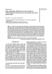
Preparation of muscle fibers and parahippocampal homogenates About 50 mg of biopsy tissue was used for isolation of saponin-permeabilized fibers. Bundles of muscle fibers containing usually 2-4 single fibers were isolated by mechanical dissection. The saponin treatment was performed by incubation of the fiber bundles in relaxing solution (composition see below) containing 50 µg/ml saponin as described in Kunz et al., 2000. For the preparation of human parahippocampal homogenates about 50 mg wet weight grey matter, obtained directly after epilepsy surgery, was homogenized two times 20 s at 8000 rpm using an ultra-turrax homogenizer T 25 (IKA, Staufen, Germany) in 0.5 ml ice cold medium containing 0.25 M sucrose, 0.5 mM EDTA and 50 mM Tris-HCl (pH = 7.4). The protein content was determined using a protein assay kit based on Peterson´s modification of the micro-Lowry method following the instructions of the manufacturer (Sigma, Deisenhofen, Germany). Respiration measurements The respiration of muscle fibers and parahippocampal homogenates was measured at 25°C using an Oroboros high-resolution oxygraph (Anton Paar, Graz, Austria) in the measurement medium containing 2 mM ADP and 10 mM glutamate + 5 mM malate as mitochondrial substrates as described previously (Kunz et al. 2000). The KCN titration of COX activity was performed in the same oxygraphic chamber in the presence of 500 µM TMPD, 1 mM ascorbate, 2 mM ADP and 0.2 µM antimycin A. Enzyme activities The activity of cytochrome c oxidase were measured spectrophotometrically as previously described (Kunz et al. 2000). The activity of citrate synthase was determined by a standard method. Determination of the flux control coefficients Using specific irreversible non-competitive inhibitors, the flux control coefficient Ci of an enzyme i can be determined experimentally according to the following equation: Ci = - (dJ/J) / (dpi/Imax) (1) where J is the flux through the pathway, pi is the concentration of a specific inhibitor of enzyme i and Imax, the maximal amount of inhibitor binding sites. The flux control coefficients of cytochrome c oxidase were determined from cyanide titration curves, respectively, using non-linear regression analysis accounting for the dissociation equilibrium of the inhibitor (Kunz et al. 2000). Determination of COX reserve capacity Inhibition plots were constructed of relative ADP-stimulated oxygen consumption with the different combinations of substrates versus percent decrease of COX activity at the same KCN concentrations, respectively. The resulting curves can be usually divided into two descending phases. The slope of the first part depends on the flux control coefficient of COX whereas the second descending portion represents the inhibition of the flux rate-limited by COX. The reserve capacities of COX (COXRmax) were determined from the intersection of the best fitting regression line through the data points of the second descending part of the inhibition plot with the ordinate axis (Villani and Attardi 1997). Results and discussion To determine the flux control coefficient of COX over oxidative phosphorylation in human saponin-permeabilized skeletal muscle fibers and digitonin-treated parahippocampal homogenates we performed titrations of the ADP-stimulated oxygen consumption rates with cyanide. The panels A and B of the figure show the averages of a series of KCN titrations of the oxygen consumption of digitonin-treated parahippocampal homogenates and saponinpermeabilized muscle fibers with the substrate combination glutamate+malate, respectively. Both titration curves are sigmoidal shaped and show considerable differences. With KCN about four-fold more inhibitor is required in brain homogenates to reach a comparable degree
of inhibition to that in muscle fibers. The averages of all experimental points were fitted to a theoretical model (Kunz et al. 2000) allowing calculation of the flux control coefficient of COX which was found to be equal to 0.12 for parahippocampal homogenates but approached almost 0.28 for muscle fibers. To determine the reserve capacity of COX (e.g. the under the experimental conditions available maximal enzyme activity) we titrated in separate experiments the TMPD+ascorbate respiration of muscle fibers and digitonin-treated parahippocampal homogenates with KCN using identical additions. This allows to measure the exact degree of COX inhibition at each used concentration of KCN and enabled us to construct the so called inhibition plots shown in Fig. 1, panels C and D (for details see Villani and Attardi 1997 and Rossignol et al. 1999). From the intercept of the regression lines of the descending part of the plot with the y-axis we determined the reserve capacities of COX in skeletal muscle fibers - COXRmax = 1.7 - and parahippocampal homogenates - COXRmax = 3.9.
Figure 1: Cyanide titrations of ADP-stimulated oxygen consumption of human parahippocampal homogenates (A) and saponin-permeabilized muscle fibers (B) and corresponding COX inhibition plots (C, D). The experimental conditions are given in Materials and Methods. Through the averages of the experimental points (± SD) in A and B theoretical curves (filled lines) were fitted (cf. Kunz et al. 2000). The inhibition plots were constructed from the relative ADP-stimulated oxygen consumption rates plotted versus the percent decrease of COX activity (TMPD+ascorbate respiration) at identical KCN concentrations.
To ascertain the reason for the differing in vivo reserve capacities of COX in human skeletal muscle and brain we determined the enzyme activities in our investigated tissue samples. In order to compare the individual activities we determined activity ratios in respect to citrate synthase (CS) - an established mitochondrial marker enzyme. The results of these determinations are summarized in the Table. In line with the approximately two-fold higher COX reserve capacity in human parahippocampal gyrus we observed about a 1.7-fold higher COX/CS ratio. Table: Enzyme pattern, flux control coefficient (Ci) and reserve capacity of COX (COXRmax) in human muscle fibers and parahippocampal gyrus specimen Muscle Citrate synthase COX COX/CS Ci (COX) COXRmax
11.8 ± 2.4 4.8 ± 0.9 0.42 ± 0.08 0.24 ± 0.07 1.9 ± 0.2
Parahippocampal gyrus 155 ± 39 88.8 ± 25.2 0.70 ± 0.19** 0.12 ± 0.05** 3.9 ± 0.6**
The activities are given as averages ± SD in U/g wwt (muscle, n=18) or U/g protein (parahippocampal gyrus, n=11). ** significant different with p < 0.01 (nonpaired two-tailed t-test) In the present report we have applied metabolic control analysis to oxidative phosphorylation in human saponin-permeabilised muscle fibers and digitonin-treated hippocampal homogenates to address the problem of tissue specificity of distribution of control in mitochondrial respiratory chain. Previous studies applying isolated mitochondria from different rat tissues including skeletal muscle and brain (Rossignol et al. 1999, 2000) showed tissue specific differences in reserve capacities and flux control of individual complexes of mitochondrial respiratory chain. In these reports differences in the amounts of inhibitors needed to titrate the activity suggested that differences in the enzyme content of isolated mitochondria might be the possible mechanism for tissue specific differences in control of oxidative phosphorylation. Here we show in permeabilized human tissue preparations that differences in the maximal activities of respiratory chain enzymes correlate to the metabolic reserve capacity and, therefore could explain the tissue dependency of control of oxidative phosphorylation. Our results predict stronger metabolic effects of mild COX activity deficits in human skeletal muscle than in brain tissue. References Kunz, W.S., Kudin, A., Vielhaber, S., Elger, C.E., Attardi, G. and Villani, G. (2000) J. Biol. Chem. 275, 27741-27745 Rossignol. R., Malgat, M., Mazat, J.-P. and Letellier, T. (1999) J. Biol. Chem. 274, 33426-33432 Rossignol, R., Letellier, T., Malgat, M., Rocher, C. and Mazat, J.-P. (2000) Biochem. J. 347, 45-53 Villani, G., and Attardi, G. (1997) Proc. Natl. Acad. Sci. USA 94, 1166-1171 Villani, G., Greco, M., Papa, S., and Attardi, G. (1998) J. Biol. Chem. 273, 31829-31836 Wallace, D.C. (1992) Ann. Rev. Biochem. 61, 1175-1212