Kathleen J. Doane and David E. Birk. Purpose. .... heterodimer,. LM609, was a gift from Dr. David Cheresh*" and was ..... Albelda SM, Buck CA. Integrins and ...
Differences in Integrin Expression During Avian Corneal Stromal Development Kathleen J. Doane and David E. Birk
Purpose. The purpose of this study was to determine whether there are changes in integrin expression associated with the spatial and temporal variations in matrix expression that occur during specific stages in corneal stromal development. Methods. Immunofluorescence techniques were used to analyze /?,-containing integrins and avj8-S localization both in situ and in cell cultures. Results. In situ, /?, and a\./3H were present with different patterns of localization, and these varied with developmental stage, /^-containing integrins were present on most cells, whereas ajfty was present on cells at the corneal-scleral epitheliomesenchyinal interface during migration of keratocyte precursors; very little asfi^ was localized in keratocytes. Keratocyt.es and undiffercntiated periocular mesenchyme cells grown in vitro also exhibited differences in localization of |3,-containing integrins and a\j8;(. All focal adhesions contained /?,, whereas a subset contained both /?, and «vj8H, indicating potential functional differences in focal adhesions. In addition, most periocular mesenchyme cells exhibited ag3rcontaining focal adhesions throughout, but the majority of keratocytes contained only peripherally located av/?.rpositive focal adhesions. The localization of both /3,-coniaining integrins and av/3:, was modulated by time allowed for attachment and spreading. Conclusions. Keratocytes and undifferentiated periocular mesenchyme cells exhibit developmental differences in integrin localization in situ. These two cell types also exhibit different patterns of av/3:, localization in vitro, possibly as a result of developmental differences in ligandbinding properties, j3,-containing integrins and aj3 ( define different types of focal adhesions, implying different functions. These differences in expression may be important in the initiation of cellular migration in (he early stages of corneal development, as well as in the transition from the undifferentiated to the differentiated keratocyte phenolype. Invest Ophthalmol Vis Sci. 1994:35:2834-2842.
V>iorneal development and the establishment of optical function require ihe interaction of avian keratocytes (corneal fibroblasts) and their immediate precursors with three different extracellular matrices (within the periocular mesenchyme, the primary siroma, and the secondary stroma), containing a variety of extracellular matrix molecules.1 During development, the neural crest-derived keratocyte precursors2"4 undergo phenotypic changes: from a stationary cell within the periocular mesenchyme to a migratory cell, which
From thi' Department of Anatomy nml Cellular Biology. Tufts University School of Medicine, Huston. Massachusetts. Supported b\ grant* from the National Institutes of Health (F.Y05I29) and Fight for Sight (GA9207-1). Submitted fin publication September 15, 199.3; revised November 23. 1993; accepted December 10. 1993. Proprietary interest category: /V. Reprint requests: K~7 Each subunit of the heterodimer has a cytoplasmic domain, a transmembrane portion, and an amino-terminal extracellular domain. The extracellular domain can bind to recep-
l m o i i j ; ; i i i w (.)|)liili.ilmulo K y K: Vi.Mial .Science. May I!)!M, Vol. :\:i. N C o | ) \ t i g h t © Association for Rr.scaich in Vision a n d O p h t h a l m o l o g y
Integrins and Corneal Development tors on other cells (cell-cell adhesion) or extracellular matrix proteins (cell-matrix adhesion). The interaction of cells with matrix via integrins is important in many functions, including migration, spreading, and differentiation.8"14 The elucidation of the interactions of keratocytes and their precursors with the surrounding extracellular matrix is important to the understanding of corneal development, growth, and repair, as well as normal corneal function. We have examined the expression of /3,-containing integrins and avj8a. Many of the matrix-binding heterodimers identified to date belong to the j8, subclass, including all known collagen receptors (a.,/8,, a^u ai/?i). Many heterodimers that bind glycoproteins such as fibronectin and laminin, contain |8, as well. Thus, /3, may associate with several different a subunits, leading to variations in ligand specificity. The vitronectin receptor (av(3-A) binds a variety of matrix molecules, and (3$ is associated only with «v except in platelets.'1"7 We examined expression of this receptor for three reasons: This heterodimer binds fibronectin, an important molecule during embryogenesis, which is present in early stages of corneal development;15 it has been shown to bind denatured type I collagen;1" type VI collagen has several sequences homologous to von Willebrand type A repeats, 1 ' indicating av/3y could bind lo this collagen, a major component of the secondary corneal stroma. In this study, j8, and /3:< (as avj8;0 integrin expression on cells at two developmental stages was studied both in vivo and in vitro. The following questions were addressed: Does the developmental stage of the cell and differences in the matrix the cell encounters result in variations in matrix receptor localization in situ? Do keratocytes and their precursors exhibit localization patterns for /3, and av/3:, in vitro similar to those in situ? These analyses indicate that integrin localization in situ is a function of developmental stage and that developmental differences in localization of these integrin subclasses are maintained in vitro, implying that these cultures will be useful as a model system in future studies. Also, these data indicate that /3|-containing integrins and etv(3:s have different functions from each other and ai different stages of development.
METHODS Cell Culture These investigations conformed to the ARVO Statement for the Use of Animals in Ophthalmic and Vision Research. Embryonic chicken corneas from stage 26 or 2 mm central corneas from stage 40 of chicken development were treated with Versene (Gibco, Grand Island, NY) with 0.25% trypsin to remove the endothelium and epithelium, and the cells were iso-
2835 lated using 200 U/nil collagenase (based on the method of Doane and Birk).IH The primary cultures were maintained initially in complete minimal essential medium (CMEM, Gibco, Grand Island, NY) containing 20% fetal bovine serum, with 50 [ig/m\ gentamicin and 2.5 Axg/ml fungizone. Subsequent medium changes used CMEM with 10% fetal bovine serum with 50 jug/ml ascorbate and antibiotics. Primary cultures were trypsinized using 0.25% trypsin in PBS and plated onto collagen-coated (Vitrogen, bovine type I collagen, Collagen Corp., Palo Alto, CA) slides. The assay involved allowing cells to attach and spread for various periods of time. Initial attachment took place overnight in CMEM with 20% serum and antibiotics, with subsequent medium changes into CMEM with 10% serum, antibiotics and 50 Mg/ m ' ascorbate. Cells were allowed to spread for 1, 3, and 7 days and were then fixed and used in immunolluorescence procedures to analyze integrin localization.
Antibodies Three different antibodies against the /?, subunit of integrin were used. One monoclonal antibody, VyEr,,1'1 was directed against the avian form of the subunit. This antibody was culture supernatant used at 10 to 25 jug/ml and was obtained from the Developmental Studies Hybridoma Bank (Department of Pharmacology and Molecular Sciences, Johns Hopkins University School of Medicine, Baltimore, MD and the Department of Biological Sciences, University of Iowa, Iowa City, IA, under contract N01-HD-6-2915 from the National Institutes of Child and Human Development). One polyclonal antibody, AB1938, was purchased from Chemicon (Temecula, CA). This rabbit antisera was directed against a synthetic peptide from the COOH terminal cytoplasmic domain of the human j8i subunit but cross-reacted with the chicken subunit. The other rabbit polycional antibody was directed against the avian /3, subunit (Chickie II, Dr. Clayton Buck, Wistar, Philadelphia, PA). Both antisera were used at a dilution of 1:100. Immunoffuorescence patterns obtained using all antibodies were identical. The antibody against the human o\.j8;H heterodimer, LM609, was a gift from Dr. David Cheresh*" and was ascites fluid used at a dilution of 1:100. This antibodyhas been well characterized by numerous investigators, and we as well as others have demonstrated that this antibody cross-reacts with the avian av/3:, heterodimer. A monoclonal antibody against vinculin, VN32, also was obtained from the Developmental Studies Hybridoma Bank, and was culture supernatant used at 10 to 25 /ig/ml. Secondary antibodies were labeled either with rhodamine, Texas red, Huorescein, or a tluorescein derivative, DTAF (Jackson Laboratories, West Grove, PA). For single-label experiments, the secondary antibody was either goat anti-rabbit igG (H +
2836
Investigative Ophthalmology & Visual Science, May 1994, Vol. 35, No. 6
L) or goat anti-mouse IgG (H + L). For double-label experiments, both these antibodies and species-specific antibodies (donkey anti-rabbit IgG; donkey antimouse IgG; goat anti-rabbit IgG; goat anti-mouse IgG; all H + L) were used. Species-specific antibodies and nonspecies-specific antibodies yielded similar results. Control experiments included normal serum from mouse and rabbit, both used at a concentration of 1:100, and an irrelevant antibody against type X collagen (found only in hypertrophic cartilage), which was used at 25 /xg/ml. Immunofluorescence Procedure For analysis of stages 26 and 28 avian corneas, whole eyes were fixed in 4% paraformaldehyde in 0.15 M phosphate buffer with 4.5% sucrose and 3% NaCl, pH 7.3. Tissue was treated with 7% sucrose in phosphatebuffered saline, pH 7.3, frozen in OCT (Tissue Tek, Miles Laboratories, Naperville, IL) and sectioned either en face (parallel to the ocular surface) or in crosssection (perpendicular to the ocular surface). To ensure that periocular mesenchyme and not epithelium was used for the en face sections, sections were taken only from the level of the lens; tissue immediately surrounding the lens that did not contain pigment granules (indicative of the pigment epithelium) was designated as periocular mesenchyme. For stage 40 avian corneas, the cornea and surrounding scleral tissue was either fixed directly, or the central cornea was obtained using a 2 mm dermal punch. Tissue was then fixed and cut as described above. Stage 43 corneas were unfixed, cryoprotected in 7% sucrose, frozen in OCT, and sectioned as above. For analysis of cells isolated and grown in vitro then allowed to attach and spread on substrate-coated slides, cells were fixed at 1, 3, and 7 days in methanol and then acetone, both at —20°C, and air dried. Nonspecific binding sites were blocked by incubation in 2% normal goat serum. Cells or cryostat sections were then incubated in primary antibody or control serum followed by a (luorescently labeled secondary antibody. Results were photographed and printed at set time exposures to facilitate comparisons between experimental and control samples, except as discussed in the figure legends. For double-label experiments, both primary antibodies were allowed to bind cells at the same time, as were both secondary antibodies. Controls were either normal rabbit serum, normal mouse serum, or an irrelevant antibody (anti-type X collagen) as previously discussed; for double-label experiments, both normal sera were used together. Slides were mounted using FITC-Guard (Testog, Inc., Chicago, IL). RESULTS In situ, /?,-containing integrins and av/?3 exhibited developmental differences in their localization pat-
terns. To determine whether developmental stage affected the distribution of fi} and av@s in situ, immunofluorescence experiments were performed using four developmental stages. Different integrin classes were compared at stage 26 (day 5), when the keratocyte precursors are stationary within the periocular mesenchyme; at stage 28 (day 5.5), when these neural crest, derivatives have initiated migration, entered the primary stroma, and begun differentiating; and at stages 40 (day 14) and 43 (day 1 7), when the keratocytes are fully differentiated and secondary stroma is present. /?] and a\./3:J exhibited differences in both tissue and cellular distribution when these developmental stages were compared. fii was present throughout both undijferentiated periocular mesenchyme and. fully differentiated corneal stroma, whereas cevfij was localized primarily on the migratoiy ker-
atocyte precursor cells. To characterize the tissue localization of j8, and avj8:J in different regions of the cornea and periocular mesenchyme, sections of stage 28 and stage 43 avian eyes were cut perpendicular to the ocular surface (Fig. 1). With the tissue oriented in this manner, the location of immunopositive cells within the cornea and periocular mesenchyme could be determined. A later stage of corneal development (stage 28) was chosen to characterize the keratocyte precursor population, which is identifiable only by its migration into the cornea. At this stage, /3, was present on cells throughout the cornea and periocular mesenchyme. However, a\j8;! was present on cells along the epitheliomescnchyinal junction subjacent to the limbal and corneal epithelium. Intense fluorescence also was observed within the epithelium of the cornea, periocular mesenchyme, and lens, as well as the corneal cndothelium and pigment epithelium. At stage 43, cells throughout the corneal stroma and corneal epithelium express /?,, whereas little fluorescence for av/3:< was discerned. This may be due to lack of this integrin or to levels below the limit of detection. This would be consistent with the low levels of «,./?., localized in en face sections (see below). /?/ xuas present in a punctate pattern in adhesive structures on cells at stage 26 and stage 40 of development, whereas av^3 was localized in a peripheral, occasionally punctate pattern only on cells within the undifferentiated
periocular mesenchyme. To visualize better the cellular distribution of adhesive structures, particularly in keratocytes, which are spread in a plane parallel to the surface of the cornea, en face sections were cut parallel to the ocular surface; stage 26 and stage 40 were compared in these experiments. To ensure that tissue was periocular mesenchyme and not epithelial, sections were taken at the level of the lens; only areas with no pigment granules were designated as periocular mesenchyme. The localization of /3, and av^s varied with developmental stage (Fig. 2). At both stages, 0, was present on most cells in a punctate array around
Integrins and Corneal Development
2837
dimer was present at very low levels, scarcely above background, subsequent to corneal stromal differentiation. In vitro, /?x-containing integrins and ajl3 exhibited developmental differences in localization patterns that can be correlated with the in situ distribution. To determine whether cells isolated from different developmental stages maintained variations in integrin expression in vitro, the localization of integrins was analyzed. Cells were isolated from stage 26 periocular mesenchyme (before the initiation of migration of keratocyte precursors) and from stage 40 (when the keratocytes are fully differentiated) and grown in vitro. These cells exhibited differences in localization of /?] and avj3s. Although all focal adhesions, which were defined by vinculin staining, contained j8]} only a subpopulation of cells contained av/?3. In addition, av@3 was present in a subset of focal adhesions, and the distribution varied as a result of developmental stage and cellular phenotype. The length of time
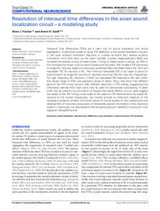
FIGURE 1. In situ, 0i and aj? s exhibited developmental differences in tissue localization patterns. Immunofluorescence experiments comparing the localization of fi\ (a, c, e) and cvv/?3 (b, d, f) in sections of anterior eyes cut perpendicular to the ocular surface revealed significant differences in localization, as well as developmental variations. At stage 28 of development (a), /?, was localized to cells within the periocular inesenchyme (PM), to partially differentiated keratocyte precursor cells within the cornea proper (C), and in the area of the endothelium, as well as within the epithelium. At a higher magnification, individual fibroblasts within the corneal stroma can be seen to label with this antibody (c, arrows). In contrast, avj93 was localized to cells within a narrow area subjacent to the epithelium within the periocular mesenchyme and cornea (b, arrows). At a higher magnification, individual cells within the corneal stroma appear to exhibit staining for this antibody, whereas cells migrating along the epithelial-mesenchymal interface are more intensely stained (d, arrows). The epithelium of the cornea, periocular mesenchyme, and lens, as well as the corneal endothelium and pigment epithelium, also stained intensely for j8,. By stage 43, /3, was present on cells within the corneal stroma and the epithelium (e), whereas little immunopositive staining for av/?3 could be discerned (f). C, cornea; L, lens; PM, periocular mesenchyme; PE, pigmented epithelium; Ep, epithelium; En, endothelium. (a, c, e) Using the polyclonal antibody Chiclde II against j8,. (b, d, f) Using the monoclonal antibody LM609 against av/?3. (a-d) Stage 28. (e, f) Stage 43. (a, b) Bar = 200 nm. (c-f) Bar = 100 urn. the cellular periphery. av/33 was present on cells within the periocular mesenchyme at stage 26 of development, both around the periphery of the cells and occasionally in a punctate fashion. However, this hetero-
FIGURE 2. In situ, 0i-containing integrins and aj3 s exhibited developmental differences in cellular localization patterns. Stage 26 and stage 40 anterior eyes were cut parallel to the ocular surface, and immunofluorescence was used to identify cells exhibiting (3, (a, b) and orvj8s (c, d). Undifferentiated cells within the periocular mesenchyme before keratocyte precursor migration exhibited immunopositive fluorescence for both /3, (a) and av^.s (c). $, was present on many cells and could be found in both a punctate, peripheral pattern of fluorescence (a, arrow) and diffusely across the cell. avi8H was present diffusely around cellular peripheries and occasionally in a bright, punctate pattern (c, see cell at large arrow). In keratocytes at stage 40, /3, was localized around the cell perimeter in a punctate pattern {b, arrow), whereas immunopositive staining for av/3» was barely above background (d). Controls (normal mouse serum, normal rabbit serum) showed little nonspecific staining (results not shown), (a, b) Monoclonal antibody V2E9 against /?,. (c, d) Monoclonal antibody LM609 against av/J3. (a, c) Stage 26. (b, d) Stage 40. Bar = 20 ^m.
2838
Investigative Ophthalmology 8c Visual Science, May 1994, Vol. 35, No. 6
cells were allowed to attach and spread on substratecoated slides also affected integrin expression. Focal adhesions, as identified by vinculin-positive immunofluorescence, all contained j3r To determine whether all focal adhesions contained j3u double-label immlinolocalization experiments were performed to identify simultaneously both /3j and a cytoplasmic component of focal adhesions, vinculin (Fig. 3). In both undifferentiated and differentiated cell types, the antibody to |£?j colocalized with all vinculin-positive focal adhesions throughout the cell. This correlates well with the in situ data, which indicates that most cells within both the fully differentiated corneal stroma and undifferentiated periocular mesenchyme expressed this subunit. (3j and avj33 localized to different sets of focal adhesions
that differed with cell type. To determine whether periocFIGURE 4. /?, and aj}5 localized to different sets of focal ular mesenchyme cells and keratocytes exhibited difadhesions that differed with cell type. /?, was localized to all ferent localization patterns for /?j and av/33, the distrifocal adhesions (see Fig. 3). These were present in both cenbution of these proteins was analyzed using doubletral and peripheral areas of the cell using the polyclonal antilabel, co-localization experiments after 3 days of body AB1938 in periocular mesenchyme cells (a) and keraspreading on substrate-coated slides (Fig. 4). In contocytes (b) 3 days after seeding. The distribution of /?, was compared with that of avj83 using the monoclonal antibody LM609 in double-label immunofluorescence. Although /S r containing focal adhesions were present throughout the cytoplasm in both cell types, o-jSy was present in a subset of focal adhesions in both periocular mesenchyme cells (c) and keratocytes (d). Some focal adhesions contained both /?, and av(3$, whereas others expressed only /?, (arrows indicate focal adhesions that contain only /3,-positive focal adhesions). In addition, the localization pattern in these two cell types was different: in keraLocytes, a^ was present primarily in a peripheral distribution, whereas on the undifferentiated cells, this integrin was present on peripheral and central focal adhesions. Not all cells expressed avj93; cells that do not contain «v/3;, are indicated by an asterisk. Photographic negatives for /?, immunofluorescence are underexposed compared to those depicting av|SH positive fluorescence, (a, b) Polyclonal antibody AB1938 against jt?1. (c, d) Monoclonal antibody LM609 against aj3 3 . (a, c) Periocular mesenchyme cells, (b, d) Keratocytes. Bar = 20 ^m. FIGURE 3. Focal adhesions all contained /S^ A monoclonal antibody to vinculin, a cytoplasmic component of focal adtrast to the co-localization of/3] with all focal adhesions hesions, was used to identify focal adhesions. Focal adhethroughout the cell, av/33 was localized to a subset of sions were seen in both peripheral (arrows) and central (arfocal adhesions. The pattern of localization of this rowheads) areas of the cell in periocular mesenchyme cells subset of av|53-containing focal adhesions was different (a) and keratocytes (b) at 3 days after seeding. Double-label immunofluorescence experiments with the polyclonal antiin periocular mesenchyme cells as compared to kerabody AB1938 against /?, in addition to anti-vinculin detertocytes. In the undifferentiated periocular mesenmined that in both periocular mesenchyme cells (c) and kerchyme cells, crv/?3 was present in focal adhesions along atocytes (d), all focal adhesions contained this subunit. the periphery of the cell, but it was also present on Arrows indicate examples of peripheral focal adhesions comany focal adhesions throughout the cell. Only a sublabeled with both antibodies; arrowheads indicate examples set of both cell types expressed av/?3. This cell populaof central focal adhesions labeled with both antibodies. Photion was examined and estimated to be between 10% tographic negatives for the /3,-positive reaction were printed and 25% of the total cell population; a higher percentoptimally; negatives depicting vinculin staining are underexage of periocular mesenchyme cells than keratocytes posed for best demonstration of focal adhesions, (a, b) expressed this heterodimer (results not shown). The Monoclonal antibody against vinculin. (c, d) Polyclonal antipattern of localization of av/?3 was different, however, body AB1938 against /?,. (a, c) Periocular mesenchyme cells. (b, d) Keratocytes. Bar = 20 nm. in keratocytes. In most cells, only peripheral focal ad-
Integrins and Corneal Development
2839
hesions contained this heterodimer, although occasional cells could be found that exhibited av/33 in both central and peripheral focal adhesions. These results can be correlated with the data from in situ immunolocalization. In the periocular mesenchyme, only a subpopulation of cells exhibited positive immunofluorescence for av/33, and there was a further subpopulation of cells that are brightly fluorescent when labeled with this antibody. In contrast, the fully differentiated corneal stroma had cells that exhibited little, if any, positive immunofluorescence using these techniques. It is possible that culturing the cells has caused expression of this heterodimer. It is also possible that the av/33 heterodimer was present at levels below the limit of detection. This variation in av/?3 localization when keratocytes and their precursors are compared implies a developmental difference in avP5 function, which is maintained in vitro. /3; expression and distribution varied with the length of time cells were allowed to attach and spread subsequent to
trypsinization. The localization of /^ 1, 3, and 7 days after seeding revealed differences between periocular mesenchyme cells and keratocytes (Fig. 5). At all time points, both cell types exhibited focal adhesions. Two differences in immunolocalization were observed between the two cell types. After being allowed to attach and spread for 1 day, periocular mesenchyme cells contained perinuclear punctate structures that appeared to be vesicles, whereas keratocytes rarely contained these structures. In contrast, 7 days after seeding several keratocytes exhibited a similar staining pattern, and few periocular mesenchyme cells contained these punctate structures. These vesicles were intracellular and were possibly indicative of /?, synthesis. These data indicate that cellular phenotype and the amount of time cells are allowed to attach and spread may modulate the expression of /^-containing integrins. av(33 expression and distribution varied with the length
of time after seeding. The localization of av/?3 1, 3, and 7 days after seeding affected expression and distribution of this subunit (Fig. 6). At 1 day after seeding, periocular mesenchyme cells had av(3a in focal adhesions and often in a diffuse, perinuclear distribution. This perinuclear fluorescence did not appear to be associated with the cell surface. Keratocytes occasionally displayed this perinuclear staining, although most positive cells exhibited small numbers of peripheral focal adhesions. After being allowed to attach and spread for 3 days, the difference in localization pattern between the two cell types became apparent; whereas periocular mesenchyme cells had focal adhesions throughout their cytoplasm that contained av@3, keratocytes had predominantly peripheral focal adhesions, which were positive for this heterodimer. After 7 days in vitro, the difference in the immunostaining pattern for av/?a is basically similar when keratocytes and peri-
FIGURE 5. #, expression and distribution varies with length of time cells are allowed to attach and spread on collagen-coated slides, /J, was localized to focal adhesions using immunofluorescence in both periocuiar mesenchyme cells (a, c, e) and keratocytes (b, d, f) from primary cultures passaged and grown for 1, 3, and 7 days on collagen-coated slides using the monoclonal antibody V2Et,. An increase in staining that appeared intracellular and vesicular in nature was present in periocular mesenchyme cells after 1 day in culture (a, arrow) and in keratocytes after 7 days (f, arrow). (a, b) 1 day after seeding, (c, d) 3 days after seeding, (e, f) 7 days after seeding, (a, c, e) Periocular mesenchyme, (b, d, f) Keratocytes. Bar = 20 pm.
ocular mesenchyme cells were compared. Few keratocytes could be found that were positive for av0.it and the level of fluorescence was apparently decreased. DISCUSSION In the present study, we have shown that keratocytes and undifferentiated cells from the periocuiar mesenchyme at earlier developmental stages exhibit differences in the localization of /^-containing integrins and «vj03. These differences are present at both the tissue and the cellular level, in vivo and in vitro, and were correlated with variations in developmental stage and cellular phenotype. This implies that integrin expression varies with the spatial and temporal changes in matrix that occur during corneal development. Several studies have indicated that changes in integrin expression are associated with migration of cells on matrix substrates and with differentiation.8"14
2840
Investigative Ophthalmology & Visual Science, May 1994, Vol. 35, No. 6
tenascin), and various proteoglycans (including decorin and the corneal-specific proteoglycan lumican) (unpublished observations).15'21"24 Virtually all the matrix components present at various times throughout development undergo spatial and temporal variations in localization. Antisense oligonucleotides have demonstrated that /?! integrins are important in neural crest migration on fibronectin and laminin.25 Although the current study does not identify the neural crestderived keratocyte precursors, it implies that these cells use av/33 during their migration into the cornea. Further study is necessary to determine what matrix protein(s) the av/53 heterodimer is interacting with at this time. The ubiquitous presence of j8] indicates that /?!-containing integrins could be functioning at this time as well. Because j8, associates with multiple a subunits, these and the relevant matrix component(s) must still be identified. As the keratocyte precursor population differentiates into a stationary cell population, little av/53 is present, indicating that j8,-containing integrins are more important in the interaction of these cells with the secondary stroma. Although the associated a subFIGURE 6.