Virchows Arch (2009) 455:133–142 DOI 10.1007/s00428-009-0807-x
ORIGINAL ARTICLE
Differences in the expression of histamine-related genes and proteins in normal human adrenal cortex and adrenocortical tumors Peter M. Szabó & Zoltán Wiener & Zsófia Tömböl & Attila Kovács & Péter Pócza & János Horányi & Janina Kulka & Peter Riesz & Miklós Tóth & Attila Patócs & Rolf C. Gaillard & András Falus & Károly Rácz & Peter Igaz
Received: 6 April 2009 / Revised: 28 May 2009 / Accepted: 18 June 2009 / Published online: 1 July 2009 # Springer-Verlag 2009
Abstract Histamine is involved in the pathogenesis of several tumors; however, there are no data on its possible involvement in human adrenocortical tumorigenesis. The expression of genes and proteins involved in the biosynthesis (histidine decarboxylase, HDC), action (histamine receptors: HRH1–HRH4), and metabolism of histamine is largely unknown both in the normal human adrenal cortex and in adrenocortical tumors. In this study, we examined the expression of histamine-related genes and proteins and histamine content in normal adrenal cortex, benign adrenocortical adenomas, and malignant adrenocortical cancer (ACC). Fifteen normal adrenals and 43 tumors were
studied. mRNA expression was examined by real time RT-PCR. Western-blotting and immunohistochemistry were used for the study of proteins. Tissue histamine content was determined by enzyme-linked immunosorbent assay. We found that all proteins involved in histamine biosynthesis and action are present both in the normal adrenal cortex and in the tumors studied. HDC expression and histamine content was highest in the normal tissues and lower in benign tumors, whereas it was significantly less in ACCs. HRH3 expression was significantly higher in ACC samples than in the other groups. Adrenocortical tumorigenesis might, thus, be characterized by reduced histamine biosyn-
P. M. Szabó : Z. Tömböl : M. Tóth : K. Rácz : P. Igaz (*) 2nd Department of Medicine, Faculty of Medicine, Semmelweis University, 1088 Budapest, Szentkirályi str. 46, Hungary e-mail:
[email protected]
P. Riesz Department of Urology, Faculty of Medicine, Semmelweis University, 1082 Budapest, Üllői str. 78/B, Hungary
Z. Wiener : P. Pócza : A. Falus Department of Genetics, Cell and Immunobiology Faculty of Medicine, Semmelweis University, 1089 Budapest, Nagyvárad square 4, Hungary
A. Patócs Molecular Medicine Research Group, Hungarian Academy of Sciences and Semmelweis University, 1088 Budapest, Szentkirályi str. 46, Hungary
A. Kovács : J. Kulka 2nd Department of Pathology Faculty of Medicine, Semmelweis University, 1091 Budapest, Üllői str. 93, Hungary
R. C. Gaillard Service d’Endocrinologie, Diabétologie et Métabolisme, Centre Hospitalier Universitaire Vaudois, 1011 Lausanne, Rue de Bugnon 46, Switzerland
J. Horányi 1st Department of Surgery, Faculty of Medicine, Semmelweis University, Üllői str. 78, 1083 Budapest, Hungary
A. Falus Inflammation Biology and Immune Genomics Research Group, Hungarian Academy of Sciences and Semmelweis University, 1089 Budapest, Nagyvárad square 4, Hungary
134
thesis; furthermore, different adrenocortical tumor subtypes may show unique histamine receptor expression profiles. Keywords Adrenal cortex . Adrenocortical tumor . Histamine . mRNA . Protein . Receptor Abbreviations ACC Adrenocortical carcinoma ACTH Adrenocorticotropin APA Aldosterone producing adenoma cAMP Cyclic AMP CPA Cortisol-producing adenoma DAO Diamino-oxydase DHEA Dehydroepiandrosterone DHEAS Dehydroepiandrosterone sulfate GAPDH Glyceraldehyde-3-phosphate dehydrogenase HDC Histidine decarboxylase HNMT Histamine-N-methyltransferase HRH1 Histamine receptor H1 HRH2 Histamine receptor H2 HRH3 Histamine receptor H3 HRH4 Histamine receptor H4 IA Hormonally inactive adenoma IFN-γ Interferon-γ IL-10 Interleukin-10 IL-12 Interleukin-12 PKA cAMP-dependent protein kinase PRKAR1A Protein kinase A regulatory-subunit type-I alpha QRT-PCR Quantitative real-time RT-PCR
Introduction The biogenic amine histamine is involved in the regulation of numerous physiological and pathological processes. Histamine has been implicated in the pathogenesis of several human tumors, e.g., malignant melanoma [1], breast [2], colon cancer [3–5], and pancreatic endocrine tumors [6]. In malignant melanoma and breast cancer tissues, histamine may act as an autocrine growth factor being secreted by the tumor cells themselves [1, 2]. Although there are some reports on the action of histamine on adrenal functioning, the relevance of histamine in normal adrenal gland physiology is not clear, and there are no data on its possible involvement in adrenal tumorigenesis. Adrenal tumors are common; their prevalence reaches up to 5–9% in pathological series [7]. More than 80% of adrenal tumors are of adrenocortical origin. The majority of these tumors are hormonally inactive benign adenomas, but cortisol- or aldosterone-producing adenomas are also found.
Virchows Arch (2009) 455:133–142
If left untreated, hormonally active benign tumors are associated with significant morbidity and mortality [8]. Adrenocortical carcinoma (ACC) is rare, and its prognosis is poor with an overall 5-year survival below 30% [9–11]. The pathogenesis of sporadic adrenocortical tumors is poorly elucidated. In this study, we attempted to characterize the expression of histamine-related genes and proteins both in normal and neoplastic adrenocortical tissues. Histamine is synthesized from the amino acid L-histidine by the single enzyme histidine decarboxylase (HDC). Its action is realized via four receptors: histamine receptor 1 (HRH1) is a major regulator of allergic responses, histamine receptor 2 (HRH2) is involved in the regulation of gastric acid secretion, and both of them are involved in many immune processes and tumorigenesis, as well. Histamine receptor 3 (HRH3) is primarily localized on the presynaptic membranes of central nervous system neurons. The recently discovered histamine receptor 4 (HRH4) is mostly involved in immune cell migration and inflammatory mediator release [12]. Two enzymes, the diamino-oxydase (DAO) and histamine-N-methyltransferase (HNMT), are involved in histamine degradation [13, 14]. Most available data on the relevance of adrenal histamine involve adrenomedullary actions. The first described action of histamine on adrenal functioning was the stimulation of catecholamine secretion that did not depend on splanchnic nerve innervation [15]. Histamine was found to be present in the adrenal gland: immunhistochemical studies of adult rat adrenal glands localized histamine to clusters of noradrenergic chromaffin cells in the medulla but failed to detect it in nerve fibers, adrenaline-containing chromaffin, and cortical cells [16]. HDC immunoreactivity has been documented both in normal human adrenomedullary chromaffin cells, as well as in human adrenomedullary tumors (phaeochromocytomas) [17]. Histamine evoked catecholamine secretion from bovine chromaffin cells via HRH1 [18]. HRH2 mRNA expression was detected in guinea pig adrenal medulla [19], but there is no evidence for expression of functional HRH3 and HRH4 receptors in chromaffin cells [20]. The expression pattern of histamine-related genes and proteins in the normal human adrenal cortex and adrenocortical tumors is unknown.
Materials and methods Tissue samples The study, approved by the Ethical Committee of the Hungarian Health Council, involved 58 tissue samples: 18 hormonally inactive adenomas (all from females, mean
Virchows Arch (2009) 455:133–142
age=49.4, range=30.8–61.8 years), eight cortisol-producing adenomas (CPA; seven females and one male, mean age= 38.3, range=24–46.8 years), nine aldosterone-producing adenomas (APA; six females and three males, mean age= 45.9, range=20.7–69.1 years), eight ACC (six females and two males, mean age=55.9, range=26.1–70.4 years), and 15 normal adrenal tissues (11 females and four males, mean age= 49.7, range=19.3–67.9 years). All patients were of Caucasian descent. All malignant adrenocortical tumors were cortisolsecreting. Only tumor samples where clinical, laboratory, and histological data were all available and clear-cut were included in our study. Normal adrenocortical tissues were obtained from adrenal glands removed during operations for kidney tumors.
135
TaqMan Fast Universal Master Mix on a 7500 Fast Real-Time PCR system according to the manufacturer’s instructions. TaqMan assays were used as follows: HDC (Hs00157914_m1), human HRH1 (Hs00911670_s1), HRH2 (Hs00254569_s1), HRH3 (Hs00200610_m1), HRH4 (Hs0222094_m1), histamine-N-methytransferase (HNMT, Hs00199373_m1), DAO (Hs00266481_m1), and glyceraldehyde-3-phosphate dehydrogenase (GAPDH, Hs02758991_g1) as internal housekeeping gene (all materials from Applied Biosystems, Foster City, CA, USA). All samples were run in triplicate. Normalized signal levels were calculated using the comparative CT (ΔΔCT) [22] method relative to GAPDH according to the manufacturer’s instructions (SDS program, Applied Biosystems, Foster City, CA, USA).
Immunohistochemistry Western-blotting Formalin-fixed and paraffin-embedded tissues were cut and mounted onto SuperFrost slides. Specimens were deparaffinized, then two slides were stained with hematoxylin–eosin and toluidine blue [21]. Immunohistochemistry was carried out on a Ventana ES automated staining system (Ventana Medical Systems, INC, Tucson, AZ, USA) according to the instructions of the manufacturer. Rabbit polyclonal antihuman HRH1 (H1R12-A), HRH2 (H2R21-A), HRH3 (H3R32-A), HRH4 (H4R41-A; Alpha Diagnostic International, San Antonio, TX), HDC (ab37291), HNMT (ab63147; Abcam plc, Cambridge, UK), and DAO (WH0001610M1; Sigma–Aldrich Corp., MO, USA) antibodies at a dilution of 1:100 and iWIEW DAB Detection Kit (Ventana Medical Systems, INC, Tucson, AZ, USA) for automatization were used. Cleaned coverslips were mounted on the slides using Tissue Tek GLC Class Coversiper with GLC Mounting Medium (Sakura Finetek Europe B.V., Zoeterwoude, The Netherlands). Immunostainings were examined by a Nikon Eclipse i80 microscope (Nikon Spot Advanced Program, Nikon, Tokyo, Japan). Normal human colon and antral stomach tissues were used as positive controls. All negative controls, performed by omitting the primary antibodies, demonstrated negligible background. RNA isolation, reverse transcription, and QRT-PCR Samples were snap frozen in liquid nitrogen immediately after adrenalectomy. Total RNA was isolated using RNeasy Lipid tissue Mini Kit (Qiagen GmbH, Hilden, Germany). RNA quality was verified by an Agilent 2100 bioanalyzer (Agilent Tech. Inc., Santa Clara, CA, USA). Samples with RNA integrity number above 7.0 were used. One microgram RNA was transcribed to cDNA using high capacity cDNA Reverse Transcription Kit (Applied Biosystems, Foster City, CA, USA). QRT-PCR was performed by
Tissue samples were lysed in lysis buffer (10 nM Tris-HCL (pH 8.0), 10 mg/ml leupeptin, 0.5 mM EGTA, 25 mM PMSF, 2% NaF, 1% Triton X-100, 2% Na-orthovanadate). Fifteen-microgram heat-denaturized and β-mercaptoethanoltreated protein samples were loaded on 10% Ready Gel TrisHCl gels (Bio-rad Lab., Hercules, CA, USA) and blotted to PVDF-membranes (Bio-rad Lab., Hercules, CA, USA). Membranes were probed with rabbit anti-human polyclonal HRH1 (H1R12-A), HRH2 (H2R21-A), HRH3 (H3R32-A), HRH4 (H4R41-A; all from Alpha Diagnostics Int., San Antonio, TX, USA), HDC (ab37291), and HNMT (ab63147; Abcam plc, Cambridge, UK) antibodies at concentrations of 1 µg/ml. Secondary peroxidase-conjugated goat polyclonal anti-rabbit IgG (Sigma–Aldrich Corp., MO, USA) antibodies were used at a concentration of 0.1 µg/ml. As loading control, rabbit anti-β-actin antibody (A5060; Sigma– Aldrich Corp., MO, USA) was applied (concentration=1 µg/ ml). Specificity of primary antibodies has been previously checked with blocking peptides [3]. Relative protein expression levels were calculated from X-ray film densitometry data normalized to the respective β-actin bands. Image analysis was done using Fluorchem 8000 image analysis platform and the Chemilmager 5500 image analysis software package (Alpha Innotech Corp., San Leandro, CA, USA). ELISA Histamine was extracted from 100 mg of homogenized sample by 0.2 M perchloric acid and 1 M potassium borate. All measurements of histamine concentration were performed using EIA Histamine Kit (Beckman Coulter, Inc., Fullerton, CA, USA) according to the manufacturers’ instructions. Absorbance was measured at 405 nm on a microplate reader (Multiskan MS, Labsystems, Finland).
136
Detection of mast cells The presence of mast cells was examined by toluidine blue staining [21] and CD117 (c-kit receptor) immunohistochemistry [23, 24] in normal adrenal tissue samples. Immunohistochemistry was performed as above.
Virchows Arch (2009) 455:133–142
Microsoft EXCEL softwares (Microsoft Inc., Redmond, WA, USA).
Results
Statistical analysis
Immunohistochemistry of histamine-related proteins in normal adrenocortical tissues
Results were analyzed by one-way ANOVA with Scheffé post hoc test using the STATISTICA 7 and
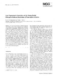
HDC was detected in all three layers of the adrenal cortex (zona glomerulosa, fasculata, and reticularis; Fig. 1a).
Fig. 1 Immunohistochemistry of histamine-related proteins in the normal adrenal cortex (representative images). a HDC, b HRH1, c HRH2, d HRH3, e HRH4, f HNMT (magnification ×20)
Virchows Arch (2009) 455:133–142
137
HRH1 immunohistochemistry showed focal staining in all adrenocortical layers (Fig. 1b). HRH2 and HRH3 were positive in the fasciculata and reticularis layers, but they were absent from the zona glomerulosa (Fig. 1c, d). HRH4 showed focal staining in the zona fasciculata (Fig. 1e). HNMT was colocalized with HDC in all three layers (Fig. 1f). DAO was undetectable (data not shown). The pattern of HDC and HNMT immunohistochemistry was dot-like cytoplasmic. HRH1 and HRH2 immunohistochemistry revealed diffuse cytoplasmic staining, whereas HRH3 and HRH4 expression was dot-like. Available literature data on HRH1 and HRH2 expression in epithelial and mesenchymal cells show similar staining patterns (e.g., basolateral cytoplasmic in gastric epithelial cells and dot-like cytoplasmic in chondrocytes) [25, 26]. Having confirmed the presence of all proteins involved in histamine physiology in the normal adrenal cortex at the protein level, we next examined their mRNA and protein expression in adrenocortical tumors.
histamine receptors were detected in adrenocortical tumor cells by immunohistochemistry (data not shown).
Expression of HDC and histamine content in adrenocortical tumors
The present study provides novel data on the expression of histamine-related genes and proteins, and histamine content in the normal human adrenal cortex and adrenocortical tumors. We demonstrated the expression of HDC, HNMT, and all histamine receptors both at the mRNA and protein levels in the normal adrenal cortex and compared these data to those observed in various benign and malignant adrenocortical tumor subtypes. We found significant differences in HDC expression, histamine content, and receptor expression profiles. Only few studies on the adrenocortical actions of histamine have been reported to date, and these presented contradictory results [27–30] that may be in part due to species differences. Yoshida et al. failed to detect any effect of histamine on basal cortisol secretion of isolated bovine adrenocortical cells; however, if co-cultured with bovine adrenomedullary cells, histamine dose-dependently induced cortisol secretion acting indirectly via the stimulation of adrenomedullary catecholamine secretion [27]. In another study, however, performed on isolated human adrenocortical cell cultures without adrenomedullary cells, histamine decreased aldosterone and cortisol and increased dehydroepiandrosterone sulfate (DHEAS) production [28]. When histamine was administered together with adrenocorticotropin (ACTH), it increased ACTH-stimulated DHEA production, decreased ACTH-stimulated cortisol and aldosterone production, and did not affect ACTH-stimulated DHEA levels [29]. The molecular mechanism of these actions was unclear, but a direct action of histamine on adrenocortical cells may be supposed. There have been no available data on the expression of histamine receptors in the normal human adrenal cortex.
HDC expression was detected in all examined tumors both at the mRNA and protein levels. Both quantitative real-time PCR and Western-blotting showed that the expression of HDC mRNA and protein was the highest in the normal adrenal cortex and benign hormonally inactive adenomas, somewhat lower in hormone-producing adenomas, whereas significantly lower expression was found in the ACC group (Fig. 2a–c). Immunohistochemical analysis revealed the presence of HDC in both benign and malignant tumor cells (Fig. 2e, f). Having proven the presence and different expression of the histamine biosynthetic enzyme HDC in adrenocortical samples, we next examined whether histamine content (by ELISA) of normal and neoplastic adrenocortical tissues was different. Parallel to the results of HDC mRNA and protein expression, histamine content was significantly lower in all tumor samples compared to normal adrenocortical tissues (Fig. 2d). Histamine receptors in adrenocortical tumors Quantitative RT-PCR and Western-blotting studies failed to demonstrate any significant differences in the expression of HRH1 (Fig. 3a, b). Expression of HRH2 was significantly lower in CPA and APA (Fig. 3c, d) and HRH4 showed significantly lower expression in CPA compared to normal adrenocortical tissues and other tumors (Fig. 3g, h). HRH3 expression was significantly higher both at the mRNA and protein levels in malignant tumors than in both normal tissues and benign tumors (Fig. 3e, f and 4). All types of
Histamine metabolizing enzymes HNMT was detected both by Western-blotting and immunohistochemistry; however, no differences in expression were found either at the mRNA or protein levels (Fig. 3i, j). DAO was undetectable both at the mRNA level and by immunohistochemistry (data not shown). Detection of mast cells We have not detected metachromatic cells characteristic for mast cells by toluidine blue staining and only very few ckit-positive cells were found by immunohistochemistry (data not shown).
Discussion
138
Virchows Arch (2009) 455:133–142
Fig. 2 Expression of HDC (a quantitative RT-PCR results expressed as ΔΔCT values relative to normal samples, b results of a representative Western-blotting experiment, c densitometric analysis of Western-blotting results using β-actin as loading control) and
histamine content (d) in adrenocortical tumors. Representative images of HDC immunohistochemistry in a cortisol-producing benign (e) and malignant (f) tumor (magnification ×20). Mean±SE. *p