liver was perfused in situ through the portal vein with 150 mL of .... (Apple Computer, Cupertino, CA) and an EPSON GT-5000 scanner ...... In Vitro Cell Dev.
Different Changes in Expression and Function of Connexin 26 and Connexin 32 During DNA Synthesis and Redifferentiation in Primary Rat Hepatocytes Using a DMSO Culture System TAKASHI KOJIMA,1,4 MASAO YAMAMOTO,2 CHIHIRO MOCHIZUKI,1 TOSHIHIRO MITAKA,1 NORIMASA SAWADA,3 1 AND YOHICHI MOCHIZUKI
In the present study, we determined in detail the changes of liver gap junctions, connexin 26 (Cx26), and connexin 32 (Cx32), during DNA synthesis and redifferentiation of hepatocytes in vitro. We used primary rat hepatocytes that expressed the liver gap junction proteins, which were cultured in the medium containing epidermal growth factor (EGF) with 2% dimethylsulfoxide (DMSO) and 1007 mol/L glucagon (a DMSO culture system), as we previously reported. In the present cultures, almost confluent hepatocytes cultured in the medium containing EGF with 2% DMSO and 1007 mol/L glucagon, underwent a nearly synchronous wave of DNA synthesis induced by the removal of 2% DMSO and 1007 mol/L glucagon, and the addition of 10 mmol/L nicotinamide, after which the DNA synthesis was completely re-inhibited by the re-addition of 2% DMSO and 1007 mol/L glucagon. During stimulation of DNA synthesis, both Cx26 and Cx32 messenger RNA (mRNAs) in hepatocytes transiently increased in the G1 phase and then markedly decreased before the onset of the S phase, while only Cx26 messenger RNA (mRNA) increased slightly in the S/M phase. Furthermore, before the onset of the S phase, a disappearance of both Cx26 and Cx32 immunoreactivities and gap junction plaques were observed. Gap junctional intercellular communication (GJIC), as measured by lucifer yellow, which indicated the function of Cx32, decreased markedly from before the onset of the S phase. GJIC measured by propidium iodide, which indicated the function of Cx26, decreased from before the onset of the S phase and then increased slightly in the S/M phase. During the re-inhibition after the stimulation of DNA synthesis, Cx32 mRNA, but not Cx26 mRNA, rapidly returned to the pretreatment control level. Cx32 immunoreactivity and gap junction plaques also recovered. However, the
Abbreviations: Cx, connexin; mRNA, messenger RNA; GJIC, gap junctional intercellular communication; PH, partial hepatectomy; cdk, cyclin-dependent; DMSO, dimethylsulfoxide; EGF, epidermal growth factor; BrdU, 5-bromo-2*-deoxyuridine; PBS, phosphate-bufferd saline. From the 1Department of Pathology, Cancer Research Institute, Sapporo Medical University School of Medicine, Sapporo, Japan; the 2Department of Anatomy, Hiroshima University School of Medicine, Hiroshima, Japan; and the 3Department of Pathology, Sapporo Medical University School of Medicine, Sapporo, Japan. Received April 4, 1997; accepted June 13, 1997. Supported by Grants-in-Aid from the Ministry of Education, Science and Culture, Japan, the Akiyama Foundation and the Japanese Society of Alternatives to Animal Experiments for Research Promotion. Address reprint requests to: Takashi Kojima, M.D., Department of Pathology, Cancer Research Institute, Sapporo Medical University School of Medicine, S.1, W.17, Chuo-ku, Sapporo 060, Japan. Fax: 81-11-615-3099. Copyright q 1997 by the American Association for the Study of Liver Diseases. 0270-9139/97/2603-0010$3.00/0
recovery of GJIC measured by lucifer yellow was later than that of Cx32 expression. These results indicated the different changes of expression and function of Cx26 and Cx32 in the hepatocytes during stimulation and re-inhibition of DNA synthesis. This culture system should be useful as a model in which to study liver gap junctions during hepatocyte growth and differentiation in vitro. (HEPATOLOGY 1997;26:585-597.) Gap junctions are intercellular membrane channels that link neighboring cells and mediate reciprocal exchanges of small molecules of less than 1,000 Da and ions, including second messengers, such as cyclic adenosine monophosphate inositol triphosphate, and Ca2/, between adjacent cells in contact.1-4 Gap junctions are composed of proteins termed ‘‘connexins (Cxs)’’.5 Both Cx26 and Cx32 are expressed in hepatocytes, and it is known that the relative ratios of Cx26 protein and messenger RNA (mRNA) in rat livers to those of Cx32 are 1:10 and 1:50, respectively.6,7 The distribution of these Cx proteins is reported to be different within the liver lobules: Cx26 preferentially localizes in the periportal zone of the lobules, whereas Cx32 appears in most hepatocytes throughout the lobules.8,9 They have been colocalized to the same gap junction plaques in hepatocytes.6,10,11 Furthermore, more recently, Kumar and Gilula12 reported that gap junction channels in the mouse liver are heterotypic channels, which dock among heteromeric connexons composed of heteromeric connexins of both Cx26 and Cx32. However, the specific functions and the regulation of expression of both Cxs in normal hepatocytes are still unclear. Gap junctional intercellular communication (GJIC) is thought to play a crucial role in cell growth and cell differentiation in multicellular organisms.13-18 For example in growth control, the growth of some transformed cells that exhibit decreased GJIC is inhibited or is slowed by transfection of specific Cxs.18 Furthermore, it is well known that in a model of liver regeneration after 70% partial hepatectomy (PH), the expression of Cx26 and Cx32 is downregulated during DNA synthesis and rapidly recovers to the normal level after inhibition of DNA synthesis.19-21 On the other hand, if the changes of GJIC or Cx genes affect cell growth or the cell cycle, they may be involved in cell cycle checkpoint regulation. More recently, Chen et al.22 reported changes in the expression of genes regulating the cell cycle, including decreases in cyclin A, D1, and D2 and in cyclin-dependent kinase 5 (cdk 5) and cdk 6 in transformed cells transfected with Cx43. However, the detailed roles of GJIC and Cx genes during the cell cycle in hepatocytes remain unclear.
585
AID
Hepa 0061
/
5P26$$1201
08-20-97 15:09:41
hepa
WBS: Hepatology
586 KOJIMA ET AL.
HEPATOLOGY September 1997
On the other hand, during cell diferentiation dramatic changes of gap junctions are also known. During keratinocyte differentiation and oval cell differentiation into hepatocytes, there is a switch in expression among Cxs.23,24 Furthermore, it was recently reported that small gap junction plaques may be closely related to cell polarity, which is one of the differentiation markers of epithelial cells.25,26 In the present study, we determined in detail the changes of liver gap junctions, Cx26 and Cx32 during DNA synthesis, and redifferentiation of hepatocytes in vitro. We used the primary cultures of rat hepatocytes highly expressing the liver gap junction proteins using a dimethylsulfoxide (DMSO) culture system, as we previously reported.11,26,27 The almost confluent hepatocytes, which were cultured in the medium containing epidermal growth factor (EGF), 2% DMSO, and 1007 mol/L glucagon, underwent a nearly synchronous wave of DNA synthesis induced by removal of 2% DMSO and addition of 10 mmol/L nicotinamide. Furthermore, the DNA synthesis was completely re-inhibited by the re-addition of 2% DMSO and 1007 mol/L glucagon. The maintenance of differentiation in 2% DMSO and the removal of DMSO to stimulate DNA synthesis using primary rat hepatocytes has been extensively studied.11,28-31 In 1985, Isom et al. first reported that adult rat hepatocytes in a chemically defined medium supplemented with EGF and 2% DMSO survived much longer and synthesized albumin much better than did cells cultured in standard serum-free medium.29 Recently, it was reported that the stimulation of DNA synthesis and proto-oncogene expression in primary rat hepatocytes using a DMSO culture system was observed by the removal of DMSO.30 Furthermore, many studies have been reported the effects of 10 mmol/L nicotinamide to the proliferation of cultured hepatocytes.32-34 The proliferation of cultured hepatocytes could be well maintained by the Dulbecco’s Modified Eagle Medium containing EGF with 10 mmol/L nicotinamide. In the present study, during the stimulation and the reinhibition of DNA synthesis of the cultured hepatocytes, liver gap junctions markedly changed and the different changes in expression and function of Cx26 and Cx32 were observed. MATERIALS AND METHODS Isolation and Culture of Rat Hepatocytes. Male Sprague-Dawley rats
(Shizuoka Laboratory Animal Center, Hamamatsu, Japan) (range, 300-400 g) were used to isolate hepatocytes by the two-step liver perfusion method of Seglen35 with some modification. Briefly, the liver was perfused in situ through the portal vein with 150 mL of Ca2/, Mg2/-free Hanks’ balanced Salt Solution supplemented with 0.5 mmol/L egtazic acid (Sigma Chemical Co., St. Louis, MO), 0.5 mg/L of insulin (Sigma), and antibiotics. After the initial brief perfusion, the liver was perfused with 200 mL of Hanks’ Balanced Salt Solution containing 40 mg of collagenase (Yakult Co., Tokyo, Japan) for 10 minutes. The isolated cells were purified by Percoll iso-density centrifugation.36 Viability of the cells by the Trypan Blue exclusion test was more than 90% in these experiments. The cells were suspended in L-15 medium (GIBCO BRL, Gaithersburg, MD) with 0.2% bovine serum albumin (Seikagaku Kogyo Co., Tokyo, Japan), 20 mmol/L HEPES (Dojindo, Kumamoto, Japan), 0.5 mg/L of insulin (Sigma), 1007 mol/L dexamethasone (Sigma), 1 g/L of galactose (Sigma), 30 mg/L of proline (Sigma), and antibiotics. The isolated hepatocytes were plated on 35-mm and 60-mm culture dishes (Corning Glass Works, Corning, NY), which were coated with rat tail collagen (500 mg of dried tendon/mL of 0.1% acetic acid),37 and placed in a 100% air incubator at 377C. Two to three hours after plating, the medium was changed to L-15 medium sup-
AID
Hepa 0061
/
5P26$$1202
08-20-97 15:09:41
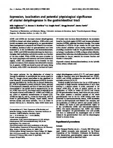
FIG. 1. Culture conditions. After hepatocytes plated on collagen-coated dishes were cultured in modified L-15 medium containing EGF for 96 hours they were cultured in the medium containing EGF with 2% DMSO and 1007 mol/L glucagon for 10 days. One set of cells was cultured with modified DMEM medium containing EGF with 10 mmol/L nicotinamide from day 10 and the cells were maintained until day 14 (condition 1). After two sets of the cells were maintained with the modified DMEM medium containing EGF with 10 mmol/L nicotinamide for 48 hours, the cells were cultured in the modified DMEM medium containing EGF with 10 mol/L nicotinamide plus 2% DMSO and 1007 mol/L glucagon until day 15 (condition 2). Culture time after treatment is calculated as 0, 3, 8, 24, 48, 72, 96, and 120 hours from day 10 after plating.
plemented with 0.2% bovine serum albumin, 20 mmol/L HEPES, 0.5 mg/L of insulin, 1007 mol/L dexamethasone, 1 g/L of galactose, 30 mg/L of proline, 20 mmol/L NaHCO3 , 5 mg/L of transferrin (Wako Pure Chemical Inc., Osaka, Japan), 0.2 mg/L of CuSO4r5H2O, 0.5 mg/L of FeSO4r4H2O, 0.75 mg/L of ZnSO4r7H2O, 0.05 mg/L of MnSO4 , 5 mg/L of Na2SeO3 , 10 ng/ mL of EGF (Collaborative Res., Lexington, MA), and antibiotics (modified L-15 medium). The cells were then placed in a humidified, 5% CO2 :95% air incubator at 377C. The medium was replaced with fresh medium every other day. After 96 hours of culture, 2% DMSO (Aldrich Chemical Co. Inc., Milwaukee, WI) and 1007 mol/ L glucagon (glucagon novo, Yamanouchi, Tokyo, Japan) were added to the modified L-15 medium.11 DMSO Shift Protocol. Hepatocytes used in DMSO shift experiments were maintained in the modified L-15 medium containing EGF with 2% DMSO and 1007 mol/L glucagon until day 10 after plating. Culture time after treatment was calculated as 0, 3, 8, 24, 48, 72, 96, and 120 hours from day 10 (Fig. 1). One set of cells was cultured in the DMEM medium (GIBCO) supplemented with 0.2% BSA, 20 mmol/L HEPES, 0.5 mg/L of insulin, 1007 mol/L dexamethasone, 1 g/L of galactose, 30 mg/L of proline, 20 mmol/L NaHCO3 , 10 ng/ mL of EGF, and antibiotics (modified DMEM medium), all of which were added 10 mmol/L nicotinamide from day 10, and the cells were maintained until day 14 (Fig. 1, condition 1). After two sets of the cells were maintained with the modified DMEM medium containing EGF with 10 mmol/L nicotinamide for 48 hours, the cells were cultured with the modified DMEM medium containing EGF with 10 mmol/L nicotinamide to which 2% DMSO and 1007 mol/L glucagon were added; they were then maintained until day 15 (Fig. 1, condition 2). Labeling Index. To examine the labeling index, immunocytochemical staining for 5-bromo-2*-deoxyuridine (BrdU) was carried out. Twenty micromoles of BrdU was added to each 35-mm dish 24 hours before fixation, after which the cells were fixed in cold absolute ethanol. Mouse anti-BrdU antibody (DAKO Co., Santa Barbara, CA) was used as the primary antibody, followed by application of the ABC method (Vectain ABC Elite kit, Vector Laboratory, Burlingame, CA). Labeled cells that had nuclei stained for BrdU were
hepa
WBS: Hepatology
HEPATOLOGY Vol. 26, No. 3, 1997
KOJIMA ET AL.
counted using a microscope (magnification 1200). More than 1,000 cells were counted per dish and three dishes were examined per experiment. Mitotic Index and Total Cell Number. To examine the mitotic index and total cell number, cells stained with hematoxylin and eosin were counted. The mitotic index is indicated as the mitotic cell number per 1,000 cells. The total cell number is the cell number per 103 mm2. Three dishes were examined per experiment. Northern Blot Analysis and Densitometry. Total RNA was extracted from the cells using the single-step thiocyanate-phenol-chloroform extraction method38 as modified by Xie and Rothblum.39 For electrophoresis, 10 mg of total RNA was loaded on 1% agarose gel containing 0.5 mg/L of ethidium bromide. Gels were capillary-blotted in 201 standard saline citrate onto a nylon membrane (HybondN; Amersham Corp., Buckinghamshire, UK) and fixed by ultraviolet light. For the detection of Cx26 mRNA, Cx32 mRNA, and b-actin mRNA, digoxigenin-labeled RNA probes were prepared from rat Cx26 complementary DNA,7 rat Cx32 complementary DNAs40 and b-actin complementary DNAs (Oncogene Science, Inc., Uniondale, NY) using an RNA labeling kit (Boehringer Mannheim, Mannheim, Germany). Hybridization, washing, and chemiluminescent detection were carried out following the DIG luminescent protocol.41 For detection of c-fos mRNA, c-myc mRNA, and rat albumin mRNA, the membranes were prehybridized in a solution containing 50% formamide, 0.9 mol/L NaCl, 0.1 mol/L NaPO4 (pH 7.4), 1% sodium dodecyl sulfate, 10 mg/mL of herring sperm DNA and 51 Denhart’s solution for 4 hours at 427C, and then hybridized overnight at 427C in the same solution with a 32P-labeled complementary DNA probe for c-fos, c-myc (Oncogene Science, Inc.), and rat albumin (a generous gift from M. Sakai). The membranes were washed twice in 21 SSC buffer containing 0.1% sodium dodecyl sulfate for 5 minutes at room temperature and twice in 21 SSC buffer containing 1% SDS for 30 minutes at 687C before exposure to film. Scanningdensitometry was performed using a Macintosh LC-520 computer (Apple Computer, Cupertino, CA) and an EPSON GT-5000 scanner (Seiko Epson, Suwa, Japan). The signals were quantified by the NIH Image 1.52 Densimetric Analysis Program (Wayne Rasband, NIH, Bethesda, MD).42 The expression of the transcripts was shown as a percent of 0-hour values (isolated hepatocytes) in the same experiment, was demonstrated as a histogram. The results are representative of three separate experiments. Western Blot Analysis. The dishes were washed twice with phosphate-buffered saline (PBS) and in 1 mL of the buffer (1 mmol/L NaHCO3 ), 2 mmol/L phenylmethylsulfonic acid (Sigma) and 2 mg/ L of leupeptin (Sigma) were added to 60-mm dishes. The cells were scraped, collected in eppendorf tubes, and then sonicated for 30 seconds. The sonicates were centrifuged at 4,500g for 10 minutes. The final pellets were resuspended in Laemmli sample buffer43 without dithiothreitol. The protein concentration of the samples was determined using a protein assay kit (Bio-Rad, Richmond, CA). Twenty micrograms of protein of each sample was treated dithiothreitol (final concentration, 100 mmol/L) for 15 minutes at room temperature and then loaded on 12.5% sodium dodecyl sulfate polyacrylamide gel (Daiichi Pure Chemicals Co., Tokyo, Japan). After electrophoretic transfer to a nitrocellulose membrane (Bio-Rad) using semi-dry blotting for 6 hours (0.65 mA/cm2), the membrane was stained with Ponceau S (Sigma) and photographed. Thereafter, the membrane was saturated overnight at 47C with a blocking buffer (25 mmol/L Tris, pH 8.0, 125 mmol/L NaCl, 0.1% Tween 20, and 4% skim milk) and was incubated with Cx3244 and Cx2645 antibodies at room temperature for 2 hours. The membrane was incubated with a horseradish peroxidase-conjugated anti-mouse or anti-rabbit immunoglobulin G (Vector Laboratories, Burlingame, CA) and 3,3*diaminobenzidine was used as a substrate. Immunofluorescence Microscopy. The cells grown on coated glass cover slips (BIOCOAT, Becton Dickinson Labware, Bedford, MA) were fixed with acetone for 30 minutes at 0207C. After rinsing with PBS, the cover slips were used for double staining for Cx32, Cx26, and BrdU. Twenty micromoles BrdU was added to the culture
AID
Hepa 0061
/
5P26$$1202
08-20-97 15:09:41
587
FIG. 2. (A), BrdU labeling index, Numbers of mitotic cells and (B) all cells in different culture conditions. (h), Condition 1; and ( ) condition 2.
medium 24 hours before the fixation. The polyclonal anti-Cx32,8 polyclonal anti-Cx2645 and monoclonal anti-BrdU (DAKO, Copenhagen, Denmark) antibodies were used as primary antibodies. The secondary antibodies were used fluorescein isothiocyanate-conjugated anti-rabbit IgG for Cx32 and Cx26 staining and fluorescein isothiocyanate-conjugated anti-mouse IgG (DAKO) for BrdU staining. All samples were examined with a Nikon Fx epifluorescence photomicroscope (Nikon, Tokyo, Japan). Immunoelectron Microscopy. The pre-embedding labeling method was used for Cx32-immunoelectron microscopy.26 The cells were fixed with 1% paraformaldehyde in 3% sucrose/0.1 mol/L PBS (pH 7.4) at 47C for 2 hours and were then permeabilized with 0.2% Triton X-100. The cells were treated at 47C for 30 minutes with a blocking buffer (4% skim milk in 0.1 mol/L PBS) and were then incubated with a monoclonal anti-rat Cx32 antibody44 overnight at 47C. After being washed three times with 3% sucrose/0.1 mol/L PBS, the cells were stained using the ABC method. Thereafter, the cells were refixed in 2.5% glutaraldehyde/0.1 mol/L cacodylate buffer (pH 7.3) overnight at 47C, postfixed in 2% osmium tetroxide in the buffer, dehydrated by graded ethanol, and embedded in situ in Epon 812, ultrathin sections were then cut in a Sorvall Ultramicrotome MT-5000. The sections were stained with uranyl acetate followed by lead citrate and examined at 60 kV with a JEM transmission electron microscope (JEOL, Tokyo, Japan).
hepa
WBS: Hepatology
588 KOJIMA ET AL.
HEPATOLOGY September 1997
in liquid nitrogen, and fractured at 01707C to 01807C in a JFD7000 freeze-fracture device (JEOL Ltd., Tokyo, Japan). Platinumcarbon replicas were made without etching. After thawing, the replicas were floated on filtered 10% sodium hypochlorite solution for 10 minutes in a Teflon dish. Replicas were washed in distilled water for 30 minutes, mounted on copper grids, and examined at 80 kV with a JEM transmission electron microscope. Measurement of GJIC. For measuring GJIC, we used the scrape loading/dye transfer method47 with some modification.27 The hepatocytes on 35-mm dishes were rinsed several times with PBS. Two or three lines were made around the center of the dish using a scalpel and 2 mL of 0.05% lucifer yellow CH (LY; Sigma) and 5 mg/mL of propidium iodide (PI; Sigma) in PBS was added. LY and PI are small molecules (443 da and 414 da, respectively) which can freely move through gap junctions from loaded cells to neighboring ones. Three minutes after the dye treatment, the cells were rinsed several times with PBS to remove excess dye. We used rhodamine dextran (10 kd; Sigma), which is known not to move through gap junctions, as a control. We immediately observed the intensity of LY and PI transfer with an Olympus inversed microscope equipped with appropriate filters (Olympus, Tokyo, Japan) and photographed it. We carried out at least three separate experiments. To measure the intensity of LY and PI, the numbers of cells labeled by LY and PI were counted on both sides of the scrape line (120-mm wide).48 The results are shown as a percent of control values demonstrated as a histogram.
FIG. 3. Photographs of phase contrast and BrdU staining of the hepatocytes cultured in conditions 1 and 2. Phase contrast (A, B, and C); BrdU staining (D, E, and F). (A and D): Day (0 hours); (B and E): 48 h in condition 1; mitotic cells (arrowheads); (C and F): 96 h in condition 2. Bars: 40 mm for (A, B, and C); 80 mm for (D, E, and F).
Freeze-Fracture Analysis. For the freeze-fracture technique,46 cells
cultured on cover slips were immersed in 40% glycerin solution after fixation in 2.5% glutaraldehyde/0.1 mol/L cacodylate buffer (pH 7.3). The specimens were mounted on a copper stage, cooled
AID
Hepa 0061
/
5P26$$1202
08-20-97 15:09:41
FIG. 4. Northern blot analysis for c-fos and c-myc of the hepatocytes cultured in conditions 1 and 2. Total RNA (10 mg/lane) was fractionated by electrophoresis in a 1% agarose-formaldehyde gel and hybridized with c-fos and c-myc cDNA probes (B). Ethidium bromide staining of ribosome RNAs before transfer to membranes.
hepa
WBS: Hepatology
HEPATOLOGY Vol. 26, No. 3, 1997
KOJIMA ET AL.
589
FIG. 5. Northern blot analysis for Cx32, Cx26, albumin, and b-actin of the hepatocytes cultured in condition 1 and 2. Total RNA (10 mg/lane) was fractionated by electrophoresis in a 1% agarose-formaldehyde gel and hybridized with digoxigenin-labeled Cx32, Cx26, b-actin cRNA probes and an albumin cDNA probe. The (A) shows ethidium bromide staining of ribosome RNAs before transfer to membranes. Scanning-densitometric analysis of the mRNA level was performed. Details are described in Materials and Methods. (B) Expression of the transcripts is shown as the percent of 0-hour values.
RESULTS BrdU Labeling Index, Mitotic Index, and Total Cell Number. We recently showed that primary rat hepatocytes can induce and maintain the gap junctional proteins Cx26 and Cx32 in serum-free modified L-15 medium containing EGF with 2% DMSO and 1007 mol/L glucagon.11,27 It is also known that 2% DMSO can inhibit the proliferation of the cultured rat hepatocytes and maintain the liver specific-functions of the
AID
Hepa 0061
/
5P26$$1202
08-20-97 15:09:41
cells long term.28,29 In our culture system, the cells were well maintained in long-term culture, and a few percent of the hepatocytes were stimulated to synthesize DNA after the addition of 2% DMSO and 1007 mol/L glucagon.27 To study the stimulation and re-inhibition of DNA synthesis of the cultured hepatocytes, we determined whether medium conditions could be established that would yield a higher BrdU labeling index using DMSO shift protocols. Hepatocytes were
hepa
WBS: Hepatology
590 KOJIMA ET AL.
HEPATOLOGY September 1997
FIG. 6. (A) Western blot analysis for Cx26 and Cx32 proteins of the hepatocytes cultured in conditions 1 and 2. Whole cells were separated by electrophoresis in 12.5% sodium dodecyl sulfate–polyacrylamide gel and transferred to a nitrocellulose membrane. After transfer, the blots were stained with antibodies against Cx26 and Cx32. (B) The lower lanes stained with Ponceau S, are shown to confirm the presence of equal amounts of protein.
labeled with BrdU 24 hours before fixation. First, we cultured hepatocytes in the modified L-15 medium containing EGF without 2% DMSO and 1007 mol/L glucagon. However, the labeling index of the cells was maximally about 20% at 48 or 72 hours after treatment, and the proliferation of the cells was not well maintained (data not shown). Some hepatocytes were cultured in the modified DMEM medium containing EGF with 10 mmol/L nicotinamide (Fig. 1, condition 1), because it was reported32-34 that the proliferation of cultured hepatocytes is well maintained by DMEM medium containing EGF with 10 mmol/L nicotinamide. In hepatocytes cultured in condition 1, the labeling index clearly increased from 48 hours after treatment and was more than 60% at 96 hours (Fig. 2A and B). Furthermore, after some of the cells were maintained with the modified DMEM medium containing EGF with 10 mmol/L nicotinamide for 48 hours, the cells were cultured with the modified DMEM medium containing EGF with 10 mmol/L nicotinamide to which 2% DMSO and 1007 mol/L glucagon were added (Fig. 1, condition 2). The labeling index of the cells markedly decreased at 96 hours in condition 2 compared with that of the cells at 48 hours in condition 1 and completely returned to the pretreatment control level (Fig. 2A and B). We also examined the numbers of mitotic cells and all cells for the hepatocytes cultured in the conditions 1 or 2 medium. In condition 1, the numbers of the mitotic cell and all cells markedly increased from 48 hours to 96 hours (Fig. 2B). In condition 2, the numbers of mitotic cells decreased from 72 hours and completely returned to the pretreatment control level at 96 hours (Fig. 2B). The total number of
AID
Hepa 0061
/
5P26$$1202
08-20-97 15:09:41
cells at 72 or 96 hours increased to slightly more than the pretreatment control level. As a result of these experiments, we chose the protocols of conditions 1 and 2 for the following study during stimulation and inhibition of DNA synthesis of the cultured hepatocytes. c-fos and c-myc mRNAs’ Expression. To examine the mRNA levels of the proliferative cells in conditions 1 and 2, the expression of proto-oncogene, c-fos and c-myc mRNAs was observed (Fig. 3). In condition 1, maximum induction of cfos mRNA was observed at 1 hour, and it returned to basal levels by 8 hours. The expression of c-myc mRNA increased from 2 to 24 hours and decreased from 48 hours. In condition 2, expression of c-fos mRNA was not observed and expression of c-myc mRNA was at the control level (0 hours). These results of the proto-oncogene expression were similar to those of Serra and Isom,30 previously reported using a DMSO culture system. We thought that the BrdU labeling index, number of mitotic cells, and the expression of c-fos and c-myc mRNAs indicated the cell-cycle phases in conditions 1 and 2; the G1 phase was from 0 to 24 hours in condition 1 and at 96 hours in condition 2, while the S/M phase was from 48 to 96 hours in condition 1 and at 72 hours in condition 2. Cx26, Cx32, and Albumin mRNAs’ Expression. We previously showed the high-expression of Cx26, Cx32, and albumin mRNAs in the hepatocytes cultured in the modified L-15 medium containing EGF with 2% DMSO and 1007 mol/L glucagon.11 In the present study, to examine the changes in expression of Cx26, Cx32, and albumin mRNAs expression during stimulation and re-inhibition of DNA synthesis of cultured hepatocytes, we performed Northern blot analysis of them in conditions 1 and 2 (Fig. 4). Four separate experiments were done and the signals were quantified by densitometer. The expression of Cx32 mRNA slightly increased until 8 hours and markedly decreased from 24 to 96 hours in condition 1. The expression returned to the control level from 72 hours in condition 2. After the expression of Cx26 mRNA rapidly increased until 8 hours, the expression markedly decreased or disappeared from 24 hours, and slightly increased from 72 to 96 hours in condition 1. The expression was not observed or was at a very low level in condition 2. The expression of albumin mRNA gradually decreased from 48 to 96 hours in condition 1 and returned to the control level at 96 hours in condition 2. Cx26 and Cx32 Proteins’ Expression. To examine the changes in expression of Cx26 and Cx32 proteins during stimulation and inhibition of DNA synthesis of the cultured hepatocytes, we performed Western blot analysis of them in conditions 1 and 2 (Fig. 5). No changes in expression of Cx26 and Cx32 proteins were observed in conditions 1 and 2. Localization of Cx26, Cx32, and BrdU. Double fluorescent immunocytochemistry was carried out in conditions 1 and 2 to examine the relationship between Cx26 and Cx32 immunoreactivities and BrdU of the cells during stimulation and re-inhibition of DNA synthesis of the cultured hepatocytes (Fig. 6). Many Cx26- and Cx32-positive macular spots and a few BrdU-positive nuclei were observed in the cells at day 10 (0 hours) (Fig. 6A and E). Cx26- and Cx32-positive spots decreased at 24 hours and disappeared from 48 to 96 hours in condition 1, at which time many BrdU-positive nuclei were observed (Fig. 6B, C, F, and G). At 96 hours in condition 2, Cx32-positive spots returned to the control level and a few Cx26-positive spots were observed (Fig. 6D and H).
hepa
WBS: Hepatology
HEPATOLOGY Vol. 26, No. 3, 1997
KOJIMA ET AL.
591
FIG. 7. Double flourescent immunocytochemistry of BrdU and Cx32 (A, B, C, and D), BrdU and Cx26 (E, F, G, and H) in the hepatocytes cultured in conditions 1 and 2. (A and E): Day 10 (0 hours); (B and F): 24 hours in condition 1; (C and G): 48 hours in condition 1; and (D and H): 96 hours in condition 2. Twenty mmol/L BrdU was added to the medium 24 hours before fixation. Round and oval white spots show BrdU-positive nuclei, and the small dots between adjacent cells show Cx32- and Cx26-positive spots. Figures are the same magnification. Bar Å 10 mm.
Changes of Gap Junction Structures or Plaques. We previously demonstrated more developed gap junction structures and plaques in hepatocytes cultured in the modified L15 medium containing EGF with 2% DMSO and 10 07 mol/
AID
Hepa 0061
/
5P26$$1202
08-20-97 15:09:41
L glucagon, using ultrastructural analysis and immunoelectron microscopy for Cx32 and freeze fracture.26 In the present study, we examined the changes of the gap junction structures and plaques during stimulation
hepa
WBS: Hepatology
AID
Hepa 0061
/
5P26$$0061
08-20-97 15:09:41
hepa
WBS: Hepatology
HEPATOLOGY Vol. 26, No. 3, 1997
KOJIMA ET AL.
and re-inhibition of DNA synthesis of cultured hepatocytes. Immunoelectron Microscopy for Cx32. We performed immunoelectron microscopy for Cx32 using DAB-reaction in the hepatocytes in condition 1 and 2 (Fig. 7A-C). DAB-positive substances decorated the gap junctions. They were also observed in close proximity to several immunoreactive cytoplasmic granular bodies in para-junctional regions, although we thought that this was artifactual as a result of using 3*, 3*-diaminobenzidine. In the cells at day 10 (0 hours), Cx32immunoreactive structures 4 mm in length or longer were observed on the plasma membrane (Fig. 7A). In the cells at 48 hours in condition 1, immunoreactive structures were never observed (Fig. 7B). In the cells at 96 hours in condition 2, small immunoreactive structures were observed (Fig. 7C). Freeze-Fracture. Further ultrastructural analysis was carried out using the freeze-fracture technique in the hepatocytes in conditions 1 and 2 (Fig. 7D-F). In the cells at day 10 (0 hours), more developed tight junctions composed of many typical strands were observed on the luminal surfaces containing many microvilli (Fig. 7D). Furthermore, typical aggregates of gap junctional membrane particles (gap junction plaques) were observed on the abluminal lateral membrane, and many small gap junction plaques (õ0.05 mm2) were observed peripherally or within the network of the tight junction strands (Fig. 7E and F). In the cells from 48 to 96 hours in condition 1, only single or no tight junction strands were observed, and neither typical gap junction plaques nor small gap junction plaques were ever observed (Fig. 7G). In the cells at 72 hours in condition 2, a few small gap junction plaques first reappeared on the luminal surfaces where tight junction strands were not observed, while typical gap junction plaques were not yet observed. Then small gap junction plaques increased peripheral to the short single fibrils of tight junctions (Fig. 7H and I). In the cells at 96 hours in condition 2, levels of tight junction strands, typical gap junction plaques, and small gap junction plaques almost returned to the pretreatment control level (Fig. 7J-7L). Changes of GJIC. As we previously reported,11 the hepatocytes in the modified L-15 medium containing EGF with 2% DMSO and 1007 mol/L glucagon after day 10 had extensive GJIC, as measured using scrape-loading/dye transfer by LY. In the present study, to examine the changes of GJIC during stimulation and re-inhibition of DNA synthesis of cultured hepatocytes, we performed scrape-loading/dye transfer using LY in the hepatocytes in conditions 1 and 2 (Fig. 8A-E and Fig. 9A). In the cells from 24 to 96 hours in condition 1, LY spread markedly decreased compared with the pretreatment control level (Fig. 8A-C and Fig. 9A). In condition 2, the extensive spreading of LY was observed in some of the cells from 72 to 96 hours, and the dye spread at 120 hours showed 75% recovery of the pretreatment control level (Fig. 8D and E and Fig. 9A).
593
On the other hand, Elfgang et al.49 recently reported the permeabilities of various Cx after transfection to human HeLa cells that were deficient in GJIC, using iontophoretically injected tracer molecules. Their results showed that LY could pass through Cx26 and Cx32 channels, while PI penetrated very poorly through Cx32 but passed through the Cx26 channel. Therefore, we performed scrape-loading/dye transfer by PI and LY in the hepatocytes highly expressing Cx32 cultured with 2% DMSO or those highly expressing both Cx26 and Cx32 cultured in 2% DMSO and 1007 mol/ L glucagon, as we previously reported.26 In the cells highly expressing both Cx26 and Cx32, not only LY spread but also PI spread were widely observed, while in the cells highly expressing Cx32 only LY spread was widely observed (Fig. 8F and G). Using the hepatocytes at day 10, which had strong permeabilities for both LY and PI, we performed scrape-loading/dye transfer by PI in the hepatocytes in condition 1 and 2 compared to LY. In the cells in conditions 1, after PI spread markedly decreased from 24 to 48 hours, it increased from 72 hours. The PI spread at 96 hours in condition 1 showed 75% recovery of the pretreatment control level (Fig. 8H and I and Fig. 9B). In the cells from 72 to 120 hours in condition 2, the PI spread was small (Fig. 8J and Fig. 9B). DISCUSSION
This is the first detailed report that examines the changes of liver gap junctions during DNA synthesis and redifferentiation in normal hepatocytes in vitro. In the hepatocytes during DNA synthesis, both Cx26 and Cx32 mRNAs transiently increased in the G1 phase and then decreased before the onset of the S phase, while only Cx26 mRNA slightly increased at the S/M phase. Furthermore, before the onset of the S phase, decreases of gap junction plaques and GJIC were observed. During re-inhibition after stimulation of DNA synthesis, Cx32 mRNA, but not Cx26 mRNA, rapidly returned to the pretreatment control level. However, the recovery of GJIC measured by LY was later than that of Cx32 mRNA. During stimulation and re-inhibition of DNA synthesis, changes of cell polarity, one of the differentiation markers of epithelial cells, was observed. Furthermore, early after the re-inhibition small gap junction plaques reappeared on the luminal surfaces before the development of tight junction strands. These results suggested that the small gap junction plaques might be closely related to cell polarity. The detailed roles of GJIC or Cx genes during DNA synthesis and differentiation in hepatocytes remain unclear. The main reason why the roles of GJIC and Cx genes during the cell cycle are not sufficiently investigated in hepatocytes is that the induction and maintenance of the gap junction proteins Cx26 and Cx32 are very difficult. In primary cultures, Spray et al.4 showed that extracellular matrix components, such as proteoglycans and glycosaminoglycans, can induce the synthesis and expression of Cx32 and that dibutyl cyclic
b FIG. 8. Immunoelectron micrographs of Cx32 (A-C) and freeze-fracture replicas (D-L) in the hepatocytes cultured in conditions 1 and 2. (A-F): day 10 (0 hours); (B and G): 48 hours in condition 1; (H and I): 72 hours in condition 2; and (C-L): 96 hours in condition 2. (A and C) In immunoelectron micrographs of Cx32, all gap junctions structures were decorated by DAB-positive substances. (D and J) Numerous tight junction strands are observed on the faces containing many microvilli. (E and K) A typical gap junction plaque is observed on the abluminal lateral membrane. (F and L) Many small gap junction plaques were observed within the network of tight junction strands (arrowheads). (G) A single tight junction strand is observed but gap junction plaques were never observed. (H) A few small gap junction plaques are observed on the luminal surfaces where tight junction strands were not observed (arrowheads). (I) Many small gap junction plaques are observed peripheral to the short single fibrils of tight junctions (arrowheads). Bars Å 200 nm for A-D and J. Bars Å 100 nm for E-L.
AID
Hepa 0061
/
5P26$$1202
08-20-97 15:09:41
hepa
WBS: Hepatology
594 KOJIMA ET AL.
HEPATOLOGY September 1997
FIG. 9. Lucifer yellow and propidium iodide distribution in the hepatocytes cultured in conditions 1 and 2. (A-E): Lucifer yellow; (F-J): lucifer yellow and propidiun iodide. (A) Day 10 (0 hours); (B) 24 hours in condition 1; (C) 48 hours in condition 1; (D) 96 hours in condition 2; and (E) 120 hours in condition 2. (F) The cells were cultured in the modified L-15 medium containing EGF with 2% DMSO at day 10. (G) The cells were cultured in the modified L-15 medium containing EGF with 2% DMSO and 1007 mol/L glucagon at day 10. (H) Forty-eight hours in condition 1; (I) 96 hours in condition 1; and (J) 96 hours in condition 2. Figures are the same magnification. Bar Å 80 mm.
AMP was shown to increase the stability of Cx32 mRNA and to delay the disappearance of gap junctions in cultured hepatocytes.14,50 Recently, Kwiatkowski et al.51 reported that dexamethasone maintained GJIC and Cx mRNA in primary
AID
Hepa 0061
/
5P26$$1202
08-20-97 15:09:41
rat hepatocytes. However, these experiments were carried out using cells in which the gap junctions disappeared within one week. Furthermore, the cell lines derived from normal rat liver expressed Cx43 and, to a lesser extent, Cx26; hepa-
hepa
WBS: Hepatology
HEPATOLOGY Vol. 26, No. 3, 1997
KOJIMA ET AL.
toma cells expressed Cx43 or were deficient in GJIC and Cx expression.52 It was not yet possible to examine the changes of liver gap junctions during DNA synthesis and differentiation of normal cultured hepatocytes. We previously showed that Cx32 in primary cultures of adult rat hepatocytes is re-expressed in the modified L-15 medium containing EGF with 2% DMSO27 and that Cx26 can also be induced by the addition of 1007 mol/L glucagon together with 2% DMSO.11 Both the proteins and the mRNA levels increased and the cells were well maintained with extensive GJIC for more than 4 weeks. This culture system may be useful for studying the regulation of gap junction proteins Cx26 and Cx32 in cultured hepatocytes under various conditions. In the present study, using cultured hepatocytes highly expressing both Cx26 and Cx32 using a DMSO culture system, we examined the changes of liver gap junctions during DNA synthesis and redifferentiation of hepatocytes. In the hepatocytes during DNA synthesis, both Cx26 and Cx32 mRNAs transiently increased in the G1 phase; the increase in the level of Cx26 mRNA was greater than that of Cx32 mRNA. Both Cx26 and Cx32 mRNAs decreased from before the onset of the S phase, while only Cx26 mRNA slightly increased at the S/M phase. Furthermore, before the onset of the S phase, decreases of both Cx26- and Cx32-positive spots and the disappearance of gap junction structures and plaques was observed. It is well known that dramatic changes of gap junctions occur after PH. It has been reported that after PH, the mRNA and protein expression of Cx26 and Cx32 decrease from before the onset of the S phase and then gradually return to the basal levels after the S phase.19 However, Kren et al.20 recently reported that mRNA expression of Cx26 and Cx32 decreased twice at 12 h and 48 h after PH. Furthermore, more recently, Neveu et al.21 reported that Cx26 mRNA expression transiently increased before the onset of the S phase and then rapidly returned to the basal levels after PH. The difference among these results from several experiments may be caused by various factors and conditions in vivo. As shown in Fig. 5 in the cultured hepatocytes, the results were almost the same in four separate experiments. On the other hand, similar to our results, an increase of Cx26 mRNA expression during DNA synthesis was observed in the S/M phase in proliferating human mammary epithelial cell culture.53 We also thought that Cx26 mRNA expression was more sensitive to the cell cycle than was Cx32 mRNA in the hepatocytes and that the regulation of Cx26 and Cx32 was different during cell proliferation.18 The changes of Cx genes after PH were regulated primarily by posttranscriptional events.20 The changes of Cx genes in this culture system may have been mainly regulated by posttranscriptional events such as the changes of Cx mRNA stability and intracellular cyclic adenosine monophosphate. However, as a decrease of albumin mRNA, which was very stable, were observed during proliferation in a DMSO culture system,30 we thought that transcriptional changes induced by various transcriptional factors also occurred. In the cultured hepatocytes, GJIC measured by LY was closely related to expression of Cx32 mRNA as we previously reported.11 In the present study, a decrease of GJIC measured by LY was observed from before the onset of the S phase and Cx32 mRNA expression decreased. Furthermore, an increase of GJIC measured by PI was observed at the S/M phase, and Cx26 mRNA expression slightly increased. As Elfgang et al.49
AID
Hepa 0061
/
5P26$$1202
08-20-97 15:09:41
595
recently reported, in this culture system PI could pass through Cx26 channels but not through Cx32 channels in the experiments using the cells highly expressing only Cx32; the cells highly expressing both Cx26 and Cx32 (Fig. 10). These results suggested that the Cx26 expression, which increased at the S/M phase, might be a functional phenomenon. On the other hand, during re-inhibition after the stimulation of DNA synthesis, Cx32 mRNA, but not Cx26 mRNA, rapidly returned to the control levels. Gap junction structures or plaques also reappeared. However, the recovery of GJIC measured by LY was later than that of Cx32 mRNA. Although GJIC in hepatocytes after partial hepatectomy could not be measured in vivo, it is possible that the recovery of GJIC might also occur later in them. Tight junctions, which were observed as networks of strands by freeze fracture, have two functions. One is a gate function that controls the paracellular pathway and the other is a fence function that separates molecules in the apical and basolateral plasma membrane domains.54 It is thought that cell polarity in epithelial cells may be detected by the fence function. In the present study, more developed tight junctions composed of many strands in the cultured hepatocyes disappeared or changed to single tight-junction strands during DNA synthesis, returning to the control levels during reinhibition after stimulation of DNA synthesis. These changes of tight junction strands in this culture system indicated that the monolayer hepatocytes, which had cell polarity, had no cell polarity during DNA synthesis, and, again, regained it during the re-inhibition.25 This suggested that re-inhibition of DNA synthesis in this culture system might indicate redifferentiation. Early after the re-inhibition or redifferentiation, small gap junction plaques reappeared on the luminal surfaces before development of tight junction strands on the same region, and these results indicated that the small gap junction plaques might be closely related to cell polarity. One of the gap junctional functions during cell proliferation that has been postulated for the channels is intercellular communication to control the passage of growth regulation signals.13,17 We also thought that intercellular communication between adjacent cells might be necessary to cause a synchronous wave of DNA synthesis in cultured hepatocytes as in proliferative hepatocytes after PH. In the present study, Cx mRNAs transiently increased in the G1 phase and markedly decreased from before the onset of the S phase. It was thought that the increase at the G1 phase may be necessary to pass cell growth regulatory signals through the gap junctional channels and that the decrease before the onset of the S phase may be necessary so as not to prevent the growth of individual cells by means of gap junctions. Although the means which only Cx26 mRNA slightly increased in the S/ M phase were unclear, it is possible that gap junctions may also be needed in proliferative cells. Furthermore, during reinhibition after stimulation of DNA synthesis, Cx32 mRNA rapidly returned to the control levels of before the cell growth arrest. This indicated that gap junctions may have an important role in cell growth arrest.18 In the present study, the dramatic changes of Cx32 and Cx26 mRNAs expression and the immnoreactivities were observed during stimulation and re-inhibition of DNA synthesis of the cultured hepatocytes, while no changes of Cx32 and Cx26 proteins expression of the whole cells were observed in Western blot. These results suggested that membrane insertion of Cx32 and Cx26 might be also affected during
hepa
WBS: Hepatology
596 KOJIMA ET AL.
HEPATOLOGY September 1997
Kyushu University, Fukuoka, Japan, for the anti-Cx26 antibody; and Dr. M. Sakai, Hokkaido University, Sapporo, Japan, for albumin cDNA. We are also grateful to Dr. S. Takahashi, Sapporo Medical University, Sapporo, Japan, for his discussion and suggestions for preparing this manuscript. We thank Ms. M. Kuwano and Ms. Y. Takahashi for technical support. We also thank Mr. K. Barrymore, Sapporo Medical University, Sapporo, Japan, for help with the manuscript. REFERENCES
FIG. 10. (A) Lucifer yellow and (B) propidium iodide distribution of the hepatocytes was measured using the scrape-loading technique, as, described in Materials and Methods. The results are shown as the percent of day 10 (0 hours) values in histograms. (h) Condition 1; and ( ) condition 2.
stimulation and re-inhibition of DNA synthesis of the cultured hepatocytes. However, the mechanisms were not yet unclear. Although we need to further examine the roles and the functions of Cxs in cultured hepatocytes, this culture system should be useful as a model in which to study liver gap junctions during hepatocyte growth and differentiation in vitro. Acknowledgment: We are grateful to Dr. B. Nicholson, State University of New York, NY, for Cx26 cDNA; Dr. D. Paul, Harvard Medical School, Boston, MA, for the anti-Cx32 antibody and Cx32 cDNA; Dr. Y. Shibata and Dr. A. Kuraoka,
AID
Hepa 0061
/
5P26$$1203
08-20-97 15:09:41
1. Sa´ez JC, Spray DC, Narin AC, Hertzberg EL, Greengard P, Bennett MVL. c-AMP increases junctional conductance and stimulates phosphorylation of the 27 KDa principal gap junction polypeptide. Proc Natl Acad Sci U S A 1986;83:2473-2476. 2. Sa´ez JC, Conner JA, Spray DC, Bennet MVL. Hepatocyte gap junctions are permeable to the second messenger, inositol 1,4,5-triphosphate and to calcium ions. Proc Natl Acad Sci U S A 1989;86:2708-2712. 3. Spray DC, Ginzberg RD, Morales EA, Gatmaitin Z, Arias IN. Physiological properties of dissociated rat hepatocytes. J Cell Biol 1986;103:135144. 4. Spray DC, Fujita M, Sa´ez JC, Choi H, Watanabe T, Hertzberg EL, Rosenberg LC, et al. Proteoglycans and glycosaminoglycans induce gap junction synthesis and function in primary liver cultures. J Cell Biol 1987; 105:541-551. 5. Haefliger JA, Bruzzone R, Jenkins NA, Gillbert DJ, Copeland NG, Paul D. Four novel members of the connexin family of gap junction proteins. J Biol Chem 1992;267:2057-2064. 6. Nicholson BJ, Dermietzel R, Teplow D, Traub O, Willecke K, Revel JP. Two homologous protein components of hepatic gap junctions. Nature 1987;329:732-734. 7. Zhang J-T, Nicholson BJ. Sequence and tissue distribution of a second protein of hepatic gap junctions, Cx26, as deduced from its cDNA. J Cell Biol 1989;109:3391-3401. 8. Sakamoto H, Oyamada M, Enomoto K, Mori M. Differential changes in expression of gap junction proteins connexin 26 and 32 during hepatocarcinogenesis in rats. Jpn J Cancer Res 1992;83:1210-1215. 9. Traub O, Look J, Dermietzel R, Bru¨mmer F, Hu¨lser D, Willecke K. Comparative characterization of the 21-KD and 26-KD gap junction proteins in murine liver and cultured hepatocytes. J Cell Biol 1989;108: 1039-1051. 10. Fujimoto K. Freeze-fracture replica electron microscopy combined with SDS digestion for cytochemical labeling of integral membrane proteins. J Cell Sci 1995;108:3443-3449. 11. Kojima T, Mitaka T, Shibata Y, Mochizuki Y. Induction and regulation of connexin26 by glucagon in primary cultures of adult rat hepatocytes. J Cell Sci 1995;108:2771-2780. 12. Kumar NM, Gilula NB. The gap junction communication channel. Cell 1996;84:381-388. 13. Loewenstein WR. Junctional intercellular communication and the control of growth. Biochim Biophys Acta 1979;560:1-65. 14. Bennett MVL, Barrio LC, Bargiello TA, Spray DC, Hertzberg E, Sa´ez JC. Gap junctions: new tools, new answers, new questions. Neuron 1991; 6:305-320. 15. Lang L, Beyer EC, Schwartz AL, Gitlin JD. Molecular cloning of a rat uterine gap junction protein and analysis of gene expression during gestation. Am J Physiol 1991;260:E787-793. 16. Mesnil M, Yamasaki H. Cell-cell communication and growth control of normal and cancer cells: evidence and hypothesis. Mol Carcinog 1993; 7:14-17. 17. Bruzzone R, White TW, Paul DL. Connections with connexins: the molecular basis of direct intercellular signaling. Eur J Biochem 1996; 238:1-27. 18. Yamasaki H, Naus CCG. Role of connexin genes in growth control. Carcinogenesis 1996;17:1199-1213. 19. Yancey SB, Easter D, Revel JP. Cytological changes in gap junctions during liver regeneration. J Ultrastruc Res 1979;67:229-242. 20. Kren BT, Kumar NM, Wang S, Gilula NB, Steer CJ. Differential regulation of multiple gap junction transcripts and proteins during rat liver regeneration. J Cell Biol 1993;123:707-718. 21. Neveu MJ, Hully JR, Babcock KL, Vaughan J, Hertzberg EL, Nicholson BJ, Paul DL, et al. Proliferation-associated differences in the spatial and temporal expression of gap junction genes in rat liver. HEPATOLOGY 1995;22:202-212. 22. Chen SC, Pelletier DB, Ao P, Boynton AL. Connexin43 reverses the
hepa
WBS: Hepatology
HEPATOLOGY Vol. 26, No. 3, 1997
23.
24. 25. 26.
27.
28. 29. 30. 31. 32. 33.
34. 35. 36.
37.
KOJIMA ET AL.
phenotype of transformed cells and alters their expression of cyclin/ cyclin-dependent kinases. Cell Growth Diff 1995;6:681-690. Guerrier A, Fonlupt P, Morand I, Rabilloud R, Audebet C, Krutovskikh V, Gros D, et al. Gap junctions and cell polarity: connexin32 and connexin43 expressed in polarized thyroid epithelial cells assemble into separate gap junctions, which are located in distinct regions of the lateral plasma membrane domain. J Cell Sci 1995;108:2609-2617. Zhang M, Thorgeirsson SS. Modulation of connexins during differentiation of oval cells into hepatocytes. Exp Cell Res 1994;213:37-42. Decaens C, Rodriguez P, Bouchaud C, Cassio D. Establishment of hepatic cell polarity in the rat hepatoma-human fibroblast hybrid WIFB9. J Cell Sci 1996;109:1623-1635. Kojima T, Yamamoto M, Tobioka H, Mizuguchi T, Mitaka T, Mochizuki Y. Changes in cellular distribution of connexins 32 and 26 during formation of gap junctions in primary cultures of rat hepatocytes. Exp Cell Res 1996;223:314-326. Kojima T, Mitaka T, Paul DL, Mori M, Mochizuki Y. Reappearance and long-term maintenance of connexin32 in proliferated adult rat hepatocytes: use of serum-free L-15 medium supplemented with EGF and DMSO. J Cell Sci 1995;108:1347-1357. Isom HC, Secott T, Georgoff I, Woodworth C, Mummaw J. Maintenance of differentiated rat hepatocytes in primary culture. Proc Natl Acad Sci U S A 1985;82:3252-3256. Isom HC, Georgoff I, Salditt-Georgoff M, Darnell-Jr JE. Persistence of liver-specific messenger RNA in cultured hepatocytes: different regulatory events for different genes. J Cell Biol 1987;105:2877-2885. Serra R, Isom HC. Stimulation of DNA synthesis and protooncogene expression in primary rat hepatocytes in long-term DMSO culture. J Cell Physiol 1993;154:543-553. Chan K, Kost DP, Michalopoulos G. Multiple sequential periods of DNA synthesis and quiescence in primary hepatocyte cultures maintained on the DMSO-EGF on/off protocol. J Cell Physiol 1989;141:584-590. Mitaka T, Sattler CA, Sattler GL, Sargent LM, Pitot HC. Multiple cell cycles occur in rat hepatocytes cultured in the presence of nicotinamide and epidermal growth factor. HEPATOLOGY 1991;13:21-30. Block GD, Locker L, Bowen WC, Peterson BE, Katyal S, Strom SC, Riley T, et al. Population expression, clonal growth, and specific differentiation patterns in primary cultures of hepatocytes induced by HGF/SF, EGF and TGFa in a chemically defined (HGM) medium. J Cell Biol 1996;132:1133-1149. Tateno C, Yoshizato K. Long-term cultivation of adult rat hepatocytes that undergo multiple cell divisions and express normal parenchymal phenotypes. Am J Pathol 1996;148:383-392. Seglen PO. Preparation of isolated rat liver cells. Meth Cell Biol 1976; 13:29-83. Kreamer BL, Staecker JL, Sawada N, Sattler GL, Hsia MTS, Pitot HC. Use of a low-speed, isodensity Percoll centrifugation method to increase the viability of isolated rat hepatocyte preparations. In Vitro Cell Dev Biol 1986;22:201-211. Michalopoulos GK, Pitot HC. Primary cultures of parenchymal liver cells on collagen membranes: morphological and biological observations. Exp Cell Res 1975;94:70-78.
AID
Hepa 0061
/
5P26$$1203
08-20-97 15:09:41
597
38. Chomczynski P, Sacchi N. Single-step method of RNA isolation by acid guanidinium thiocyanate-phenol-chloroform extraction. Anal Biochem 1987;162:156-159. 39. Xie W, Rothblum LI. Rapid, small-scale RNA isolation from tissue culture cells. Biotechniques 1991;11:325-327. 40. Paul D. Molecular cloning of cDNA for rat liver gap junction protein. J Cell Biol 1986;103:123-134. 41. Ho¨ltke HJ, Sagner G, Kessler C, Scmitz G. Sensitive chemiluminescent detection of digoxigenin-labeled nucleic acids: a fast and simple protocol and its applications. Biotechniques 1992;12:104-113. 42. Masters DB, Griggs CT, Berde CB. High sensitivity quantification of RNA from gels and autoradiograms with affordable optical scanning. Biotechniques 1992;12:902-911. 43. Laemmli UK. Cleavage of structural proteins during the assembly of the head of bacteriophage T4. Nature 1970;227:680-685. 44. Goodenough DA, Paul D, Jesaitis L. Topological distribution of two connexin 32 antigenic sites in intact and split rodent hepatocyte gap junctions. J Cell Biol 1988;107:1817-1824. 45. Kuraoka A, Iida H, Hatae T, Shibata Y, Itoh M, Kurita T. Localization of gap junction proteins, connexins 32 and 26, in rat and guinea pig liver as revealed by quick-freeze, deep-etch immunoelectron microscopy. J Histochem Cytochem 1993;41:971-980. 46. Yamamoto M, Toyota T, Kataoka K. Electron microscope observations on the formation of primitive villi in rat small intestine with special reference to intercellular junctions. Arch Histol Cytol 1992;55:551-560. 47. El-Fouly MH, Trosko JE, Chang CC. Scrape-loading and dye transfer: a rapid and simple technique to study gap junctional intercellular communication. Exp Cell Res 1987;168:422-430. 48. Pepper MS, Montesano R, EL Aoumari A, Gros D, Orci L, Meda P. Coupling and connexin 43 expression in microvascular and large vessel endothelial cells. Am J Physiol 1992;262:C1246-C1257. 49. Elfgang C, Eckert R, Lichtenberg-Frate´ H, Butterweck A, Traub O, Klein RA, Hu¨lser DF, et al. Specific permeability and selective formation of gap junction channels in connexin-transfected Hela cells. J Cell Biol 1995;129:805-817. 50. Sa´ez JC, Gregory WA, Watanabe T, Dermietzel R, Hertzberg EL, Reid L, Bennett MVL, et al. c-AMP delays disappearance of gap junctions between pairs of rat hepatocytes in primary culture. Am J Physiol 1989; 257:C1-C11. 51. Kwiatkowski AP, Baker TK, Klauning JE. Comparison of glucocorticoidmediated changes in the expression and function of rat hepatocyte gap junctional proteins. Carcinogenesis 1994;15:1753-1757. 52. Neveu MJ, Sattler CA, Sattler GL, Hully JR, Hertzberg EL, Paul DL, Nicholson BJ, et al. Differences in the expression of connexin genes in rat hepatomas in vivo and in vitro. Mol Carcinog 1994;11:145-154. 53. Lee SW, Tomasetto C, Paul D, Keyomarsi K, Sager R. Transcriptional downregulation of gap junction proteins blocks junctional communication in human mammary tumor cell lines. J Cell Biol 1992;118:12131221. 54. Gumbiner BM. Breaking through the tight junction barrier. J Cell Biol 1993;123:1631-1633.
hepa
WBS: Hepatology