Differential Effects of Costimulatory Pathway Modulation on Corneal Allograft Survival Martin P. Watson,1 Andrew J. T. George,1 and Daniel F. P. Larkin2,3 PURPOSE. T lymphocytes have a central role in allograft rejection. On engagement of the T cell receptor by antigenic peptide-major histocompatibility complex (MHC) complex, a second “costimulatory” signal is critical to full T-cell activation or downregulation. In this study, the effect on corneal allograft survival of modulation of the costimulatory molecules programmed death-1 (PD-1) and inducible costimulatory (ICOS) molecule was examined. These molecules are known to modulate, respectively, negative or positive T-cell activation signals. METHODS. A dimeric PD-L1 immunoglobulin (Ig) fusion protein was generated to stimulate the inhibitory receptor PD-1, and a monoclonal antibody was used to block ICOS. The effect of PD-1 engagement and ICOS blockade on lymphocyte activation by in vitro T-cell proliferation and the effect on orthotopic corneal allograft survival in BALB/c mice were determined. RESULTS. Both reagents demonstrated T-cell inhibition in vitro. PD-L1.Ig treatment of BALB/c mice prolonged fully MHC-mismatched C3H donor corneal allograft survival, with a median survival time (MST) of 21 days. This was significantly prolonged compared to isotype control protein-treated recipients (MST 13 days, P ⬍ 0.003). Allograft survival in BALB/c recipients treated with anti-ICOS antibody showed no prolongation of survival compared with the isotype control antibody (MST, 12 days in both groups). CONCLUSIONS. Augmented ligation of the PD-1 negative costimulatory molecule significantly prolongs corneal allograft survival. However, in contrast to findings in other allograft models, signaling through the positive costimulatory molecule ICOS appears to be less important in allogeneic rejection of cornea. (Invest Ophthalmol Vis Sci. 2006;47:3417–3422) DOI: 10.1167/iovs.05-1597
C
ornea is the most commonly transplanted tissue and the only treatment for many blinding corneal disorders. Despite a relative degree of immune privilege, all reports indicate that rejection is the most frequent cause of corneal graft failure. Corneal recipients at perceived high risk of allograft rejection are still reliant on systemic immunosuppression, the adverse effects of which are not justified in many patients in whom vision is normal in the contralateral eye.1,2 In this way,
From the 1Department of Immunology, Hammersmith Hospital, Imperial College London, London, United Kingdom; 2Moorfields Eye Hospital, London, United Kingdom; and the 3Institute of Ophthalmology, University College London, London, United Kingdom. Supported by Research Training Fellowship 071527/Z/03/Z from the Wellcome Trust (MPW) and by a Research Development Fellowship (AJTG) from the Biotechnology and Biological Sciences Research Council. Submitted for publication December 15, 2005; revised February 27, 2006; accepted June 6, 2006. Disclosure: M.P. Watson, None; A.J.T. George, None; D.F.P. Larkin, None The publication costs of this article were defrayed in part by page charge payment. This article must therefore be marked “advertisement” in accordance with 18 U.S.C. §1734 solely to indicate this fact. Corresponding author: Daniel F. P. Larkin, Moorfields Eye Hospital, City Road, London EC1V 2PD, UK;
[email protected]. Investigative Ophthalmology & Visual Science, August 2006, Vol. 47, No. 8 Copyright © Association for Research in Vision and Ophthalmology
particularly in corneal transplantation, there is a requirement for more specific immunomodulatory interventions that would allow the opportunity of transplantation to those patients to whom it is currently unjustified or denied. T lymphocytes are known to have a central role in rejection of grafts, including corneal allografts.3,4 After interaction of antigen-major histocompatibility complex (MHC) with the Tcell receptor (TCR)–CD3 complex, which gives specificity to the immune response, costimulation through one or more additional receptor–ligand pairs has been shown to be necessary for full activation of naive T cells.5 After engagement of the TCR by the allogeneic peptide–MHC molecule complex, a second costimulatory signal is critical for T-cell activation to full effector function. The best-studied costimulatory interactions are those of CD80 and/or CD86 molecules on antigenpresenting cells (APCs) with CD28 or CTLA-4 on the T-cell surface6 and CD40 engagement of its transiently expressed ligand CD154 on the T-cell surface. A positive stimulus to T-cell activation results from each of these costimulatory interactions, with the exception of CTLA-4 ligation by CD80 or CD86, which results in a negative signal. A second tier of costimulatory molecules includes inducible costimulatory (ICOS) molecule and the programmed death molecule (PD-1). ICOS is the third member of the CD28 family and is upregulated on activated T cells.7–9 ICOS does not interact with the ligands for CD28 and CTLA4 but interacts with its own ligand (ICOSL). It has been shown to maintain or modulate the immune responses in primed and memory T cells, and stimulation of ICOS in response to TCR activation augments T cell proliferation.7 Blockade of the ICOS–ICOSL pathway is effective in suppressing the function of recently activated helper T cells, resulting in inhibition of IL-4 and IFN␥ production.10 As opposed to the CD28-CD80/CD86 pathway, ICOS–ICOSL interaction may be required at a later stage in the immune response and predominate over CD28 for inducing secondary immune responses.8 Several studies have examined modulation of the ICOS pathway in transplantation. Blockade with anti-ICOS antibody combined with cyclosporine (Cys) or anti-CD154 antibody not only suppressed intragraft T-cell activation and cytokine expression but prevented an occurrence of chronic rejection and induced long-term acceptance of cardiac allograft in mice.11 These findings suggest that the ICOS– ICOSL pathway is important in the development of acute and chronic allograft rejection. Others have shown that the blockade of the ICOS–ICOSL pathway can prolong the allograft survival of liver,12 islet cells,13 and other tissues.14 –16 PD-1 (CD279) is an inhibitory receptor expressed on activated T, B, and myeloid cells.17 PD-1 is a member of the immunoglobulin (Ig) superfamily, shares a 23% identity with CTLA-4 and has two ligands: PD-L1 and -L2. In vitro experiments demonstrate that PD-1 can inhibit T- and B-cell activation.18,19 PD-1⫺/⫺ mice have shown that PD-1 serves as a negative regulator for immune responses as these mice develop lymphoproliferative and autoimmune diseases, demonstrating a role in lymphocyte deactivation and tolerance.20,21 Defects in positive thymocyte selection found in transgenic mice that constitutively overexpress PD-1 on CD4⫹CD8⫹ thymocytes resolve on elimination of PD-L1, but not PD-L2, ex3417
3418
Watson et al.
pression; this indicates that PD-1:PD-L1 interactions are important in development of the T-cell repertoire.22 Systemic treatment of mouse cardiac allograft recipients with PD-L1 and PD-L2 fusion proteins alone did not prolong allograft survival, although therapy with PD-L1.Ig and Cys significantly enhanced survival. This remained significant in comparison with PD-L2.Ig and Cys, or Cys alone.23 PD-L1.Ig in combination with antiCD154 antibody has also been shown to extend survival of pancreatic islet allografts.24 In keeping with the known importance of both CD28 and CD154 ligation in primary T-cell activation, it has already been demonstrated that blockade of these pathways individually25,26 and in combination27 prolongs corneal graft survival. This reported extended corneal survival was not indefinite—perhaps a parallel to the observation that in vascularized organ models, CD28 or CD154 pathway blockade usually fails to prevent graft loss because of chronic rejection.24 It is known that these costimulatory molecules are less important in the generation and maintenance of effector and/or memory T-cell functions.20,21 Accordingly, modulation of the additional PD-1 and ICOS costimulatory pathways may provide the necessary second signals for complete T-cell activation. In the work described herein, we examined the role of these pathways in the allogeneic response to a cornea. The results show that ligation of PD-1, but not blockade of signaling through ICOS, diminished graft infiltration by alloreactive mononuclear cells and promoted graft survival.
METHODS Construction and Purification of PD-L1.Ig and HuIgG1Fc Fusion Proteins Recombinant PD-L1.Ig fusion protein and the control HuIgG1Fc protein were produced as secreted proteins from Chinese hamster ovary (CHO) cells. Briefly, the cDNA fragment encoding the extracellular domain (amino acids 19-237) of murine PD-L1 was polymerase chain reaction (PCR) amplified from a pAXEF vector (kind gift from Gordon Freeman, Dana Farber Cancer Institute, Boston, MA) that contained the full PD-L1 cDNA sequence (GenBank AF233517; http://www.ncbi. nlm.nih.gov/Genbank; provided in the public domain by the National Center for Biotechnology Information, Bethesda, MD). The sense and antisense primers 5⬘-CGGGGTACCTTTACTATCACG GCTC-3⬘ and 5⬘CTAGTCTAGACCTGTTCTGTGGAGG-3⬘ were designed and include the restriction sites KpnI and XbaI, respectively. The PCR product was cloned into the mammalian expression vector signal pIg (Invitrogen, Carlsbad, CA) upstream of the Fc portion of human IgG1Fc already contained within the vector. The ligated vector (PD-L1.Ig) and original vector (HuIgG1Fc) were each transfected into the CHO cell line, using a lipophilic transfection reagent (Lipofectamine; Invitrogen) according to the manufacturer’s instructions. Transfectants were selected in 1 mg/mL G418 and subsequently subcloned to generate a stable transfectant. Cells were maintained in serum-free CD-CHO medium (Invitrogen). PD-L1.Ig and HuIgG1Fc in the culture supernatant were each purified by passing through a protein G-Sepharose column (SigmaAldrich, Dorset, UK), and bound protein was eluted with 100 mM glycine-HCl (pH 2.7) and immediately neutralized with 1 M Tris-HCl (pH 9.0). The eluted protein was concentrated with a centrifuge (Vivaspin; Sartorius, Epsom, UK). The correct size of PD-L1.Ig and HuIgG1Fc was determined by a reducing sodium dodecyl sulfate– polyacrylamide gel electrophoresis, with PD-L1.Ig purchased from R&D Systems (Oxfordshire, UK) as a standard.
Quantification of PD-L1.Ig by ELISA A 96-well microtiter plate (Nalge Nunc International, Roskilde, Denmark) was coated with 10 g/mL rat anti-mouse PD-L1 antibody (eBioscience, San Diego, CA) prepared in 0.1 M Na2HPO4 (pH 9.6). After overnight incubation at 4°C, the plate was washed with PBS 0.1%
IOVS, August 2006, Vol. 47, No. 8 Tween and blocked with 2% bovine serum albumin (Sigma-Aldrich) for 1 hour at 37°C. Plates were washed again, and the standard (PD-L1.Ig; R&D Systems) added in 100 L volumes to each well in duplicate; incubated for 1 hour at 37°C and washed again. The secondary antibody, biotinylated anti-human IgG (Zymed, South San Francisco, CA) at 1:1000 dilution was then added and incubated for 1 hour at 37°C. After three washes, 100 L of streptavidin/HRP (1:10,000; Dako, Glostrup, Denmark) in PBS 0.1% Tween was added to each well for 30 minutes at 37°C. The plate was washed in PBS 0.1% Tween and once in PBS, and 100 L TMB substrate (Sigma-Aldrich) was added and incubated at room temperature. The reaction was stopped by the addition of 50 L of 1 M H2SO4. Plates were read at 450 nm on a microplate reader.
Antibodies The rat anti-mouse ICOS mAb (7E.17G9) and control rat IgG2b (eB149/ 10H5) were purchased from eBioscience and used in both in vitro and in vivo experiments.
Mixed Lymphocyte Reaction Splenocytes from BALB/c mice were incubated with a mixture of anti-CD16/CD32, anti-CD8 (eBioscience) and anti-MHC class II (BD Biosciences, Erembodegem-Aalst Belgium) for 20 minutes at a concentration of 1 g/107 cells/100 L of RPMI-1640. Cells were washed and incubated with goat anti-rat IgG– coated beads (Dynal, Bromborough, UK) for 20 minutes at two beads per cell. The cells were then negatively selected and incubated with a further round of beads. Soluble anti-CD3 (eBioscience) at 1 g/mL was then added to the purified CD4⫹ T cells and plated in a 96-well plate at 5 ⫻ 105 cells per well at 37°C for 24 hours before coculture. This step was necessary to upregulate the ICOS molecule on the T cell. The preactivated T cells were then cocultured with irradiated (60 Gray) splenocytes (1 ⫻ 105 cells/ well) from C3H mice that had been dendritic cell (DC) enriched by incubating them in lipopolysaccharide (LPS; Sigma-Aldrich) at 10 g/mL for 48 hours before coculture. The cells were cultured for 4 days, and proliferation was measured by a 16-hour pulse with 1 Ci [3H]thymidine (GE Healthcare, Little Chalfont, UK).
Lymphocyte Proliferation Assay Testing of the functional capacity of PD-L1.Ig was as previously described,22 with minor modifications. In brief, a flat-bottomed, 96-well plate was coated with anti-CD3 (eBioscience) at 2 g/mL in 50 L of PBS (pH 7.2) at 37°C for 3 hours. Various doses of PD-L1.Ig or control HuIgG1Fc in 50 L PBS were then added into the plates and kept at 4°C overnight. The plates were washed with 100 L PBS per well twice before the splenocytes were seeded. Splenocytes (3 ⫻ 105/well) were cultured in 200 L RPMI-1640 (Invitrogen) supplemented with 100 U/mL penicillin, 100 g/mL streptomycin, 2 mM 2-mercaptoethanol, and 10% fetal calf serum at 37°C. For the last 16 hours of cells culture, cells were pulsed with 1 Ci [3H]thymidine per well. [3H]thymidine uptake in triplicate cultures was measured by a liquid scintillation counter at 4 days.
Animals Experiments were conducted in inbred adult female BALB/c mice (H-2d; Harlan Olac, Bicester, UK) as graft recipients. Female C3H/He (H-2k; Harlan Olac) mice were used as allogeneic donors. These are fully mismatched for MHC and multiple minor histocompatibility antigens. Animals were maintained in a specific pathogen-free facility and treated in accordance with the United Kingdom government regulations for care of experimental animals and with the ARVO Statement for the Use of Animals in Ophthalmic and Vision Research.
Orthotopic Corneal Transplantation and Definition of Graft Rejection Murine corneal transplantation was performed in the right eye of all animals (age, 6 –10 weeks) as originally described by Zhang et al.28
IOVS, August 2006, Vol. 47, No. 8
Lymphocyte Costimulation and Corneal Graft Rejection
with some modifications.27 A 2.5-mm diameter donor corneal graft was sutured into a 2-mm recipient corneal bed with one continuous 11-0 suture (Ethicon, Somerville, NJ). At the end of the procedure chloramphenicol ointment (Martindale Pharmaceuticals, Brentwood, UK) was applied to the cornea and the eyelids sutured closed with 7-0 Vicryl (Ethicon) to protect the cornea for the initial 48 hours. At that time and every 2 days thereafter the eye was examined by operating microscope. Technically satisfactory grafts had corneal opacity grade 0 to 1 at all times until the corneal suture was removed on day 7. All other graft recipients and those with intraocular hemorrhage or cataract were excluded from further analysis. The onset of graft rejection was diagnosed when corneal opacity increased to grade 3 (no iris vessels visible) in a graft previously transparent after transplantation. Grading of corneal opacity was as previously described29 with minor modifications, as follows: 0, completely transparent cornea; 1, minimal corneal opacity, but iris clearly visible; 2, moderate corneal opacity, iris vessels still visible; 3, moderate corneal opacity, pupil margin but not iris vessels visible; and 4, complete corneal opacity, pupil not visible. Grading of graft transparency was undertaken by an examiner masked to the treatment group of each animal.
3419
RESULTS Effect of PD-L1.Ig on In Vitro Activation of T-Cells For testing the functional effect of PD-L1.Ig on T-cell activation in vitro, splenocytes from BALB/c were stimulated with platebound anti-CD3 and PD-L1.Ig. Cell proliferation was measured by [3H]thymidine incorporation after 4 days. As shown in Figure 1a, anti-CD3 induced proliferation, which was markedly and dose-dependently inhibited by the inclusion of PD-L1.Ig.
Effect of Anti-ICOS Antibody on Activation of T-Cells In Vitro To confirm that anti-ICOS antibody induced blockade, it was necessary to preactivate T cells to upregulate ICOS before coculture. CD4⫹ T cells purified from BALB/c mice were preactivated with anti-CD3 for 24 hours before coculture with dendritic cell– enriched splenocytes taken from C3H mice. Cell proliferation was measured by [3H]thymidine incorporation after 4 days. This mixed lymphocyte reaction in preactivated T
In Vivo Treatment of Graft Recipients PD-L1.Ig and the control HuIgG1Fc proteins were administered to recipient animals by intraperitoneal injection of 100 g on days 0, 2, 3, 5, and 10 after transplantation, as described by Gao et al.24 Anti-ICOS antibody and the corresponding isotype control rat IgG2b were administered at the same concentration but on days 0, 2, 4, 7, and 10 after transplantation, as described by Kosuge et al.14 Control and functional protein groups were contemporaneous to allow random allocation of graft recipients to treatment groups and masked examination of grafts after transplantation.
Immunohistochemistry Rejection was confirmed by end-point histology in all graft recipients. To maintain corneal integrity for cryosectioning, whole eyes were removed on the day of observed onset of rejection. The tissue was placed in optimal cutting temperature (OCT) embedding matrix (CellPath, Powys, UK), snap frozen in liquid nitrogen, and stored at ⫺70°C. Tissue was the cryosectioned and fixed for 10 minutes in 50% acetone and 50% methanol before staining with hematoxylin and eosin or immunohistochemistry. Cryostat sections were first blocked for 20 minutes with 3% rabbit serum (Sigma-Aldrich) and then incubated with a 1:10 dilution of rat anti-mouse CD45 (BD Biosciences) for 1 hour. Sections were then washed and incubated with 1:50 dilution of rabbit anti-rat-peroxides (Dako). The chromogen 3,3⬘-diaminobenzidine tetrahydrochloride (DAB enhanced liquid substrate system; Sigma-Aldrich) was then applied before hematoxylin counterstaining. Positive controls were sections of mouse spleen. Negative controls were sections incubated with rat IgG2b as the primary antibody.
Scoring of Positive Staining in Tissue Sections Positive-stained cells in immunohistochemical sections were counted in three adjacent light microscope fields in the center of the corneal graft, under 500⫻ objective magnification. Counts were made in three nonserial sections of each specimen in three corneas from each treatment group.
Statistical Analysis Actuarial graft survival was analyzed by the Kaplan-Meier survival method.30 The log-rank test was used to examine for significant difference between the groups. Student’s t-test was used to analyze immunohistochemistry data and corneal opacity time. P ⬍ 0.05 was defined as statistically significant; only significant probabilities are shown. Standard deviation is represented by error bars.
FIGURE 1. PD-L1.Ig inhibited in vitro activation of T cells (a). Splenocytes from BALB/c mice were seeded in a 96-well plate coated with 2 g/mL anti-CD3 and graded doses of PDL1.Ig or control HuIgG1Fc. Cell proliferation was measured by [3H]thymidine incorporation after 4 days. Anti-ICOS inhibited in vitro activation of T cells (b). Purified CD4⫹ T cells from BALB/c mice were preactivated with 1 g/mL anti-CD3 and seeded in a 96-well plated for 24 hours before they were cocultured with DC-enriched splenocytes from C3H mice. At the time of coculture, graded doses of anti-ICOS antibody were added, and cell proliferation was measured by [3H]thymidine incorporation after 4 days. Both figures are representative of three independent experiments.
3420
Watson et al.
IOVS, August 2006, Vol. 47, No. 8 that blockade of this pathway in isolation had no effect on graft survival.
Duration of Corneal Graft Opacity before Diagnosis of Graft Rejection The interval from the initial detection of corneal opacity (grades 1 or 2) until diagnosis of rejection (grades 3 or 4) varied between groups. Whereas in the control allograft recipients this interval was 2.7 ⫾ 1.0 days, it was significantly prolonged to 11.3 ⫾ 5.0 days in PD-L1.Ig treated animals (P ⬍ 0.008), indicating a slower tempo to observed rejection onset (Fig. 3).
Graft-Infiltrating Cells In specimens from untreated, isotype control–treated, or antiICOS antibody–treated BALB/c mice that rejected corneal allografts, there was a high number of infiltrating CD45⫹ inflammatory cells in the graft and in the anterior chamber. In contrast, those allografts treated with PD-L1.Ig had a significantly reduced cellular infiltrate, and the anterior chamber was almost devoid of inflammatory cells (Figs. 4, 5). This result suggests that triggering negative signals through PD-1 not only inhibits the activation of T cells but also plays a key role in preventing activated T cells from migrating into the peripheral tissues.
DISCUSSION
FIGURE 2. Extension of actuarial corneal allograft survival by administering 100 g PD-L1.Ig by intraperitoneal injection on days 0, 2, 3, 5, and 10 after transplantation. (a) Modulation of the PD-1:PD-L1. MST in untreated BALB/c recipients was 12 days. Allograft survival in PD-L1.Igtreated (MST 21 day) mice was significantly prolonged compared with the control HuIgG1Fc-treated recipients (MST, 13 days; P ⬍ 0.003). (b) Intraperitoneal injection of 100 g anti-ICOS on days 0, 2, 4, 7, and 10 after transplantation. Modulation of the ICOS:ICOSL pathway. AntiICOS-treated mice had no effect on survival (MST, 12 days) with survival time similar to both the untreated recipients and those treated with rat IgG2b (MST, 13 days; P ⬎ 0.2).
Previous studies in our laboratory and others have provided clear evidence that reduction or elimination of costimulatory signaling through CD2826 and/or CD15425,27 can, in the absence of any other intervention, prolong the survival of rat and mouse corneal allografts. These approaches in several other transplant models have resulted in similar effects in prevention of rejection, although not tolerance induction.31–33 However, in contrast with reported extended survival of vascularized organ and islet allografts in other models,12–16 we did not find prolonged survival of corneal allograft after treatment with anti-ICOS antibody. Although the antibody used was functionally active in blocking T-cell proliferation in vitro, it remains possible that the dose administered in vivo was insufficient to reduce the allogeneic response after corneal transplantation as determined by graft-infiltrating cell numbers and actuarial sur-
cells showed proliferation that was markedly and dose-dependently inhibited by the inclusion of anti-ICOS (Fig. 1b).
Corneal Allograft Survival Actuarial graft survival in recipient groups is illustrated in Figure 2. Sygeneic grafts in BALB/c recipients (n ⫽ 6) remained transparent throughout a 100-day observation period. Survival of C3H donor corneas in unmodified BALB/c recipients ranged from 11 to 14 days (median survival time [MST] 12 days, n ⫽ 6). Using this transplantation model, we determined the impact of stimulating the PD-1 receptor on the T cell with the soluble ligand PD-L1.Ig. The MST of allografts in BALB/c mice (n ⫽ 6) treated with PD-L1.Ig was 21 days. This was significantly prolonged (P ⬍ 0.003), compared with either untreated or human IgG1Fc treated recipients (n ⫽ 7, MST 13 days; Fig. 2a). In contrast, allograft survival in BALB/c recipients treated with anti-ICOS antibody (n ⫽ 8) was found to be similar to survival in those treated with the isotype control (n ⫽ 8), with both groups having an MST of 12 days (P ⬎ 0.2; Fig. 2b). This clearly suggests that the ICOS pathway has no significant functional role in corneal allograft rejection, at least to the extent
FIGURE 3. Progression from early to advanced corneal opacity. The interval from the initial detection of corneal opacity (grades 1, 2) until rejection (grade 3, 4) was recorded. The time was significantly prolonged (* P ⬍ 0.008) in PD-L1.Ig-treated recipients compared with all other groups.
IOVS, August 2006, Vol. 47, No. 8
Lymphocyte Costimulation and Corneal Graft Rejection
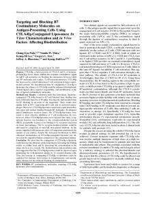
FIGURE 4. Immunohistochemistry of graft-infiltrating CD45⫹ cells in representative corneal graft sections. (a) Syngeneic graft and allograft in (b) untreated, (c) PD-L1.Ig-treated, (d) HuIgG1Fc-treated, (e) antiICOS antibody–treated, or (f) rat IgG2b-treated BALB/c recipient. Magnification, ⫻100.
vival. As in murine cardiac grafts,11 it may be necessary to combine anti-ICOS antibody with a calcineurin inhibitor or anti-CD154 antibody treatment to observe a more significant in vivo effect. In contrast to the experimental approach that reduces T-cell activation by inhibiting positive costimulatory molecule signaling, augmented ligation of the downregulatory, costimulatory ligand PD-1 is a converse method. The two earlier reports on cardiac and islet transplantation models demonstrated that PD-L1.Ig treatment alone did not extend heart or islet graft survival, but did so when administered with Cys23 and with anti-CD154 antibody,24 respectively. As with ICOS modulation, cornea differs from other allografts in PD-1 modulation. In our experiments, PD-L1.Ig administration without any additional intervention significantly reduced alloreactive graft infiltrates and extended corneal allograft survival. We found that engagement of PD-1 with the dimeric recombinant fusion protein, consisting of the extracellular domain of PD-L1 and the Fc portion of human IgG1, at the time of anti-CD3 stimulation inhibited T-cell proliferation in vitro. This finding, along with the effects of the fusion protein on corneal allografts in vivo, is consistent with the reports of other groups using this recombinant protein and supports the growing consensus of the role of PD-1 as a negative regulator of T-cell activation.24,34 One possibility for the difference between our data and the findings in these other studies could be the potential differences in PD-L1.Ig fusion proteins used. Although the fusion protein used in the experiments described was prepared in our laboratory, others have the same PD-L1.Ig construct design using the extracellular portion of the ligand PD-L1 fusing it to HuIgG1Fc.23,24 Subudhi et al.35 have also
3421
compared PD-L1.Ig developed in different laboratories and demonstrated consistent in vivo results. PD-1 ligation triggers negative regulation in lymphocytes and appears to play an important role in the regulation of peripheral tolerance.21 A noteworthy feature of the PD-1 costimulatory pathway is a very broad expression of both the receptor and its PD-L1 and -L2 ligands in lymphoid and nonlymphoid tissue in comparison to the better characterized negative costimulation pathway: the CTLA4 receptor and its CD80/86 ligands.34,36 PD-1 is induced on activated T, B, and myeloid cells,37 whereas PD-1 ligands are constitutively expressed in parenchymal tissues and induced on activated lymphocytes, APCs, and endothelium. We have found that PD-L1 is expressed on activated mouse corneal endothelial cells (manuscript in preparation), suggesting a role for PD-L1 in downregulating T-cell responses and the maintenance of immune privilege in the anterior chamber. Therefore a possible explanation for delayed corneal allograft rejection after treatment with PD-L1.Ig alone may be a synergistic effect of the endogenous PD-L1 expressed on the endothelium of the donor and residual recipient cornea with systemically administered PD-L1.Ig. The exogenous PD-L1.Ig may function at the regional lymph node in suppressing T-cell activation, which alone may not be sufficient to prolong graft survival. However, alloreactive T cells that are not suppressed by PD-L1.Ig and migrate to the anterior chamber may then be suppressed by endogenous PD-L1 expressed on graft endothelium. Although each mechanism alone may be insufficient to prolong graft survival, an additive effect may result from the combination in vivo. Finally, it is noteworthy that the C3H (H-2k)3 BALB/c (H2d) donor–recipient strain combination used in these experiments is a high-responder allogeneic combination, with median corneal graft survival in unmodified graft recipients of 12 days. In contrast, reported survival times in BALB/c recipients of C57BL/6 (H-2b) and C57BL/10 (H-2b) donor corneas were 5 weeks38 and 38 days, respectively.39 Irrespective of the immunogenetic basis of the variation in survival in combinations with different histocompatibility mismatches, experimentation in a high-responder combination is one strategy for evaluating immunomodulatory agents with greatest therapeutic potential. This may be one reason underlying the failure to find an effect on graft survival after ICOS modulation and may explain why, even though graft survival was prolonged, all grafts were rejected in PD-L1.Ig-treated recipients by 40 days. Combination with reagents blocking other costimulatory pathways such as CD28 and with subtherapeutic doses of calcineurin inhibitors in further studies would be of clear interest.
FIGURE 5. Graft-infiltrating cell counts at ⫻500 magnification. The mean ⫾ SD of positive cells counted in three corneas in each group is shown. The number of positive cells was significantly lower in the syngeneic (**P ⬍ 0.001) and PD-L1.Ig-treated groups (*P ⬍ 0.001) compared with the other treatment groups.
3422
Watson et al.
In summary, these results indicate significant therapeutic potential for reagents ligating PD-1 but not, at least on the basis of the preliminary experiments reported, ICOS in corneal transplantation. There is no clear explanation for some divergence of these findings from those reported in other transplantation models. However, as in any such comparison of corneal transplantation to that of other tissues, possible influences of fundamental differences in corneal transplantation, such as the predominance of indirect allorecognition and multiple mechanisms contributing to relative immune privilege, must be borne in mind.
Acknowledgments The authors acknowledge the assistance of Nicola Rogers in the construction of the fusion protein PD-L1.Ig.
References 1. Reis A, Reinhard T, Voiculescu A, et al. Mycophenolate mofetil versus cyclosporin A in high risk keratoplasty patients: a prospectively randomised clinical trial. Br J Ophthalmol. 1999;83:1268 –1271. 2. George AJT, Larkin DFP. Corneal transplantation: the forgotten graft. Am J Transplant. 2004;4:678 – 685. 3. Larkin DFP, Alexander RA, Cree IA. Infiltrating inflammatory cell phenotypes and apoptosis in rejected human corneal allografts. Eye. 1997;11:68 –74. 4. Larkin DFP, Calder VL, Lightman SL. Identification and characterization of cells infiltrating the graft and aqueous humour in rat corneal allograft rejection. Clin Exp Immunol. 1997;107:381–391. 5. Bromley SK, Iaboni A, Davis SJ, et al. The immunological synapse and CD28-CD80 interactions. Nat Immunol. 2001;2:1159 –1166. 6. Walunas TL, Lenschow DJ, Bakker CY, et al. CTLA-4 can function as a negative regulator of T cell activation. Immunity. 1994;1:405– 413. 7. Hutloff A, Dittrich AM, Beier KC, et al. ICOS is an inducible T-cell co-stimulator structurally and functionally related to CD28. Nature. 1999;397:263–266. 8. Coyle AJ, Gutierrez-Ramos JC. The expanding B7 superfamily: increasing complexity in costimulatory signals regulating T cell function. Nat Immunol. 2001;2:203–209. 9. Yoshinaga SK, Whoriskey JS, Khare SD, et al. T-cell co-stimulation through B7RP-1 and ICOS. Nature. 1999;402:827– 832. 10. London CA, Lodge MP, Abbas AK. Functional responses and costimulator dependence of memory CD4⫹ T cells. J Immunol. 2000;164:265–272. 11. Ozkaynak E, Gao W, Shemmeri N, et al. Importance of ICOSB7RP-1 costimulation in acute and chronic allograft rejection. Nat Immunol. 2001;2:591–596. 12. Guo L, Li XK, Funeshima N, et al. Prolonged survival in rat liver transplantation with mouse monoclonal antibody against an inducible costimulator (ICOS). Transplantation. 2002;73:1027–1032. 13. Nakamura Y, Yasunami Y, Satoh M, et al. Acceptance of islet allografts in the liver of mice by blockade of an inducible costimulator. Transplantation. 2003;75:1115–1118. 14. Kosuge H, Suzuki J, Gotoh R, et al. Induction of immunologic tolerance to cardiac allograft by simultaneous blockade of inducible co-stimulator and cytotoxic T-lymphocyte antigen 4 pathway. Transplantation. 2003;75:1374 –1379. 15. Harada H, Salama AD, Sho M, et al. The role of the ICOS-B7h T cell costimulatory pathway in transplantation immunity. J Clin Invest. 2003;112:234 –243. 16. Taylor PA, Panoskaltsis-Mortari A, Freeman GJ, et al. Targeting of inducible costimulator (ICOS) expressed on alloreactive T cells down-regulates graft-versus-host disease (GVHD) and facilitates engraftment of allogeneic bone marrow. Blood. 2005;105:3372– 3380. 17. Ishida Y, Agata Y, Shibahara K, Honjo T. Induced expression of PD-1, a novel member of the immunoglobulin gene superfamily, upon programmed cell death. EMBO J. 1992;11:3887–3895. 18. Latchman Y, Wood CR, Chernova T, et al. PD-L2 is a second ligand for PD-1 and inhibits T cell activation. Nat Immunol. 2001;2:261– 268.
IOVS, August 2006, Vol. 47, No. 8 19. Okazaki T, Maeda A, Nishimura H, Kurosaki T, Honjo T. PD-1 immunoreceptor inhibits B cell receptor-mediated signaling by recruiting src homology 2-domain-containing tyrosine phosphatase 2 to phosphotyrosine. Proc Natl Acad Sci USA. 2001;98: 13866 –13871. 20. Nishimura H, Nose M, Hiai H, Minato N, Honjo T. Development of lupus-like autoimmune diseases by disruption of the PD-1 gene encoding an ITIM motif-carrying immunoreceptor. Immunity. 1999;11:141–151. 21. Nishimura H, Honjo T. PD-1: an inhibitory immunoreceptor involved in peripheral tolerance. Trends Immunol. 2001;22:265– 268. 22. Keir ME, Latchman YE, Freeman GJ, Sharpe AH. Programmed death-1 (PD-1):PD-ligand-1 interactions inhibit TCR-mediated positive selection of thymocytes. J Immunol. 2005;175:7372–7379. 23. Ozkaynak E, Wang L, Goodearl A, et al. Programmed death-1 targeting can promote allograft survival. J Immunol. 2002;169: 6546 – 6553. 24. Gao W, Demirci G, Strom TB, Li XC. Stimulating PD-1-negative signals concurrent with blocking CD154 co-stimulation induces long-term islet allograft survival. Transplantation. 2003;76:994 – 999. 25. Qian Y, Boisgerault F, Benichou G, Dana MR. Blockade of CD40CD154 costimulatory pathway promotes survival of allogeneic corneal transplants. Invest Ophthalmol Vis Sci. 2001;42:987–994. 26. Comer RM, King WJ, Ardjomand N, Theoharis S, George AJ, Larkin DFP. Effect of administration of CTLA4-Ig as protein or cDNA on corneal allograft survival. Invest Ophthalmol Vis Sci. 2002;43: 1095–1103. 27. Ardjomand N, McAlister JC, Rogers NJ, Tan PH, George AJ, Larkin DFP. Modulation of costimulation by CD28 and CD154 alters the kinetics and cellular characteristics of corneal allograft rejection. Invest Ophthalmol Vis Sci. 2003;44:3899 –3905. 28. Zhang EP, Schrunder S, Hoffmann F. Orthotopic corneal transplantation in the mouse: a new surgical technique with minimal endothelial cell loss. Graefes Arch Clin Exp Ophthalmol. 1996;234: 714 –719. 29. Sonoda Y, Streilein JW. Impaired cell-mediated immunity in mice bearing healthy orthotopic corneal allografts. J Immunol. 1993; 150:1727–1734. 30. Kaplan EL, Meier P. Nonparametric estimation from incomplete observations. J Am Stat Assoc. 1958;53:457– 481. 31. Larsen CP, Alexander DZ, Hollenbaugh D, et al. CD40-gp39 interactions play a critical role during allograft rejection. Suppression of allograft rejection by blockade of the CD40-gp39 pathway. Transplantation. 1996;61:4 –9. 32. Larsen CP, Elwood ET, Alexander DZ, et al. Long-term acceptance of skin and cardiac allografts after blocking CD40 and CD28 pathways. Nature. 1996;381:434 – 438. 33. Kenyon NS, Chatzipetrou M, Masetti M, et al. Long-term survival and function of intrahepatic islet allografts in rhesus monkeys treated with humanized anti-CD154. Proc Natl Acad Sci USA. 1999;96:8132– 8137. 34. Freeman GJ, Long AJ, Iwai Y, et al. Engagement of the PD-1 immunoinhibitory receptor by a novel B7 family member leads to negative regulation of lymphocyte activation. J Exp Med. 2000; 192:1027–1034. 35. Subudhi SK, Zhou P, Yerian LM, et al. Local expression of B7–H1 promotes organ-specific autoimmunity and transplant rejection. J Clin Invest. 2004;113:694 –700. 36. Carter L, Fouser LA, Jussif J, et al. PD-1:PD-L inhibitory pathway affects both CD4(⫹) and CD8(⫹) T cells and is overcome by IL-2. Eur J Immunol. 2002;32:634 – 643. 37. Agata Y, Kawasaki A, Nishimura H, et al. Expression of the PD-1 antigen on the surface of stimulated mouse T and B lymphocytes. Int Immunol. 1996;8:765–772. 38. Yamagami S, Dana MR, Tsuru T. Draining lymph nodes play an essential role in alloimmunity generated in response to high-risk corneal transplantation. Cornea. 2002;21:405– 409. 39. Vitova A, Filipec M, Zajicova A, Krulova M, Holan V. Prevention of corneal allograft rejection in a mouse model of high risk recipients. Br J Ophthalmol. 2004;88:1338 –1342.