Differential Expression of Matrix Metalloproteinases and Their Tissue Inhibitors at the Advancing Pterygium Head Nick Di Girolamo,1 Denis Wakefield,1 and Minas T. Coroneo2 PURPOSE. Pterygia are a proliferative and inflammatory growth of limbal epithelial stem cell origin, characterized by corneal tissue invasion and extensive matrix remodeling including the destruction of Bowman’s layer (BL). The purpose of this study was to determine the expression of matrix metalloproteinases (MMPs) and tissue inhibitors of MMPs (TIMPs) at the advancing pterygium edge. METHODS. Formalin-fixed, paraffin-embedded whole eyes (n ⫽ 11) with pterygia attached, were serially sectioned and analyzed immunohistochemically to determine the spatial distribution of four MMPs and three TIMPs. Tear samples were collected from other patients with pterygia (n ⫽ 11) and displayed by gelatin zymography. RESULTS. Collagenase-1 was expressed by pterygium epithelial cells, corneal stromal fibroblasts and pterygium fibroblasts that had migrated between the epithelium and BL at the advancing pterygium edge. Collagenase-3 and gelatinases A and B were detected in all pterygia, intensely staining columnar epithelial cells directly adjacent to the denatured BL. In addition, gelatinase A immunoreactivity was observed on BL. Immunoreactivity for TIMP-1 and -3 paralleled that of the gelatinases, with more intense staining in epithelial cells and fibroblasts where BL was absent. TIMP-2 was faintly detected in pterygium epithelial cells but intensely stained pterygium fibroblasts. Gelatinase B was the most abundant gelatinolytic enzyme present in tears, elevated approximately twofold in eyes with pterygia versus the contralateral control eyes. CONCLUSIONS. This investigation is the first to identify the expression pattern of MMPs and TIMPs at the advancing pterygium edge in specimens of human eyes and in tears derived from patients with pterygia. These enzymes may be responsible for the destruction of BL, and their pattern of differential expression suggests that each may play a selective role in the pathogenesis of pterygia. (Invest Ophthalmol Vis Sci. 2000;41:4142– 4149)
P
terygia represent an invasion of a wing of altered ocular surface tissue into the normal cornea.1 Although the pathogenesis is still poorly understood, it has been proposed that pterygia originate at the limbus and involve the activation and proliferation of altered basal limbal epithelial stem cells that migrate and dissolve Bowman’s layer (BL).2 Despite the lack of knowledge regarding the pathogenesis of pterygia, epidemiologic evidence suggests that exposure to UV-irradiation may be an initial trigger in the development of this lesion.3 Although there exists extensive literature on pterygia, controversy still surrounds the relative roles of the pterygium fibroblast and epithelial cell in the pathogenesis of this disease. Recently, we have demonstrated the importance of the pterygium epithelial cell in the development of pterygia, because
From the 1Inflammation Research Unit, School of Pathology, University of New South Wales; and the 2Department of Ophthalmology, Prince of Wales Hospital, Sydney, New South Wales, Australia. Supported by the National Health and Medical Research Council of Australia. Submitted for publication April 19, 2000; revised August 2, 2000; accepted August 14, 2000. Commercial relationships policy: N. Corresponding author: Nick Di Girolamo, Inflammation Research Unit, School of Pathology, The University of New South Wales, Sydney 2052, Australia.
[email protected]
this cell type was found to express abundant levels of matrix metalloproteinases (MMPs) in resected tissue specimens and in vitro, in a setting of inflammation.4 Lee et al.5 have also observed a marked upregulation of MMPs in pterygium fibroblasts compared with normal conjunctival fibroblasts. In addition, cytokines and growth factors, such as basic fibroblast growth factor (bFGF), platelet-derived growth factor (PDGF), transforming growth factor (TGF)-, and tumor necrosis factor (TNF)-␣, have been localized to pterygium cells.6 Therefore, it is not unreasonable to propose that both cell types may act in concert to degrade BL and other connective tissue structural components. MMPs are a family of zinc-dependent endopeptidases capable of denaturing most components of the extracellular matrix.7 These enzymes share common structural and functional elements and are involved in physiological8,9 and pathologic processes,10 including cancer,11 arthritis,12 and inflammatory diseases of the eye.13,14 They are broadly divided into four groups according to substrate specificity and include, the collagenases, gelatinases, stromelysins, and the membrane-type MMPs. MMPs are regulated at multiple levels including transcription, secretion, activation, and inhibition. The latter accomplished by naturally occurring proteins called tissue inhibitors of MMPs (TIMPs).15 The balance between the levels of activated enzymes and free TIMPs determines the overall MMP activity. Maintenance of this equilibrium is essential, and any
4142
Investigative Ophthalmology & Visual Science, December 2000, Vol. 41, No. 13 Copyright © Association for Research in Vision and Ophthalmology
MMPs and TIMPs at the Invading Pterygium Edge
IOVS, December 2000, Vol. 41, No. 13 TABLE 1. Ocular Tissue Used for Histochemical and Zymographic Analysis Donor
Age/Sex
Location/Eye
Cause of Death
1 2 3 4 5 6 7 8 9 10 11 12 13 14 15
81/F 48/M 62/M 71/M 83/M 67/M 58/M 69/M 44/F 60/M 78/F 53/M 49/F 28/M 60/M
N/R N/R N/R N/R N&T/R N/R N/L N&T/L T/L N/L N/R N/R N/R N/R N/L
Intracranial hemorrhage Multiple injuries Myocardial infarct Respiratory failure Aortic aneurysm Respiratory failure Carbon monoxide poisoning Heart disease Glioblastoma Cardiac arrest Cardiac arrest
Histological examination by an ocular pathologist revealed no other ocular abnormalities. Other disease complications included diabetes (donor 1), emphysema (donor 4), depression (donor 7), and osteoarthritis (donor 10). Tissue from patients 12 through 15 were fresh surgical specimens from living patients. N, nasal; T, temporal; R, right; L, left.
disturbance in the balance is a critical determinant of proteolysis and tissue invasion. The purposes of this study were to determine the expression profile of MMPs and TIMPs in pterygia with intact underlying corneal stroma, paying particular attention to the advancing edge. In addition we analyzed the relative contribution of these proteins by both the pterygium fibroblast and epithelial cells, and finally we determined the gelatinolytic profile of tears derived from patients with pterygia.
MATERIALS
AND
METHODS
Donor Ocular Tissue Whole eye specimens (n ⫽ 11) were obtained from the Department of Anatomic Pathology, Prince of Wales Hospital, Sydney, Australia. All tissue specimens were formalin fixed and paraffin embedded. To determine the activation status of the MMPs, fresh pterygium specimens were obtained from surgery and immediately placed in organ culture for 48 hours in serumfree Eagle’s minimum essential medium (EMEM, Trace Biosciences, Sydney, Australia). The supernatants were collected and stored at ⫺70°C for zymographic analysis. Only eyes with primary pterygia and no signs of other ocular pathology or abnormality were obtained for this study. Patient details are summarized in Table 1. All research protocols and patients were treated in accordance with the tenets of the Declaration of Helsinki.
Collection of Tears from Patients with Pterygia Unstimulated tear fluid (approximately 1–5 l) was harvested from patient eyes with primary pterygia (n ⫽ 11) and control subjects (n ⫽ 8) by placing a 10-l capillary tube in the inferior cul-de-sac of each eye. Because only small volumes could be extracted from an individual eye, protein standardization was not possible, because this assay would require most, if not all
4143
TABLE 2. Tear Fluid from Patients with Pterygia Patient
Age/Sex
Location/Eye
Tears (l)
DJ JP SD NH NM SC PP CH DW RW JL
40/M 28/M 40/F 60/F 32/M 38/F 29/M 32/M 66/M 22/F 44/M
N/R N/L N/L N/R N/L N/L N/L N/L N/L N/R N/L
1.5 1.3 1.5 2.7 3.0 3.5 1.2 1.3 3.0 2.5 4.5
All pterygia were localized on the nasal side of the left (L) or the right (R) eye. The same volume of tears from both the pterygia affected and the control contralateral eye from the one subject was analyzed by zymography. N, nasal.
the tear specimen. In addition, different volumes were often collected between the left and right eyes of the same patient, and diluted tear fluid often resulted in inaccurate protein measurements and weak or undetectable gelatinolytic bands in the zymograms. Tears were stored frozen at ⫺70°C until used in zymography. Tears from the contralateral eyes (without pterygia) served as a control. All patients had unilateral lesions. The patients’ details are summarized in Table 2. Control subjects included three women and five men, with a mean age of 33 years.
Immunohistochemical Analysis Whole human eyes with pterygia attached were serially sectioned (2– 4 m) and histologically evaluated by staining with H&E or sirius red, then processed immunohistochemically as previously described.4 Briefly, sections were deparaffinized in xylene, rehydrated through decreasing graded ethanol, quenched for endogenous peroxidase, and then incubated with preimmune serum from the secondary host species. Antigen retrieval using proteinase K digestion13 was required before the addition of the TIMP-2 antibody (Ab). Tissue sections were incubated with mouse primary monoclonal Abs (see Table 3 for source and dilution) overnight at 4°C, then extensively washed in 0.05 M Tris-buffered saline
TABLE 3. Antibodies Used for Immunohistochemical Analyses Antibody
Source
Dilution
Clone
Collagenase-1 Collagenase-3 Gelatinase A Gelatinase B TIMP-1 TIMP-2 TIMP-3 Mouse IgG1
ICN ORP ICN ICN ICN ICN ORP Dako
1:500 1:25 1:20 1:100 1:50 1:50 1:50 1:50
41-1E5 181-15A12 42-5D11 56-2A4 147-6D11 67-4H11 136-13H4 DAK-G01
All Abs were titrated using a range of concentrations, until optimal dilutions were established for the formalin-fixed ocular tissue. These Abs are directed against human antigens, show no cross-reactivity (as specified by the manufacturer), and are all IgG1 subclass mouse monoclonal Abs. Dako, ICN Biomedicals (Sydney, Australia) Oncogene Research Products (ORP; Cambridge, MA).
4144
Di Girolamo et al.
IOVS, December 2000, Vol. 41, No. 13
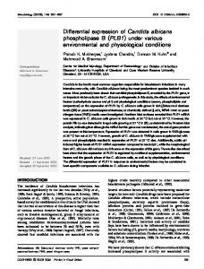
FIGURE 1. Histologic features of human pterygia. Pterygium tissue was sectioned and stained with H&E (A, B, and C) or with sirius red (D, E), with (D) or without (E) polarized light to reveal typical representative histopathologic features of this lesion. (A, B, and C) Three contiguous composite photographs that show the pterygium body (A) extending to the advancing pterygium edge (C). No disease was observed at the central cornea (inset, C). Arrows: Regions of intact BL; (ⴱ) regions where BL has become denatured. Typically, these are also areas where pterygium fibroblasts accumulate (f). Other features include areas of denatured or elastotic matrix (dm) where blood vessels (v) are prominent. cs, corneal stroma; e, epithelium. Original magnification: (A, B, and C), ⫻500; (D, E), ⫻640.
(pH 7.6), before the addition of a biotinylated goat antimouse secondary Ab. Sections were again washed, incubated for 1 hour with horseradish peroxidase-conjugated streptavidin (Dako, Carpinteria, CA), and the immunoreactivity revealed by adding 3-amino-9-ethylcarbazole (Sigma, Sydney, Australia). Control reactions included, incubating sections with an isotype Ab (see Table 3), omitting the primary Ab or adding preimmune serum. Sections were counterstained with hematoxylin.
Gelatin Substrate Zymography Zymography was performed as previously described.4,13,14 Briefly, tear samples were thawed and an equal volume of tears from eyes with and without pterygia were diluted with nonreducing sample buffer (10% sodium dodecyl sulfate [SDS], 4% sucrose, 0.25M Tris-HCl [pH 6.8], with 0.1% bromophenol blue) and loaded without boiling under nonreducing conditions. After electrophoresis, the gels were washed twice for 30 minutes each in 2.5% Triton X-100 (Sigma). Gels were rinsed and incubated overnight at 37°C in substrate buffer (50 mM Tris-HCl [pH 7.4], 10 mM CaCl2, and 0.02% NaN3), and stained (Coomassie Blue R-250; Bio-Rad, Sydney, Australia). Enzymatic activity was identified as clear zones in a blue-stained background. A low-range molecular weight standard (Bio-Rad) was run in adjacent lanes. MMP identity and activity were verified by running a sample of conditioned media derived from phorbol myristate acetate (PMA, Sigma)–stimulated pterygium epithelial cells (a potent enhancer of MMP expression).13 The addition of EDTA (10 mM final) and 1,10-phenanthroline (1 mM final; Sigma) completely abolished the gelatinolytic activity. Photographs were taken (665 film Polaroid; Cambridge, MA) and the negative exposures scanned with a densitometer
(Hoefer Scientific, San Francisco, CA) to obtain semiquantitative data.
RESULTS Histopathologic Features of the Invading Pterygium Histologic features of pterygia included a multilayered proliferating squamous epithelium, (Figs. 1A, 1B) which decreased in thickness at the advancing edge (Fig. 1C) where the pterygium encroached on the normal cornea. Behind the advancing head there was an accumulation of fibrous and/or elastotic connective tissue matrix (Fig. 1A, dm). These regions usually contain inflammatory cells (micrographs not shown4) and blood vessels (Fig. 1A, v) that may provide both stimulatory signals and nutrients for the proliferating and invading tissue mass. A characteristic feature of pterygia is the loss of BL, a natural barrier that separates the corneal epithelium from the underlying stroma. This structure is normally attached to the epithelial basement membrane (Fig. 1A, arrow) and is destroyed at the pterygium leading edge (Figs. 1A, 1B, 1C, asterisks) by an as yet unknown mechanism. In regions void of BL or where BL has detached from the underlying epithelium, clusters of pterygium fibroblasts accumulate (Figs. 1B, through 1E, f). Dissolution of BL was evident directly adjacent to pterygium epithelial cells (Figs. 1D, 1E, arrows), where BL was more irregular and fragmented (Figs. 1D, 1E, asterisks). The central cornea appeared normal, with intact BL and the absence pterygium fibroblasts (Fig. 1C, inset).
IOVS, December 2000, Vol. 41, No. 13
MMPs and TIMPs at the Invading Pterygium Edge
4145
FIGURE 2. Immunohistochemical localization of collagenases and gelatinases in the invading pterygia. Pterygium tissue was serially sectioned and stained for collagenase-1 (A, B, and C), collagenase-3 (D, E, and F), gelatinase A (G, H, and I), and gelatinase B (J, K, and L). Insets: Immunoreactivity at the central cornea with the respective Abs. Three consecutive photographs were taken along the length of the pterygium and a composite image compiled for each Ab. Red staining was regarded as specific immunoreactivity. Sections were counterstained with hematoxylin. Control samples included omitting the primary Ab (micrographs not shown) and incubating the tissue sections with an isotype control Ab (see Figs. 3J, 3K, 3L). Similar results were obtained with all other pterygium tissue specimens analyzed. The labels and abbreviations used in this figure are identical with those in Figure 1. Original magnification, ⫻500.
Expression of Collagenase-1 and -3 at the Leading Pterygium Edge Having identified the invading pterygium edge in all tissue specimens, we sectioned whole human eyes serially and analyzed them immunohistochemically to determine the expression profile of MMPs and TIMPs. Initially, tissue sections were stained for the collagenases. Immunoreactivity for collagenase-1 (MMP-1) was abundant, intensely staining blood vessels and detected along the entire length of the pterygium, predominantly within the pterygium epithelium as well as in pterygium fibroblasts (Figs. 2A, 2B, 2C). Reactivity for this protease was diminished toward the central cornea (Fig. 2C, inset). In
addition, collagenase-1 was expressed by some stromal fibroblasts and often associated with the stromal connective tissue, suggesting that it may be sequestered on specific matrix components. In contrast to collagenase-1, collagenase-3 (MMP-13) was differentially expressed within the same pterygium specimen. This enzyme stained more intensely the basal and columnar epithelium, particularly in the regions of fragmented BL, adjacent to pterygium fibroblasts, which themselves contained some immunoreactivity (Figs. 2D, 2E, 2F). Collagenase-3 was also localized to vascular endothelial cells (Fig. 2D, v) and some inflammatory cells (micrographs not shown). Faint staining was detected in stromal fibroblasts, none was matrix associ-
4146
Di Girolamo et al.
ated, and minimal reactivity was detected at or near the central cornea (Fig. 2F, inset).
Differential Expression of Gelatinases A and B at the Pterygium Leading Edge Gelatinase A (MMP-2) immunoreactivity was intense, particularly in the basal columnar epithelial cells, adjacent to regions of denatured BL (Figs. 2G, 2H, 2I). However, only faint staining for this basement membrane– degrading enzyme was noted in superficial epithelial layers and in pterygium fibroblasts, with some reactivity in the central corneal epithelium (Fig. 2I, inset). This enzyme was not expressed by the vascular endothelium or corneal stromal fibroblasts, nor was it associated with the corneal stromal matrix (as was collagenase-1). Of note, gelatinase A staining was also found specifically associated with BL, in regions adjacent to the pterygium (Fig. 2G, arrow), but not at the central cornea. In regions where BL was fragmented (Figs. 2G, 2H, 2I, asterisks), gelatinase A immunoreactivity was diffuse, and BL became indistinguishable from the stromal matrix. Additional immunoreactivity was found within the fibrous degenerative matrix of the pterygium body (Fig. 2G). Gelatinase B (MMP-9) staining was similar to that of gelatinase A, most abundant in the basal epithelium. However, pterygium fibroblasts, inflammatory cells, and the vascular endothelium also showed staining. In contrast to gelatinase A, gelatinase B was not found associated with BL or in the fibrous connective tissue, nor was it detected in stromal fibroblasts (Figs. 2J, 2K, 2L). Rarely was this proteinase detected at the central cornea. When it was, however, the intensity of staining was usually less than in the pterygium (Fig. 2L, inset).
Expressions of TIMPs 1 to 3 at the Pterygium Leading Edge TIMP-1 expression closely resembled that of collagenase-1, in that all pterygium epithelial cells contained some immunoreactivity for this inhibitor. Similarly, the intensity of staining was greatest in the more basal epithelium, particularly in the vicinity of the denatured BL. Additional TIMP-1 signal was associated with pterygium fibroblasts and fibrous bundles of the pterygium body and was faintly detected in some stromal fibroblasts (Figs. 3A, 3B, 3C). Weak epithelial cell reactivity was demonstrated at the central cornea (Fig. 3C, inset). TIMP-2 reactivity was weak or undetectable in the pterygium epithelium, corneal stromal fibroblasts, and vascular endothelium but was identified on pterygium fibroblasts (Fig. 3D, 3E, 3F) in 8 of 11 specimens. TIMP-3 staining paralleled that of TIMP-1, detected in most pterygium epithelium, and diminished in intensity from basal to superficial and from pterygium to corneal epithelium. Pterygium fibroblasts were distinctly immunoreactive, as were vascular endothelial cells and inflammatory cells (Fig. 3G, 3F, 3I). Regions of specific connective tissue staining by TIMP-3 were also observed in most specimens (micrograph not shown).4 This pattern of MMP and TIMP staining was generally observed with all tissue specimens analyzed.
Secretion of Latent and Active MMPs from Pterygium Tissue Of the four MMPs immunolocalized at the advancing pterygium edge (Fig. 2), latent and active forms of three of these enzymes were detected by gelatin zymography (Fig. 4). Progelatinase B (92 kDa) and progelatinase A (72 kDa) were detected in all four
IOVS, December 2000, Vol. 41, No. 13 pterygium tissue specimens. In addition, the active form of each gelatinase was displayed as a lower molecular weight species (gelatinase B, 83 kDa; gelatinase A, 66 kDa). Although other MMPs do not efficiently denature gelatin in this assay system, procollagenase-1 was detected as a faint doublet at 57 and 54 kDa, along with its proteolytically active species which migrated to 45 kDa.
Gelatinase B in Tears from Eyes with Pterygia Basal tear fluid was collected from patients with pterygia to determine the gelatinolytic content. Two prominent gelatinolytic activities were revealed: a 92-kDa and a 125-kDa band corresponding to gelatinase B and gelatinase B/TIMP complex, respectively. Densitometric analysis of these bands established that on average, gelatinase B and the gelatinase B/TIMP complex were elevated at least 1.7- and 1.35-fold respectively in eyes with pterygia compared with the control eyes from the same patients (Fig. 5A). Previously, we identified a similar 125-kDa gelatinolytic band in aqueous humor samples from patients with uveitis as an MMP-9/TIMP complex by Western blot analysis.14 Collagenase-1 was not detected by this method, and gelatinase A was only faintly detected in 3 of 11 samples of tear fluid from eyes with pterygia. Although, gelatinase B and the 125-kDa gelatinase/TIMP complex were detected in the tears of control subjects, densitometrically there was no significant difference in the levels of this enzyme between the left and right eyes of an individual (Fig. 5B).
DISCUSSION Whereas previous studies used resected pterygium tissue4 or in vitro assays of cultured pterygium epithelial cells4 and fibroblasts,5 in the present study we used whole human eyes with attached pterygia to determine the expression of MMPs and TIMPs along the entire invading edge. Several novel observations were recorded including the differential expression of MMPs and TIMPs within the same tissue specimen along the length of the lesion, the expression of collagenase-3, the localization of gelatinase A to BL, the relative contribution of MMPs and TIMPs by the pterygium fibroblast compared with the pterygium epithelial cell, the production of active and latent MMPs by pterygium tissue, and the detection of increased levels of gelatinase B in tears from patients with pterygia. A characteristic feature of pterygia is the loss of BL, a natural collagenous barrier that separates the epithelium from the underlying stroma. The mechanism responsible for its destruction in pterygia is currently unknown. In the present study, the overexpression of MMPs and TIMPs by pterygium epithelial cells in regions without BL was a significant finding and suggests that these proteins may contribute to the matrix remodeling and BL destruction. The absence of BL also coincided with the presence of MMP- and TIMP-expressing pterygium fibroblasts. However, the staining intensity for any given MMP or TIMP (with the exception of TIMP-2) was greatest in pterygium epithelial cells when compared with pterygium fibroblasts. In a recent study, Dushku et al.16 identified the expression of six different MMPs in altered limbal basal epithelial cells, whereas collagenase-1 was the only proteinase found in pterygium fibroblasts. Taken together, these data suggest that the epithelial cell is the more likely candidate involved in the dissolution of BL. However, the potential involvement of
IOVS, December 2000, Vol. 41, No. 13
MMPs and TIMPs at the Invading Pterygium Edge
4147
FIGURE 3. Immunohistochemical localization of the TIMPs in the advancing pterygia. Pterygium tissue was serially sectioned and stained for TIMP-1 (A, B, and C), TIMP-2 (D, E, and F), TIMP-3 (G, H, and I), or an isotype control Ab (J, K, and L). Insets represent immunoreactivity near or at the central cornea, with the respective Abs. Three consecutive photographs were taken along the length of the pterygium and a composite image compiled for each Ab. Red staining was regarded as specific immunoreactivity. Sections were counterstained with hematoxylin. Other controls included omitting the primary Ab or incubating tissue sections with preimmune serum (micrographs not shown). These results are representative of all other pterygium tissue specimens analyzed. The labels and abbreviations used in this figure are identical with those in Figure 1. Original magnification, ⫻500.
pterygium fibroblasts cannot be ruled out, because the assay used in the present study does not quantify enzyme production. In keep with the hypothesis that the pterygium epithelial cell may be the cell involved in the initial breakdown of BL, histologic examination of ocular tissue identified regions of fragmented BL directly beneath the epithelium (Fig. 1E). Despite the specific localization of MMPs and TIMPs in pterygia, MMP staining does not necessarily translate to MMP activity. Furthermore, the Abs used in this study do not discriminate between active and latent MMPs. In situ zymography, a technique used to determine enzymatic activity in fresh-frozen tissue sections, has recently been developed to address these important issues.17,18 However, this assay could not be applied
to the archival paraffin-embedded tissue used in the present study. We have overcome this limitation in our study by demonstrating the presence of both active and latent MMPs as secreted proteins from pterygium specimens. Thus, it is likely that at least some of the MMP immunostaining (Fig. 2) represents proteolytically active molecules. Another significant observation recorded in this study was the specific localization of MMPs and TIMPs on matrix components. Although matrix-associated TIMPs may function to inhibit connective tissue proteolysis.19 matrix-bound MMPs may actively degrade these structural proteins.20 –22 It is tempting to speculate that the abundant expression of both enzymes and inhibitors is a reason that these benign lesions can develop
4148
Di Girolamo et al.
over decades. In contrast, it has been reported that malignant tumors develop rapidly as a consequence of MMP overexpression and TIMP downregulation.23 Of interest, staining for gelatinase A was found to be associated with BL and in elastotic regions, whereas its structurally and functionally related molecule gelatinase B was not. Both gelatinases A and B contain fibronectin-like domains that mediate their matrix-binding capacity.24 Although these proteinases were initially characterized based on their type IV collagenolytic activity, data from other studies suggest that gelatinase A acts as an interstitial collagenase, due to its ability to cleave native type I collagen.25 In this regard, gelatinase A is a likely candidate involved in the fragmentation of BL. Perhaps it is not unreasonable to postulate that the activity of MMPs and TIMPs are essential in the development of pterygia; previous investigators have localized the same family of proteins in destructive corneal disorders18,26,27 and in migrating and regenerating epithelial cells in a rat model of corneal wounding.28 Although the principal function of TIMPs is to inhibit MMP activity,15 paradoxically, at least TIMP-2 is involved in the activation of progelatinase A.29 TIMPs also possess growth factor–like activity, and in this capacity they may be involved in pterygium proliferation; a recent study reported for corneal epithelial cells.30 Other functions attributed to TIMP-1 and TIMP-2 include, suppression31,32 or in the case of TIMP-3, induction of apoptosis,33 a process recently described in pterygia.34 TIMPs have also been reported to inhibit angiogenesis,32,35 a function unlikely to be applicable to pterygia, because these lesions characteristically contain an extensive vascular supply.4,36 The data presented in this investigation do not exclude the possible involvement of UV irradiation1,3,37,38 in the pathogenesis of pterygia. The increased expression of MMPs and TIMPs by basal pterygium epithelial cells (Figs. 2, 3) may be explained by the peripheral light–focusing effect,1,37,38 which predicts that peripheral light entering the eye laterally has a focusing effect of approximately 20-fold at the limbus (the usual site of pterygia). Therefore, the basal epithelial cells may be activated as they are struck from behind by the focused light.
IOVS, December 2000, Vol. 41, No. 13
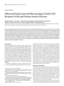
FIGURE 5. Gelatinolytic activity in tears derived from patients with pterygia. (A) Tear samples from four patients with pterygia were displayed by zymography. Equal volume of tears from eyes with pterygia (lanes 2, 4, 6, and 8) and the corresponding control eyes (lanes 1, 3, 5, and 7) were loaded. Lanes 1 and 2: patient DJ (1.5 l of tears); lanes 3 and 4: patient SD (1.5 l of tears); lanes 5 and 6: patient NM (3.0 l of tears); lanes 7 and 8: patient SC (3.5 l of tears). Conditioned media from PMA stimulated pterygium epithelial cells served as a positive control (lane 9). This gelatinolytic profile is representative of the tear fluid from all patients studied. (B) Tear samples from four control subjects were displayed by zymography. Equal volume of tears from left eyes (lanes 1, 3, 5, and 7) and right eyes (lanes 2, 4, 6, and 8) were loaded. Lanes 1 and 2: subject 1 (1.0 l of tears); lanes 3 and 4: subject 2 (1.0 l of tears); lanes 5 and 6: subject 3 (2.5 l of tears); lanes 7 and 8: subject 4 (2.7 l of tears). The same gelatinolytic profile was observed with the other four control subjects.
Although other investigators have identified gelatinase B in tears derived from patients with corneal graft failure39 and ocular rosacea,40 this is the first study to document gelatinase B levels in tears from patients with pterygia (Fig. 5). Furthermore, levels of this protease were greater (1.7-fold) in eyes with pterygia than in the contralateral control eyes, perhaps indicative of an abnormality on the ocular surface. Despite the detection of gelatinase B and an MMP/TIMP complex, no collagenase-1 and no active MMPs were observed, whereas gelatinase A was detected in only three tear samples. The recent development of synthetic inhibitors of MMP activity has provided a potentially new therapeutic strategy for the treatment of cancer, which may be applicable to pterygia. The same agents have been applied to the eye to prevent corneal ulceration and reduce retinal neovascularization41,42 or administered subcutaneously to prevent experimental autoimmune uveoretinitis.43 Anti-MMP strategies, in conjunction with surgical techniques, may be of future therapeutic use to reduce the rate of recurrence, severity of inflammation, tissue invasion, proliferation, and angiogenesis associated with pterygia.
Acknowledgments The authors thank Tina Liakos for her assistance in the collection of tear samples. FIGURE 4. Matrix metalloproteinase activity in pterygia. Fresh pterygia was obtained from surgery and immediately placed in organ culture for 48 hours. Supernatants from four different pterygium tissue specimens (patients 12–15, see Table 1) were displayed by gelatin-substrate zymography (lanes 2 through 5). A molecular weight protein ladder was run in parallel (lane 1). All samples demonstrated a similar profile of MMP activity, including the presence of pro- and active gelatinase B, gelatinase A, and collagenase-1.
References 1. Coroneo MT, Di Girolamo N, Wakefield D. The pathogenesis of pterygia. Curr Opin Ophthalmol. 1999;10:282–288. 2. Dushku N, Reid TW. Immunohistochemical evidence that human pterygia originate from an invasion of vimentin-expressing altered limbal epithelial basal cells. Curr Eye Res. 1994;13:473– 481.
IOVS, December 2000, Vol. 41, No. 13 3. Cameron ME. Geographic distribution of pterygia. Trans Ophthalmol Soc Aust. 1962;22:67– 81. 4. Di Girolamo N, McCluskey PJ, Lloyd A, Coroneo MT, Wakefield D. Expression of MMPs and TIMPs in human pterygia and cultured pterygium epithelial cells. Invest Ophthalmol Vis Sci. 2000;41: 671– 679. 5. Lee S-B, Li D-Q, Gunja–Smith Z, Liu YQ, Tan DTH, Tseng SCG. Increased expression and activity of MMP-1 and MMP-3 by cultured pterygium head fibroblasts [ARVO Abstract]. Invest Ophthalmol Vis Sci. 1999;40(4):S334. Abstract nr 1768. 6. Kria L, Ohira A, Amemiya T. Immunohistochemical localization of basic fibroblast growth factor, platelet derived growth factor, transforming growth factor- and tumor necrosis factor-␣ in the pterygium. Acta Histochem. 1996;98:195–201. 7. Massova I, Kotra LP, Fridman R, Mobashery S. Matrix metalloproteinases: structures, evolution, and diversification. FASEB J. 1998;12:1075–1095. 8. Stetler–Stevenson WG. Matrix metalloproteinases in angiogenesis: a moving target for therapeutic intervention. J Clin Invest. 1999; 103:1237–1241. 9. Ray JM, Stetler–Stevenson WG. The role of matrix metalloproteinases and their inhibitors in tumour invasion, metastasis and angiogenesis. Eur Respir J. 1994;7:2062–2072. 10. Stetler–Stevenson WG. Dynamics of matrix turnover during pathologic remodeling of the extracellular matrix. Am J Pathol. 1996; 148:1345–1350. 11. Lampert K, Machein U, Regina M, Conca, W, Peter HH, Volk B. Expression of matrix metalloproteinases and their tissue inhibitors in human brain tumors. Am J Pathol. 1998;153:429 – 437. 12. Firestein GS, Paine MM. Stromelysin and tissue inhibitor of metalloproteinases gene expression in rheumatoid arthritis synovium. Am J Pathol. 1992;140:1309 –1314. 13. Di Girolamo N, Lloyd A, McCluskey PJ, Filipic M, Wakefield D. Increased expression of matrix metalloproteinases in vivo in scleritis tissue and in vitro in cultured human scleral fibroblasts. Am J Pathol. 1997;105:653– 666. 14. Di Girolamo N, Verma MJ, McCluskey PJ, Lloyd A, Wakefield D. Increased matrix metalloproteinases in the aqueous humor of patients and experimental animals with uveitis. Curr Eye Res. 1996;15:1060 –1068. 15. Gomez DE, Alonso DF, Yoshiji H, Thorgeirsson UP. Tissue inhibitors of metalloproteinases: structure, regulation and biological functions. Eur J Cell Biol. 1997;74:111–122. 16. Dushku N, John MK, Schultz GS, Reid TW. Pterygia pathogenesis: corneal invasion by matrix metalloproteinase (MMP) expressing altered limbal basal stem cells and activation of fibroblasts [ARVO Abstract]. Invest Ophthalmol Vis Sci. 2000;41(4):S451. Abstract nr 2388. 17. Galis ZS, Sukhova GK, Libby P. Microscopic localization of active proteases by in situ zymography: detection of matrix metalloproteinase activity in vascular tissue. FASEB J. 1995;9:974 –980. 18. Zhou L, Sawaguchi S, Twining SS, Sugar J, Feder RS, Yue BYJT. Expression of degradative enzymes and protease inhibitors in corneas with keratoconus. Invest Ophthalmol Vis Sci. 1998;39: 1117–1124. 19. Fariss RN, Apte SS, Olsen BR, Iwata K, Milam AH. Tissue inhibitor of metalloproteinase-3 is a component of Bruch’s membrane of the eye. Am J Pathol. 1997;150:323–328. 20. Dahlen B, Shute J, Howarth P. Immunohistochemical localisation of matrix metalloproteinases MMP-3 and MMP-9 within the airways in asthma. Thorax. 1999;54:590 –596. 21. Allen JA, Docherty AJP, Barker PJ, Huskisson NS, Reynolds JJ, Murphy G. Binding of gelatinases A and B to type-1 collagen and other matrix components. Biochem J. 1995;309:299 –306. 22. Davis V, Persidskaia R, Regen–Baca L, et al. Matrix metalloproteinase-2 production and its binding to the matrix are increased in abdominal aortic aneurysms. Arterioscler Thromb Vasc Biol. 1998; 18:1625–1633.
MMPs and TIMPs at the Invading Pterygium Edge
4149
23. Ara T, Kusafuka T, Inoue M, Kuroda S, Fukuzawa M, Okada A. Determination of imbalance between MMP-2 and TIMP-2 in human neuroblastoma by reverse-transcription polymerase chain reaction and its correlation with tumor progression. J Pediatr Surg. 2000; 35:432– 437. 24. Shipley JM, Doyle GA, Fliszar CJ, et al. The structural basis for the elastolytic activity of the 92-kDa and 72-kDa gelatinases: role of the fibronectin type II-like repeats. J Biol Chem. 1996;271:4335– 4341. 25. Aimes RT, Quigley JP. Matrix metalloproteinase-2 is an interstitial collagenase. J Biol Chem. 1995;270:5872–5876. 26. Kenney MC, Chwa M, Alba A, Saghizadeh M, Huang Z-S, Brown DJ. Localization of TIMP-1, TIMP-2, TIMP-3, gelatinase A and gelatinase B in pathological human corneas. Curr Eye Res. 1998;17:238 –246. 27. Brown D, Chwa M, Escobar M, Kenney MC. Characterization of the major matrix degrading metalloproteinase of human corneal stroma: evidence for an enzyme/inhibitor complex. Exp Eye Res. 1991;52:5–16. 28. Ye HQ, Azar DT. Expression of gelatinases A and B, and TIMPs 1 and 2 during corneal wound healing. Invest Ophthalmol Vis Sci. 1998;39:913–921. 29. Strongin AY, Collier I, Bannikov G, Marmer BL, Grant GA, Goldberg GI. Mechanism of cell surface activation of 72-kDa type IV collagenase. J Biol Chem. 1995;270:5331–5338. 30. Saika S, Kawashima Y, Okada Y, et al. Recombinant TIMP-1 and -2 enhance the proliferation of rabbit corneal epithelial cells in vitro and the spreading of rabbit corneal epithelium in situ. Curr Eye Res. 1998;17:47–52. 31. Guedez L, Stetler–Stevenson WG, Wolff L, et al. In vitro suppression of programmed cell death of B cells by tissue inhibitor of metalloproteinase-1. J Clin Invest. 1998;102:2002–2010. 32. Valente P, Fassina G, Melchiori A, et al. TIMP-2 over-expression reduces invasion and angiogenesis and protects B16F10 melanoma cells from apoptosis. Int J Cancer. 1998;75:246 –253. 33. Baker AH, Zaltsman AB, George SJ, Newby AC. Divergent effects of tissue inhibitor of metalloproteinase-1, -2, or -3 overexpression on rat vascular smooth muscle cell invasion, proliferation, and death in vitro. J Clin Invest. 1998;101:1478 –1487. 34. Tan DTH, Tang WY, Liu YP, Goh H-S, Smith DR. Apoptosis and apoptosis related gene expression in normal conjunctiva and pterygium. Br J Ophthalmol. 2000;84:212–216. 35. Anand–Apte B, Pepper MS, Voest E, et al. Inhibition of angiogenesis by tissue inhibitor of metalloproteinase-3. Invest Ophthalmol Vis Sci. 1997;38:817– 823. 36. Seifert P, Sekundo W. Capillaries in the epithelium of pterygium. Br J Ophthalmol. 1998;82:77– 81. 37. Coroneo MT, Muller–Stolzenburg NW, Ho A. Peripheral light focusing by the anterior eye and the ophthalmohelioses. Ophthalmic Surg. 1991;22:705–711. 38. Kwok LS, Coroneo MT. A model for pterygium formation. Cornea. 1994;13:219 –224. 39. Barro CD, Romanet J-P, Morel F. Gelatinase concentration in tears of corneal-grafted patients. Curr Eye Res. 1998;17:174 –182. 40. Afonso AA, Sobrin L, Monroy DC, Selzer M, Lokeshwar B, Pflugfelder SC. Tear fluid gelatinase B activity correlates with IL-1␣ concentration and fluorescein clearance in ocular rosacea. Invest Ophthalmol Vis Sci. 1999;40:2506 –2512. 41. Schultz GS, Strelow S, Stern GA, et al. Treatment of alkali-injured rabbit corneas with a synthetic inhibitor of matrix metalloproteinases. Invest Ophthalmol Vis Sci. 1992;33:3325–3331. 42. Das A, McLamore A, Song W, McGuire PG. Retinal neovascularization is suppressed with a matrix metalloproteinase inhibitor. Arch Ophthalmol. 1999;117:498 –503. 43. Wallace GR, Whiston RA, Stanford MR, Wells GMA, Gearing AJH, Clements JM. The matrix metalloproteinase inhibitor BB-1101 prevents experimental autoimmune uveoretinitis (EAU). Clin Exp Immunol. 1999;118:364 –370.