Childs Nerv Syst (2010) 26:293–303 DOI 10.1007/s00381-009-1016-2
ORIGINAL PAPER
Differential expression profiling between atypical teratoid/rhabdoid and medulloblastoma tumor in vitro and in vivo using microarray analysis Hsin-I Ma & Chung-Lan Kao & Yi-Yen Lee & Guang-Yuh Chiou & Lung-Kuo Tai & Kai-Hsi Lu & Chi-Shuan Huang & Yi-Wei Chen & Shih-Hwa Chiou & Ing-Chan Cheng & Tai-Tong Wong
Received: 15 September 2009 / Published online: 10 November 2009 # Springer-Verlag 2009
Abstract Objectives Atypical teratoid/rhabdoid tumor (AT/RT) and medulloblastoma (MB) are the most malignant primary brain tumors in early childhood. AT/RT is frequently misdiagnosed as primitive neuroectodermal tumor/medulloblastoma. The biological features and clinical outcomes of AT/RT and MB are extremely different. In this study, we used microarray as a platform to distinguish these two tumors with the definitive diagnostic marker as well as the profiling of expression genes. Methods In order to clarify the pathogenesis and find the biological markers for AT/RT, we established a derivative AT/RT primary cell culture. The differential profiling between AT/RT and MB were analyzed by using microarray method.
Results With the use of the microarray method, we demonstrated that 15 genes were significantly changed (at least 5-fold in upregulation and 1/5-fold in downregulation) between AT/RT and MB in tissues and cell lines. The quantitative reverse transcription-polymerase chain reaction analyses further confirmed that mRNA expression levels of SERPINI1 and osteopontin were highly expressed in AT/ RT cells and tissues than those in MB. Importantly, our microarray result suggested that AT/RT presents the stemness-like pattern and expression profiling of embryonic stem cells as well as high mRNA expressions of Oct-4, Nanog, Sox-2, and c-Myc. Conclusions Our study demonstrated the differential gene expression profiling between AT/RT and MB. Based on the microarray findings, AT/RTs present embryonic stem-like
Chung-Lan Kao, Yi-Yen Lee, and Guang-Yuh Chiou contributed equally to this work. H.-I. Ma (*) Department of Neurological Surgery, Tri-Service General Hospital, National Defense Medical Center, Taipei, Taiwan e-mail:
[email protected]
C.-L. Kao Department of Physical Medicine & Rehabilitation, Taipei Veterans General Hospital, Taipei, Taiwan
C.-L. Kao : Y.-Y. Lee : C.-S. Huang : Y.-W. Chen : S.-H. Chiou Institute of Clinical Medicine, National Yang-Ming University, Taipei, Taiwan
G.-Y. Chiou : L.-K. Tai : Y.-W. Chen : S.-H. Chiou Department of Education and Research, Taipei Veterans General Hospital, Taipei, Taiwan
G.-Y. Chiou : L.-K. Tai : K.-H. Lu : S.-H. Chiou : I.-C. Cheng Genomic Center, National Yang-Ming University, Taipei, Taiwan K.-H. Lu : C.-S. Huang Cheng Hsin General Hospital, Taipei, Taiwan
Y.-Y. Lee : T.-T. Wong Division of Pediatric Neurosurgery, The Neurological Institute, Taipei Veterans General Hospital, Taipei, Taiwan
294
gene recapitulation and further provide novel insights into their underlying biology. Keywords Atypical teratoid/rhabdoid tumor (AT/RT) . Medulloblastoma (MB) . Microarray . Comparative genomic hybridization (CGH)
Introduction Primary central nervous system atypical teratoid/rhabdoid tumor (AT/RT) and medulloblastoma (MB) are extremely malignant neoplasms found in infancy and children [1–3]. The patients with AT/RT demonstrate a rapid deterioration of clinical progress and are associated with worse clinical outcomes than those with primitive neuroectodermal tumors (PNET)/MB despite aggressive surgical and adjuvant radiochemotherapy [4, 5]. AT/RTs were misdiagnosed as PNET/MB because they share indistinguishable gross, radiographic, and histopathological features [2, 6–8]. However, medulloblastoma and AT/RT present differences in chromosomal and genetic alterations. Previous studies have demonstrated isochromosome 17q in approximately one third of MB and not observed in AT/RT. Other studies further found that a subset of AT/RT contains a deletion of chromosome 22, an unbalanced 9;22 translocation leading to a loss of 22q11 [9, 10]. Some study also suggested that the rearrangement of chromosomes 6 and 11 and a reciprocal translocation on (12;22)(q24.3;q11.2–12) are detected in AT/RT tumors [9–12]. Subsequently, the absence of INI1 gene (hSNF5/INI1 gene) on 22q11.2 was responsible for the oncogenesis of AT/RT [13, 14]. These inconsistent findings indicated that somatically acquired chromosome abnormalities were not consistent enough for AT/RT. Complementary DNA microarray assays have been used to identify prognostic markers and genes of interest for therapeutic targets recently. In our previous study, osteopontin was found to be upregulated in AT/RT compared with DAOY metastatic medulloblastoma cell lines in vitro [15]. Therefore, the more gene-based information of AT/RT should be provided by the high-throughput screening of transcriptome-approached investigation. Microarray technology allows rapid visualization of molecular targets in thousands of tissue specimens at a time, either at the DNA, RNA, or protein level [16–19]. The technique of microarray facilitates rapid translation of molecular discoveries to clinical applications, especially in the exploration of tumor progression, identification of predictive, or prognostic factors and validation of newly discovered genes as diagnostic targets [16–19]. Microarray and transcriptome analysis are ideally suitable for genomicbased diagnostic and drug target discovery as well as therapeutic targets [16–19]. Because AT/RT is still a rather
Childs Nerv Syst (2010) 26:293–303
unfamiliar pathological entity, investigating the pathogenesis and discovery of the potential diagnostic markers for AT/RT become emergent issues in clinical diagnosis. The goal of this study is to apply the techniques of microarray and bioinformatics analysis to AT/RT and medulloblastoma in vitro as well as in vivo. Moreover, in order to validate our microarray findings in these two tumors, we further analyze these samples by using quantitative real-time reverse transcription-polymerase chain reaction (RT-PCR).
Materials and methods Tumor cell culture This research followed the tenets of the Declaration of Helsinki and had been reviewed by the Institutional Review Board and Human Research Committee at Taipei Veterans General Hospital. All samples were obtained after informed consent from the patients. Human astrocyte cell line (SVG12; American Type Culture Collection) and DAOY metastatic medulloblastoma cell lines were used in this study. The tumor tissues of AT/RT were dissected into 2–3-mm segments and digested by collagenase A (Liberase, Roche). The suspension cell was washed with phosphate buffered saline (pH 7.2) and treated with 0.025% trypsin in Hank’s balanced salt solution for 15 min at 37°C and then passed through a 30-μm mesh nylon screen. The filtrate was centrifuged (800×g, 5 min), and the resulting cell pellet was resuspended and seeded into a T75 flask (Corning, USA). Cultures were grown in Dulbecco’s modified Eagle medium containing 10% heat-inactivated fetal bovine serum, 2 mM glutamine, penicillin (100 U/ml), and streptomycin (100 μg/ml). The cells were incubated at 37°C in 5% CO2. Within 5 days, culture preparations were nearly confluent and were passaged 1:4. The cells then were layered with Matrigel, a commercial preparation of murine basement membrane (Collaborative Research Becton Dickinson). Microarray and bioinformatics analysis Total RNA was extracted from cells using Trizol reagent (Life Technologies, Bethesda, MD, USA) and the Qiagen RNAeasy (Qiagen, Valencia, CA, USA) column for purification. Total RNA was reverse-transcribed with Superscript II RNase H-reverse transcriptase (Gibco BRL) to generate Cy3- and Cy5-labeled (Amersham Biosciences Co., Piscataway, NJ, USA) cDNA probes for the control and treated samples, respectively. The labeled probes were hybridized to a cDNA microarray containing 10,000 gene clone immobilized cDNA fragments. Fluorescence intensities of Cy3 and Cy5 targets were measured and scanned
Childs Nerv Syst (2010) 26:293–303
separately using a GenePix 4000B Array Scanner (Axon Instruments, Burlingame, CA, USA). Data analysis was performed using GenePix Pro 3.0.5.56 (Axon Instruments, USA) and GeneSpring GX 7.3.1 software (Agilent, Palo Alto, CA, USA). The average-linkage distance was used to assess the similarity between two groups of gene expression profiles as described below [20]. The difference in distance between the two groups of sample expression profiles to a third was assessed by comparing the corresponding average linkage distances (the mean of all pair-wise distances (linkages) between members of the two groups concerned). The error of such a comparison was estimated by combining the standard errors (the standard deviation of pair-wise linkages/the square root of the number of linkages) of the average-linkage distances involved [20]. Classical multidimensional scaling (MDS) was performed using the standard function of the R program to provide a visual impression of how the various sample groups are related. Comparative genomic hybridization and spectral karyotyping Metaphase spreads from normal human lymphocytes were prepared using standard protocols [21]. The slides were aged for 2 to 3 days before denaturation at 72°C was performed using 70% formamide/2× standard sodium citrate, followed by dehydration in a graded series of ethanol. The slides were treated with proteinase K at a concentration of 0.1 μg/ml in 20 mM Tris (pH 7.5)/2 mM CaCl2 before hybridization. Nick-translated, biotin-labeled tumor DNA and digoxigenin-labeled normal DNA were coprecipitated with excess unlabeled human Cot-1 DNA, denatured, and hybridized to the normal metaphase slides preparations. The comparative genomic hybridization (CGH) procedure was similar to published standard protocols [21]. Ten to 12 images were captured using a microscope equipped with an automatic filter wheel and an 83,000 filter set with single bandpass exciter filters for ultraviolet fluorescein isothiocyanate (490 nm), 4′,6diamidino-2-phenylindole (360 nm), and rhodamine (570 nm). Spectral karyotyping (SKY) was performed following the previously published protocol [22]. The SKYPaints were denatured, pre-annealed, and hybridized to the denatured slides for 24 h at 37°C. Posthybridization washes and detections were conducted according to the manufacturer’s instructions. Spectral images were acquired and analyzed using a spectral bioimaging system attached to a microscope. Generation of a spectral image was achieved by obtaining approximately 100 frames of the same image that differed from each other only in optical path difference. The images were stored in a computer for further analysis by using appropriate software. For every chromosomal region, identity was determined by measuring
295
the spectral emission at that point. Regions at which sites for rearrangement or translocation between different chromosomes occurred were visualized by a change in the display color at the point of transition. Pseudocolor classifications were made to aid in the delineation of specific structural aberrations in which the display color of different chromosomes would have appeared quite similar. Quantitative real-time RT-PCR Relative quantification by real-time PCR was performed using SYBR green detection of PCR products in real time using the LightCycler (Roche Molecular Systems, Alameda, CA, USA). Quantification in the unknown samples was performed by the LightCycler Relative Quantification Software version 3.3 (Roche Molecular Systems, Alameda, CA, USA). Briefly, total RNA (1 μg) of each sample was reverse-transcribed in 20 μl using 0.5 μg of oligo dT and 200 U Superscript II RT (Invitrogen, Carlsbad, CA, USA). Amplification was carried out in a total volume of 20 μl containing 0.5 μM of each primer, 4 mM MgCl2, 2 μl LightCycler™ FastStart DNA Master SYBR green I (Rouche Diagnostics GmbH, Mannheim, Germany), and 2 μl of 1:10 diluted cDNA. In each experiment, the human β2-microglobulin (B2M) housekeeping gene was amplified as a reference standard. Oct-4 primers were designed: Oct-4f, 5′-ACCGAGTGAGAGGCAACC-3′ (GenBank accession no. NM_002701), and Oct-4A, 5′-GTGGAGAGCAACTCC GATG-3′. Other target genes primers were as follows: osteopontin (f), 5′-TGAGAGCAATGAGCATTCCGATG-3′ (nt 822-844, GenBank accession no. J04765), osteopontin (r), 5′-CAGGGAG TTTCCATGAAGCCAC-3′ (nt 1196-1175); MMP2 (f), 5′-GATCTTCTTCTTCAAG GACCGG-3′ (nt 1741-1762, GenBank accession no. NM_004530), MMP2 (r), 5′-TTGGGAAAGCCAGGATCCAT-3′ (nt 2100-2081); SOX-2 (f), 5′-CGAGTGGAA ACTTTTGTCGGA-3′ (GenBank accession no. NM_003106), SOX-2 (r), 5′-TGTGC AGCGCTCGCAG-3′; Nanog (f), 5′-ATTCAGGACAGCCC TGATTCTTC-3′ (GenBank accession no. NM_024865), Nanog (r), 5′-TTTTTGCGACACTCTTCTCT GC-3′; Myc (f), 5′-GGAACGAGCTAAAACGGAGCT-3′ (GenBank accession no. NM_002467), Myc (r), 5′-GGCCTTTTCA TTGTTTTCCAACT-3′; ABCG2 (f), 5′-CATGTACTGGCG AAGAATATTTGGT-3′ (GenBank accession no. NM_ 004827) ABCG2 (r), 5′-CACGTGATTCTTCCACAAGCC3′; Bmi1 (f), 5′-AAATGCTGGA GAACTGGAAAG-3′ (GenBank accession no. NM_ 005180), Bmi1 (r), 5′CTGTGG ATGAGGAGACTGC-3′; and beta-catenin (f), 5′CCAGCCGACACCAAGAAG-3′ (GenBank accession no. NM_001098209), beta-catenin (r), 5′-CGAATCAATCCAA C AG TAGCC-3′. Reactions were prepared in duplicate and heated to 95°C for 10 min followed by 40 cycles of denaturation at 95°C
296
for 10 s and annealing at 55°C for 5 s and extension at 72°C for 20 s. All PCR reactions were performed in duplicate. Standard curves (cycle threshold values versus template concentration) were prepared for each target gene and for the endogenous reference (B2M) in each sample. The relative fold of differential expression was the ratio of the normalized value of each sample (AT/RT and medulloblastoma HTB-186) to the normalized values of the controls (astrocyte SVG12). To confirm the specificity of the PCR reaction, PCR products were electrophoresed on a 1.2% agarose gel.
Childs Nerv Syst (2010) 26:293–303
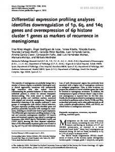
revealed by CGH (data not shown) and SKY showed gain on chromosomes 1p11p32, 1q21q32; 2p11p21, 2q11q21, 2q24q37; 3p21p26; 4q25; 5p14, 5q14, 5q23, 5q33q35; 6q15, 6q22; 7p11p22, 7q11q22; 8p12, 8p22, 8p23, 8q13q24; 10p11p13, 10q11q21; 11p12p15; 12p11, 12q11q24; 13q14q21, 13q31q32; 16q23; 20p12p13, 20q11q13; and 21q11.2q22 and loss on chromosomes 1p36.3; 2p24p25; 3p12; 6p23p25; 13q21q22, 13q32q34; 14q11q13, 14q22q32; 15q11q21; 18q11q23; and 22q11.2q13 (Fig. 1d). The phenomenon of the deletion of chromosome q-arm was obviously observed (Fig. 1d). The data are consistent with CGH and SKY results of the parent tumor.
Statistics All data were presented as mean ± standard deviation (SD). One- or two-way analysis of variance was employed to determine whether the results had statistical significance. P value less than 0.05 was considered significant differences. The statistics software used in this study was Sigma Stat 3.0.1 (SPSS Inc., USA).
Results Establishment and characterization of primary culture for AT/RT This strain of AT/RT cells (Fig. 1) could pass stably for more than 25 passages and retain morphological and immunochemical features of the parent tumor confirming by serial tumor markers indicated in our previous study [21]. The features of AT/RT culture cell were the presence of large, pale, bland cells designated as “rhabdoid” cells (Fig. 1b, c). Rapidly dividing rhabdoid tumor cells dominated the culture (Fig. 1b). The findings of electrical microscope showed the compact rhabdoid-like, large round cells with vesicular nuclei, prominent nucleoli, and abundant fibrillary-like structure and cytoplasmal organelles, including endoplasmic reticulum (Fig. 1c). Immunohistochemistry results of the AT/RT cells were compared with paraffin sections of the parent tumor [21]. Cell doubling time of AT/RT cell culture was 21.3 h, which were estimated by in vitro growth curve of AT/RT. To further investigate in vivo tumorigenic potential, AT/RT 106 cells were injected into the right subcutaneous flank of severe combined immune deficiency (SCID) mice. After 30 days of implantation, a 5×5×5-mm tumor mass was found in the right flank of those SCID mice. Immunohistochemical studies further demonstrated positive immunoreactivity for vimentin, neuron-specific enolase, and epithelial membrane antigen, same as the findings in AT/RT cells (data not shown). Giemsa banding revealed aneuploidy of chromosome numbers in this AT/RT cell line. Copy number karyotyping
The differential profiling between AT/RT and medulloblastoma in vitro and in vivo We analyzed the expressions of 22K genes using Affymetrix Hu6800 GeneChip microarray for the differential gene display of AT/RT cells, DAOY MB, and astrocyte SVG12 cell line in triplex experiments. Based on the differences of the mean expression intensity values of each gene, a total of 970 significant genes differed in their expression levels among astrocyte SVG-12 cells, DAOY cells, and AT/RT by at least 2-fold in upregulation and 1/2-fold in downregulation (P