CCL 6) were from stock culture of the American Type Culture Collec- phate buffer (pH 7.4) for 1 h at room temperature, washed with tion (Rockville, MD).
EXPERIMENTAL CELL RESEARCH ARTICLE NO.
226, 80–89 (1996)
0205
Differentiation-Associated Antimicrobial Functions in Human Colon Adenocarcinoma Cell Lines MARIE FRANC¸OISE BERNET-CAMARD, MARIE HE´LE`NE COCONNIER, SYLVIE HUDAULT,
AND
ALAIN L. SERVIN1
CJF INSERM 94-07 Pathoge´nie Cellulaire et Mole´culaire des Microorganismes Ente´rovirulents, Faculte´ de Pharmacie, Universite´ de Paris XI, F-92296 Chaˆtenay-Malabry, France
teropathogens [2], intestinal epithelium has developed several systems of defenses. A layer of mucus secreted by the goblet cells covers the epithelium and contains inhibitory products against bacteria, including lysozyme, lactoferrin, and lactoperoxidase [3]. Secretory immunoglobulin A (IgAs) that binds both to antigens on the bacterial surface and to mucins are produced by mucosa-associated lymphoid tissue (MALT) in specialized cell follicles called Peyer’s patches containing microfold cells (M cells) [4]. The entrapment of bacteria within the mucus, coupled with peristalsis, results in the rapid expulsion of bacteria from the intestine. During the last 10 years, the chemical antimicrobial system of defenses involving antimicrobial proteins and peptides located in the skin, polymorphonucleated neutrophils (PMNs), macrophages, and tracheal and airway cells has been largely documented [for reviews 5–8]. Mouse small intestinal cryptdins are structurally diverse antimicrobial peptides abundant in Paneth cells located in the basal portion of intestinal crypts, subjacent to the zone of intestinal epithelial cell division [9]. The peptides named defensins are characterized by nine highly conserved amino acids, including six invariant cysteine residues which constitute a unique disulfide motif. The three disulfides stabilize tertiary conformation consisting predominantly of bsheet. It has been recently demonstrated that the human defensins previously found in PMNs [6, 8] are present in the Paneth cells of the human intestine [10, 11]. Enzymes, such as lysozyme, which participate in host defense are the products of the Paneth cells containing numerous apically located eosinophilic secretory granules. Moreover, several gut antimicrobial peptides possessing activities against enteropathogens have been recently purified from the pig small intestine [12–14]. However, the precise cell localization of these gut peptides is not currently documented. Similarities have been found for the mechanisms of action of the gut antimicrobial peptides [15] and the PMN antimicrobial peptides [16]. We examine here the expression of a panel of antimicrobial components, such as bacteriolytic enzymes and
We report that the enterocytic cells of the HT-29 glc0// cell subpopulation strongly expressed two antimicrobial enzymes: the lysozyme and a1-antitrypsin. Moreover, we found that 20 to 30% of these cells expressed positive immunoreactivity using the mAbs directed against the gut porcine PR-39 and cecropin P1 antimicrobial peptides, but did not express immunreactivity against the human antimicrobial polymorphonucleated neutrophil-associated HNP 1-3 defensin and the Xenopus skin magainin. The HT-29 glc0// cell subpopulation develops bacteriolytic activity against the enterovirulent diffusely adhering C1845 Escherichia coli characterized by dramatic alterations of the bacterial cell, suggesting lysis, and bacterial death. In contrast, no expression of immunoreactivity against the antimicrobial peptides and no C1845 bacterial alteration were found in the cultured human embryonic undifferentiated INT407 cells and the colon adenocarcinoma T84 crypt cells. The development of the bacterial alteration and the expression of the antimicrobial components were examined as a function of the cell differentiation using the Caco-2 cell line which spontaneously differentiates in culture. We found that the bacterial alteration and the expression of the PR39 immunoreactivity are differentiation-associated events. Altogether, our results suggest that in the intestine the enterocytes could develop antimicrobial defenses participating in the protection of the gut epithelium against enterovirulent microorganisms. q 1996 Academic Press, Inc.
INTRODUCTION
The gastrointestinal tract is lined by an epithelium with a single layer of cells of different phenotypes: fluid-transporting, mucosecreting, enteroendocrine, and M and caveola cells [1]. For its protection against en1 To whom reprint requests should be addressed at UPS Faculte´ de Pharmacie Paris XI, CJF INSERM 94-07, F-92296 ChaˆtenayMalabry, France. Fax: (33.1) 46.83.56.61.
80
0014-4827/96 $18.00 Copyright q 1996 by Academic Press, Inc. All rights of reproduction in any form reserved.
AID
ECR 3216
/
6I10$$$161
06-04-96 18:05:43
ecl
AP: Exp Cell
ANTIMICROBIAL FUNCTIONS IN COLON CANCER CELL LINES
antimicrobial peptides, in the cultured human intestinal INT407, T84 , HT-29, and Caco-2 cells. For this purpose, we examined by indirect immunofluorescence staining at the apical surface the expression of the bacteriolytic enzymes lysozyme and a1-antitrypsin, the human antimicrobial PMN-associated HNP 1-3 defensin, and the antimicrobial peptides PR-39 and cecropin P1 previously found in porcine gut [12–14]. We report that the colon adenocarcinoma HT29 glc0// and HT29-FU cell subpopulations express antimicrobial functions which develop as a function of the cell differentiation in the Caco-2 cells. Moreover, we observe that the diffusely adhering Escherichia coli (E. coli) which infect the cultured human colon adenocarcinoma HT-29 and Caco-2 cells [17, 18] recognizing the decay accelerating factor as receptor [19], are subjected to bacteriolytic activities when they are associated with the brush border of randomly distributed cells over the monolayers. MATERIALS AND METHODS Cell culture. Six populations of cultured human intestinal cell lines were used. INT407 cells (human embryonic intestine; ATCC CCL 6) were from stock culture of the American Type Culture Collection (Rockville, MD). T84 cells [20], a model of colon crypt cells [21], were from K. Dharmsathaphorn (University of California, San Diego, CA). Caco-2 cells [22] and parental, mainly undifferentiated HT-29 cells referred to as HT-29 Std, were from Dr. Jorgen Fogh (Sloan Kettering Memorial Cancer Center, Rye, NY) [21]. HT-29 glc0// [23] and HT29-FU [24] subpopulations were from Dr. Alain Zweibaum (INSERM U178, Villejuif, France). Caco-2 and HT-29 glc0// are models of mature enterocyte of the small intestine [25, 26]. HT29-FU cells are a mixed population of enterocyte-like cells (80%) and goblet cells (20%) secreting mucus of colonic immunoreactivity [27]. INT407 cells were cultured in DMEM supplemented with 1% nonessential amino acids and 10% inactivated (30 min, 567C) fetal calf serum (Boehringer, Mannheim, Germany) at 377C in a 5% CO2/ 95% air atmosphere on glass coverslips, which were placed in sixwell Corning tissue culture plates (Corning Glass Works, Corning, NY). Cells were used for adherence assays at confluence, i.e., after 4 days. T84 cells were routinely grown in Dulbecco’s modified Eagle’s minimal essential medium (DMEM) (50%) and Ham’s F12 (50%) supplemented with 2 mM glutamine, 50 mM Hepes, 1% nonessential amino acids, and 10% inactivated (30 min, 567C) fetal calf serum (Boehringer-Mannheim, Germany) at 377C in a 10% CO2/90% air atmosphere on glass coverslips which were placed in six-well Corning tissue culture plates (Corning Glass Works). Cells were seeded at a concentration of 106 cells/cm2. Cells were used at late postconfluence, i.e., after 10 days. HT-29 and Caco-2 cells were routinely grown in DMEM (25 mM glucose) (Eurobio, Paris, France), supplemented with 10% (HT-29 Std, HT-29 glc0//, HT29-FU) or 20% (Caco-2) inactivated (30 min, 567C) fetal calf serum (Boehringer), and 1% nonessential amino acids (Caco-2). For adhesion assay, monolayers of Caco-2 cells and HT-29 subpopulations were prepared on glass coverslips which were placed in six-well Corning tissue culture plates (Corning Glass Works). Cells were seeded at a concentration of 105 (HT-29 glc0// and HT29-FU) and 7.4 1 104 (Caco-2) cells/cm2. For maintenance purposes, cells were passaged weekly using 0.25% trypsin in Ca2/Mg2/-free phosphate-buffered saline (PBS) containing 0.53 mM EDTA. Maintenance of the cells and all experiments were carried out at 377C in a
AID
ECR 3216
/
6I10$$$161
06-04-96 18:05:43
81
10% CO2/90% air atmosphere. The culture medium was changed daily. Differentiated cells were used for adherence assays at late postconfluence, i.e., after 15 days (Caco-2) and 20 days (HT-29) in culture. Caco-2 cells were also used at confluence (5 days, undifferentiated cells), at postconfluence (7 days when differentiation commences, and 10 days when differentiation develops), and at late postconfluence (15 days, when differentiated is completed). Bacterial strain and culture conditions. The diffusely adhering E. coli (DAEC) C1845 [27] was grown on CFA-agar containing 1% Casamino Acids (Difco Laboratories, Detroit, MI), 0.15% yeast extract, 0.005% magnesium sulfate, and 0.0005% manganese chloride in 2% agar for 18 h at 377C. Cell infection conditions. The cell monolayers, prepared in sixwell Corning tissue culture plates (Corning Glass Works), were infected apically by a fixed concentration of E. coli as previously described [28]. Briefly, the cell monolayers were washed twice with phosphate-buffered saline (PBS) and infected with 108 CFU cells/ml of E. coli suspended in the culture medium. The plates were incubated at 377C in 10% CO2/90% air for 3 h. The monolayers were then washed five times with sterile PBS. E. coli C1845 adherence assay was performed in the presence of 1% mannose to block the nonspecific attachment of the bacteria by type 1 fimbriae to the cells. Each adherence assay was conducted in triplicate in three successive cells passages. Scanning electron microscopy. For scanning electron microscopy, the intestinal cells were grown on glass coverslips and in six-well Corning tissue culture plates. After the bacterial adhesion assay, cells were fixed with 2.5% (v/v) glutaraldehyde in 0.1 M sodium phosphate buffer (pH 7.4) for 1 h at room temperature, washed with phosphate buffer, postfixed for 30 min with 2% (w/v) OsO4 in the same buffer, washed three times with the same buffer, and dehydrated in a graded series (30 to 100%) of ethanol. Cells were dried in a critical-point dryer (Balzers CPD030) and coated with gold. The specimens were then examined with a Jeol electron microscope. Determination of the surviving bacteria. The total number of adhering bacteria was determined by a quantitative binding assay involving the incubation of a fixed concentration of E. coli (108 CFU/ ml), metabolically labeled by the addition of 14C-acetic acid (Amersham, 94 mCi/mmol; 100 mCi/10 ml tube) [18]. After contact with the cells, the level of bacterial adhesion was evaluated by liquid scintillation counting after dissolution of adhering bacteria and intestinal cells in a 0.2 N NaOH solution. Each adherence assay was conducted in duplicate with three successive cell passages. Quantitative determination of the surviving adherent E. coli was conducted. After contact between the cells and the bacteria as described under cell infection conditions, the cell monolayer was lysed by adding 0.5 ml of H2O to release adhering bacteria. The surviving bacteria were then quantitated by plating serial dilutions on L agar and counting colonies arising after 24 h of incubation at 287C. Experiments were run in duplicate and repeated at least three times. Antibodies. The ascites fluid containing antibody HBB 2/614/88 against human sucrase-isomaltase (SI) was a gift from H. P. Hauri (Biocenter of the University of Basel, Switzerland). The ascites fluid containing antibody 4H3 against human dipeptidylpeptidase IV (DPP IV) was obtained from S. Maroux (Centre de Biochimie et de Biologie Mole´culaire, Marseille, France). The ascites fluid containing antibody HBB2/45 against aminopeptidase neutre (ApN) was from A. Quaroni (Cornell University, Ithaca, NY). MAb 517 antibody against carcinoembryogenic antigen (CEA) was obtained from A. Le Bivic (URA 179 CNRS, Marseille, France). MAb D1-1 against human defensins HPN1-3 was from T. Ganz (Will Rogers Pulmonary Laboratory, University of California, Los Angeles). Mabs PR485-4 against the gut porcine antimicrobial PR-39 peptide and P1-415-1 against gut porcine antibacterial cecropin P1 peptide were from Mabtech AB (Stockholm, Sweden). Polyclonal rabbit M-3654 antibody against magainins I and II was from Sigma (St. Louis, MO). Polyclonal rabbit
ecl
AP: Exp Cell
82
BERNET-CAMARD ET AL.
A O99 antibody against human lysozyme and rabbit polyclonal A012 antibody directed against human a1-trypsin (a1AT) were from Dako (Denmark). Indirect immunofluorescence. Indirect immunofluorescence was performed on unpermeabilized cells [29]. Cell layers were prepared after cells were grown on glass coverslips, and monolayers were washed three times with PBS and fixed for 10 min at room temperature in 3.5% paraformaldehyde in PBS. Preparations were fixed for 10 min at room temperature in 3.5% paraformaldehyde in PBS. Cell monolayers were incubated with specific primary antibody for 30 min at room temperature, washed, and then incubated with their respective secondary fluorescein-conjugated antibody. Primary antibodies were diluted 1:50–200 in PBS (anti-SI HBB 2/614/88, 1:200; anti-DPP IV 4H3, 1:50; anti-ApN HBB2/45, 1:50; anti-CEA 517, 1:50; anti-HNP 1-3, 1:50 and 1:100; anti-PR-39, 1:50 and 1:100; anti-cecropin P1, 1:50 and 1:100; anti-magainin, 1:50; anti-a1AT: 1:50, all in 2% BSA-PBS). Secondary antibodies were either fluorescein- or rhodamine-conjugated goat anti-mouse IgG from Immunotech (Luminy, France) and fluorescein-conjugated goat anti-rabbit IgG from Institut Pasteur Productions (Paris, France), used at a dilution of 1:20 in 2% BSA-PBS. In all cases, no fluorescent staining was observed when the primary antibody was omitted. Immunolabeling was examined using a Leitz Aristoplan microscope with epifluorescence. All photographs were taken on Kodak T-MAX 400 black and white film (Eastman Kodak Co., Rochester, NY).
RESULTS
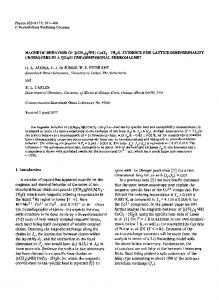
Expression of Antibacterial Components in Cultured Human Colon Adenocarcinoma Cells In the intestine a variety of antimicrobial functions have been localized in the Paneth cells of the crypts of Lieberku¨hn throughout the small intestine and proximal colon [9]. They promote the maintenance of mucosal defense barriers through production of antimicrobial enzymes such as lysozyme [30–34], phospholipase A2 [35–37] and a1-antitrypsin (a1-AT) [38, 39], and antimicrobial peptides such as defensins [10, 11]. We examined the expression of Paneth cell- and gut-associated antimicrobial components in cultured human intestinal cells. For this purpose, expression of several antimicrobial enzymes and peptides was examined by indirect immunofluorescence staining using monoclonal and polyclonal antibodies directed against lysozyme, a1-AT, HNP 1-3 defensins, peptide PR-39, cecropin P1, and magainin. The differentiated HT-29 glc0// cells present a highly ordered brush border decorated by hydrolases such as DPP IV (Fig. 1). These cells expressed lysozyme at a high level and a1-AT in a diffuse pattern. Randomly distributed intense patches of positive immunoreactivity were observed for the gut antimicrobial peptides PR-39 and cecropin P1; thus, not all the cells expressed these gut antimicrobial peptides. These results correlate well with the previous observation showing that randomly distributed cells develop lytic activity against adhering bacteria. In contrast, no HNP 1-3 defensin and magainin immunoreactivity was found.
AID
ECR 3216
/
6I10$$$161
06-04-96 18:05:43
FIG. 1. Expression of antimicrobial components in HT-29 glc0// cell subpopulations. In A, observation of brush border by scanning electron microscopy. In B and C, interference-contrast micrographs of cells (B) and mosaic pattern of positive DPP IV immunoreactivity (C). Intense diffuse immunoreactivity of lysozyme (D) and a1-AT (E). Randomly distributed patches of positive PR-39 (F) and cecropin P1 (G) immunoreactivity. Absence of immunoreactivity for defensins HNP 1-3 (H) and magainin (I). Notice that extinction of the cecropin P1 immunoreactivity was obtained by preincubating the mAb P1415-1 against cecropin P1 peptide with the purified peptide obtained from Sigma (not shown).
We examined if other cultured human intestinal cells expressed antimicrobial components (Table 1). As for the HT-29 glc0// cells, the enterocytic cells in the fully differentiated HT29-FU cell subpopulation showed positive immunoreactivity for the gut antimicrobial peptide PR-39. In the undifferentiated INT407 cells ex-
ecl
AP: Exp Cell
83
ANTIMICROBIAL FUNCTIONS IN COLON CANCER CELL LINES
TABLE 1 Expression of Antimicrobial Enzymes and Peptides and Differentiation-Associated Markers by Human Cultured Intestinal INT407, T84 , HT-29 glc0//, and HT29-FU Cells T84
HT-29 glc0//a
HT29-FUb
Negative Negative Negative Positive
Negative Positive Weakly positive Positive
Negative Positive Weakly positive Positive
Negativec Positivec Weakly positivec Positivec
Positive Weakly positive Negative Negative Negative Negative
Positive Positive Negative Negative Negative Negative
Positive Positive Positive Positive Negative Negative
Positivec ND Positivec ND Negativec Negativec
INT407 Differentiation markers Sucrase-isomaltase Dipeptidyl peptidase IV Aminopeptidase neutre CEA Antimicrobial components Lysozyme a-Antitrypsin Porcine gut PR-39 Porcine gut cecropin P1 PMNs HNP 1–3 defensin Magainin
Note. ND, not determined. a Enterocytic HT-29 glc0// cells [25] after 20 days in culture. b Mixed HT29-FU cell subpopulation after 20 days in culture (80% of enterocytic cells and 20% cells secreting mucus of colonic immunoreactivity [26]). c Immunoreactivity in enterocytic cells.
pressing the CEA but not DPP IV, SI, and ApN, the lysozyme was expressed by several cells, whereas the expression of a1-AT appeared localized at the cell junctions. In contrast, no expression of PR-39 antimicrobial peptides was found. In T84 cells expressing DPP IV, ApN, and CEA but not SI, the lysozyme and a1-AT were expressed in a diffuse pattern. In contrast, no expression of the PR-39 and cecropin P1 antimicrobial peptides was found. Alteration of E. coli Adhering to the Cultured Human HT-29 glc0// Cell Subpopulations When examined by scanning electron microscopy, numerous E. coli C1845 attaching to the brush border of HT-29 glc0// cells appeared to be altered (Fig. 2). Different degrees of bacterial cell surface alterations were concomitantly observed. Flattening of the bacterial cell and multiple surface bundle formations suggested the removal of large portions of surface-associated components (Fig. 2A). Several bacteria were extensively damaged, resulting in a dramatic disorganization of the bacterial cell (Fig. 2B). The bacteria attaching to the cells in the mixed HT29-FU cell subpopulation were also altered (Figs. 2D and 2E). It was particularly interesting to note that the bacteria associated with the mucus secreted by the goblet cells appeared also altered. In contrast, the adhering E. coli C1845 appeared not altered in INT407 and T84 cells (not shown). In a blinded review of randomly obtained scanning electron micrographs, we were able to correctly quantify the number of adhering bacteria presenting alter-
AID
ECR 3216
/
6I10$$$161
06-04-96 18:05:43
ation of their morphology (Fig. 3). The blinded analysis of random photomicrographs obtained shows that the number of altered bacteria increased during the time course of cell–bacteria contact (Fig. 3A). We quantified the total adhering bacteria and the surviving adhering bacteria after 1 and 3 h of contact with the fully differentiated HT-29 glc0// cells. After 1 h, the adhering bacteria were totally viable, whereas in contrast the number of viable bacteria after 3 h of incubation decreased significantly (Fig. 3B). No bacterial alteration (not shown) or bacterial cell death was observed when the cells are undifferentiated (6.7 { 0.3 log CFU/ml adhering bacteria after 1 h of incubation and 6.4 { 0.5 log CFU/ml surviving adhering bacteria). Expression of the Gut Antimicrobial Components by Human Colon Adenocarcinoma Cells as a Function of Cell Differentiation As summarized in Table 1, we observed that the gut PR-39 antimicrobial peptides were expressed by the fully differentiated HT-29 glc0//, HT29-FU cell subpopulations, whereas no expression occurs in the undifferentiated INT407 cells. This result strongly suggests that the intestinal cell differentiation plays a role in the expression of these gut antimicrobial components. This point was examined (Fig. 4) using the Caco-2 cell line which spontaneously differentiates in culture [22] and which represents currently the best model to study in vitro the intestinal cell organization, functions, and differentiation [1, 26, 40, 41]. The state of cell differentiation was evaluated by immunofluorescent staining of the cells with antibody di-
ecl
AP: Exp Cell
84
BERNET-CAMARD ET AL.
FIG. 2. Scanning electron microscopy of the cultured human intestinal HT-29 glc0// and HT29-FU cell subpopulations infected apically with E. coli C1845 showing different states of bacterial cell alteration. (A to C) HT-29 glc0// cells. (D and E) HT29-FU cells. In A, flattened adhering bacteria (1), flattened bacteria in which bundling commences (2), bacteria enterely bundled (3). Highly damaged bacteria dramatically disrupted (B). Note that bacterial alteration occurs in randomly distributed cells over the cell monolayer, whereas in adjacent cells adhering bacteria are not altered. In D and E, observation of alteration of bacteria associated with the brush border (D) and the secreted mucus (E) in HT29-FU cell subpopulation. M, mucus.
rected against the differentiation-associated marker SI [42] (Figs. 4B, 4G, and 4L). Expression of two antimicrobial components, i.e., lysozyme (Figs. 4C, 4H, and 4M) and PR-39 peptide (Figs. 4D, 4I, and 4N), was examined in Caco-2 cells as a function of the days in culture. Lysozyme was expressed by the confluent undifferentiated, recently differentiated, and fully differentiated cells. However, it was noticed that the number of positive cells increased during the differentiation time course, suggesting that expression of lysozyme could partially be related to the cell differentiation. No expression of PR-39 peptide was found in confluent undifferentiated cells. Discrete expression of PR-39 peptide appeared in areas of the recently differentiated cell layer (5% of the total cells). Positive expression of PR-39 peptide was found in fully differentiated cells and positive cells represent 20 to 30% of the total number of cells.
AID
ECR 3216
/
6I10$$$161
06-04-96 18:05:43
We examined the development of the bacterial alteration by scanning electron microscopy as a function of the days in culture (Figs. 4E, 4J, and 4O). No bacterial alteration was observed for bacteria associated with the undifferentiated cells (Fig. 4E). When the cells were recently differentiated the bacterial alteration commences (Fig. 4J). Marked bacterial cell alteration was observed in fully differentiated cells at late postconfluency (Fig. 4O). Altogether, these results strongly suggest that the lysis of bacteria and the expression of the gut antimicrobial peptide PR-39 is a differentiationassociated event in the cultured colon adenocarcinoma cells. DISCUSSION
It was recently postulated that antimicrobial components could regulate the bacterial concentration in the
ecl
AP: Exp Cell
ANTIMICROBIAL FUNCTIONS IN COLON CANCER CELL LINES
FIG. 3. Quantification of the adhering bacteria to human HT-29 glc0// cells presenting morphological alteration and determination of the surviving adhering bacteria. (A) In a blind review of randomly obtained scanning electron micrographs at low and high magnifications, the number of adhering bacteria presenting alteration of their morphology was determined. For this, 50 random micrographs were examined, obtained from 14 monolayers resulting from five experiments: 10 micrographs from 1 h and 125 bacteria examined, 24 from 3 h and 440 bacteria examined, 9 from 6 h and 69 bacteria examined, 7 from 9 h and 103 bacteria examined. (B) The total of adhering bacteria was determined using 14C-radiolabeled C1845 E. coli. The surviving adhering bacteria were quantitated by plating after lysis of the cell monolayer by H2O. Each adherence assay was conducted in triplicate with two successive cell passages (SD, not shown were less than 5%). Statistical analysis between total adhering bacteria and viable adhering bacteria was performed with a Student test: no significant difference is observed after 1 h, whereas a significant difference (P õ 0.01) was observed after 3 h.
upper part of the intestine and protect the mucosa against enterovirulent microorganisms participating in the mucosal defense barriers [12]. In the human intestine, antimicrobial enzymes are present in the specialized Paneth cells [30–39], located at the base of the crypt; they are involved in the defense of the gut
AID
ECR 3216
/
6I10$$$161
06-04-96 18:05:43
85
epithelium against pathogenic microorganisms. Other Paneth cell-associated substances participate in the regulation of the intestinal microflora or in the inhibition of the intestinal colonization by enterovirulent bacteria. Several antimicrobial peptides are specific of the mouse—these are referred to as cryptdins [43–46]. Others, related to the defensin family, are present in mammalian neutrophils [6, 8, 47–50], macrophages [51], and airway cells [7, 52, 53]. A human gene has been found to express the human defensin 5 (HD-5) in Paneth cells [10] which also express mRNA for another human defensin HD-6 [11]. The Xenopus skin antimicrobial magainin [54] was also expressed in the Xenopus Paneth cells [55]. It is interesting to notice that several lines of evidence suggest that the lysozyme as well as antimicrobial peptides are secreted from the Paneth cells into the lumen and act synergistically. It is known that the undifferentiated parental HT-29 Std cell line expresses lysozyme [56], whereas a1-AT is expressed by Caco-2 cells [38, 39]. The observation that the human intestinal HT-29 cell subpopulations and the Caco-2 cells express positive immunoreactivity using mAbs against the antimicrobial PR-39 and cecropin P1 peptides is of interest. PR-39 and cecropin P1 have been isolated from the entire porcine intestine [12–14]. In consequence, the cellular source of PR-39 and cecropin P1 remains to be determined. Indeed, the cells from which these peptides have been isolated comprise the different cell phenotypes of the gut epithelium, including enterocytes and Paneth cells, and contain many leukocytes, including neutrophils and macrophages. Shi et al. [57], who have recently isolated a proline–arginine-rich antibacterial peptide from neutrophils analoguous to PR-39, underlined that it is possible that the gut PR-39 isolated from pig intestinal preparations is derived from leukocytes. Agerberth et al. [58], using a PCR probe derived from the PR-39 gene to screen a human bone marrow cDNA library, obtained information for another human peptide antibiotic designated FALL-39. RNA blot analyses disclosed that the gene for FALL-39 is expressed mainly in human bone marrow and testis. Gudmundsson et al. [59] recently observed PR-39-specific RNA in wild and domesticated pig bone marrow and no expression in 10 different tissues including 6 from the gastrointestinal tract. Moreover, these authors firmly localized the porcine PR39 gene to chromosome 13, whereas the signal for FALL39 was observed on human chromosome 3. The pig chromosome 13 and human chromosomes 3 and 21 share homologies. Authors underlined that it is interesting that the PR39 and FALL39 genes, which both belong to the cathelin family, map to homologous segments in pigs and human, respectively. Our results suggested that the antimicrobial peptides PR39 and cecropin P1 could in fact be produced by the
ecl
AP: Exp Cell
86
BERNET-CAMARD ET AL.
FIG. 4. Expression of lysozyme and gut antimicrobial PR-39 peptide and development of the bacteriolytic activity as a function of the cell differentiation in Caco-2 cells. Interference-contrast micrographs of Caco-2 cells at Day 5 (A), Day 9 (F), and Day 15 (K) in culture. Micrographs of immunofluorescence labeling showing the growth-related expression of SI in Caco-2 cells at Day 5 (B), Day 9 (G), and Day 15 (L) in culture. Observation that lysozyme was expressed in undifferentiated (C), recently differentiated (H), and fully differentiated (M) Caco-2 cells. Note that the number of positive cells increase as a function of the day in culture. Absence of PR-39 immunoreactivity in undifferentiated cells (D); appearance of localized positive-immunoreactivity in random areas of the cell layer (I); increase of PR-39 positiveimmunoreactivity in fully differentiated cells (N). Scanning electron micrographs showing adhesion of the C1845 E. coli to Caco-2 cells at Day 5 (E), Day 9 (J), and Day 15 (O) in culture. In undifferentiated Caco-2 cells at confluency (E), adhering bacteria were mostly not altered, although isolated flattened bacteria are observed (arrow). In Caco-2 cells at postconfluency (J), bacteria adhere to the sparsely distributed MV, and for some bacteria cells marked flattening of bacterial surface was observed (arrow). At late postconfluency (O), bacteria adhere to the well-organized brush border with dense MV and for a majority of bacteria dramatic bacterial cell alterations are observed.
enterocytes. Indeed, neither alteration of adhering bacteria nor expression of antimicrobial peptides were found in the T84 cells, a model for colon crypt epithelium
AID
ECR 3216
/
6I10$$$161
06-04-96 18:05:43
[21]. In contrast, alteration of adhering bacteria and the antimicrobial peptides PR-39 or cecropin P1 were observed in a subpopulation of the cultured colon ade-
ecl
AP: Exp Cell
ANTIMICROBIAL FUNCTIONS IN COLON CANCER CELL LINES
nocarcinoma HT-29 glc0//, HT29-FU, and Caco-2 cells. These cells have been useful tools to study functions of mature enterocytes and mucous-secreting cells and to analyze the intestinal cell differentiation process [1, 26]. Currently, no antimicrobial activities have been reported for normal enterocytes and cultured colon adenocarcinoma enterocytic cells. That holds true also for the cultured colon adenocarcinoma cell subpopulations expressing the mucosecreting phenotype. The expression of antimicrobial peptides in these cells may not necessary reflect expression in normal cells of the human intestine. Considering the malignant characteristics of the HT-29 and Caco-2 cells, it is tempting to speculate that the antimicrobial activity could be due to abnormal Paneth cell-like functions expressed by these cells in culture. A recent report suggests that the human colon adenocarcinoma Caco-2 cells could be pluripotent [60]. The xenographs of adenocarcinoma Caco-2 cells in the nude mouse express both goblet cell, enterocytic and endocrine cell differentiation, presumably resulting from induction by stromal components. However, observation of cells secreting lysozyme, an antimicrobial enzyme normally produced by the Paneth cells, is not sufficient to conclude that the Caco-2 cells are able to differentiate into Paneth cells, since these cells normally produce this enzyme. Moreover, it has not been currently documented that cell phenotypes other than fluid-transporting could be established from this cell line and a recent report demonstrates that only cell clones expressing at different levels the characteristics of mature enterocyte of the small intestine could be established from the Caco-2 cell line [61]. Despite these considerations, the observation that the enterocytic adenocarcinoma cells HT-29 subpopulations and the Caco-2 cell line produced the antimicrobial peptides PR-39 and cecropin P1 and not the PMNs defensins HNP 1-3 found in mammalian Paneth cells [10, 11] supports the idea that enterocytes in parallel with Paneth cells could participate in the defenses of intestinal mucosa against pathogenic microorganisms. Our results are also consistent with the development of the antimicrobial activity as a function of the cell differentiation. Indeed, no bacterial lysis and no expression of antimicrobial peptides PR-39 and cecropin P1 were found in the undifferentiated embryonic INT407 cells. In contrast, the bacterial alteration occurred in fully differentiated enterocyte-like cells of HT-29 glc0// and HT29-FU subpopulations. Using the Caco-2 cells, which spontaneously differentiate in culture [22], we found that the bacterial alteration did not occur when the Caco-2 cells were undifferentiated, i.e., at confluency, and the bacterial alteration was most efficient when full differentiation was reached at late postconfluency. Moreover, expression of the PR-39 peptide in Caco-2 cells develops as a function of cell differ-
AID
ECR 3216
/
6I10$$$161
06-04-96 18:05:43
87
entiation: expression appeared when differentiation commenced and increased along with further differentiation. This point is of interest. Agerberth et al. [14] have isolated from the pig intestine three antibacterial peptides, two of which are derived from factors already known as associated with other functions [62]. The first peptide is the gastric inhibitory polypeptide (7-42) (GIP 7-42) and the second is the diazepam-binding inhibitor (32-46) (DBI 32-46). The active antimicrobial fragment from the GIP 1-42, i.e., the GIP 7-42, appears to be produced from the action of a dipeptidyl aminopeptidase [14]. It is well documented that intestinal hydrolases [63–65] such as dipeptidyl aminopeptidases [66] are targeted to the apical domain of the enterocytic cells during the cell differentiation process. Bactenecins, the defense polypeptides of bovine neutrophils, are generated from precursor molecules [67, 68]. Since we observed that the enterocyte-associated antimicrobial peptide PR-39 appeared apically localized when the cultured intestinal cells were fully differentiated, we hypothetize that it could be generated from a precursor by the cleaving activity of a differentiation-associated brush border hydrolase. In conclusion, the present report focuses on a novel function of the cultured human colon adenocarcinoma cells, i.e., the expression of antimicrobial peptide, suggesting that the enterocyte could participate with the Paneth cells in the mucosal defense against enterovirulent microorganisms by the production of active antimicrobial components. This work was supported by Institut National de la Sante´ et de la Recherche Me´dicale, the Fondation pour la Recherche Me´dicale, and the Association pour la Recherche sur le Cancer. M.F.B. was supported by Ministe`re de la Recherche, de la Technologie et de l’Espace (doctoral fellowship).
REFERENCES 1. Kedinger, M. (1994) in Small Bowel Enterocyte, Culture and Transplantation (Campbell, F. C., Ed.), pp. 1–31, RG Landes, Austin. 2. Falkow, S., Isberg, R. R., and Portnoy, D. A. (1992) Annu. Rev. Cell Biol. 8, 333–363. 3. Neutra, M. R., and Forstner, J. F. (1987) in Physiology of the Gastrointestinal Tract (Johnson, L. R., Ed.), 2nd ed., pp. 975– 1009, Raven Press, New York. 4. Kraehenbuhl, J. P., and Neutra, M. (1992) Physiol. Rev. 72, 853–879. 5. Boman, H. G. (1991) Antibacterial peptides: Key components needed in immunity. Cell 65, 205–207. 6. Lehrer, R. I., Ganz, T., and Selsted, M. E. (1991) Cell 64, 229– 230. 7. Sherman, M. P., and Ganz, T. (1992) Annu. Rev. Physiol. 54, 331–350. 8. Lehrer, R. I., Lichtenstein, A. K., and Ganz, T. (1993) Annu. Rev. Immunol. 11, 105–128.
ecl
AP: Exp Cell
88
BERNET-CAMARD ET AL.
9. Madara, J. L., and Trier, J. S. (1987) in Physiology of the Gastrointestinal Tract (Johnson, L. R., Ed.), pp. 1209–1249, Raven Press, New York. 10. Jones, D. E., and Bevins, C. L. (1992) J. Biol. Chem. 267, 23216– 23225. 11. Jones, D. E., and Bevins, C. L. (1993) FEBS Lett. 315, 187– 192. 12. Lee, J-Y., Boman, A., Chuanxin, S., Andersson, M., Jo¨rnvall, H., Mutt, V., and Boman, H. G. (1989) Proc. Natl. Acad. Sci. USA 86, 9159–9162. 13. Agerberth, B., Lee, J-Y., Bergman, T., Carlquist, M., Boman, H. G., Mutt, V., and Jo¨rnvall, H. (1991) Eur. J. Biochem. 202, 849–854. 14. Agerberth, B., Boman, A., Andersson, M., Jo¨rnvall, H., Mutt, V., and Boman, H. G. (1993) Eur. J. Biochem. 216, 623–629. 15. Boman, H. G., Agerberth, B., and Boman, A. (1993) Infect. Immun. 61, 2978–2984. 16. Lehrer, R. I., Barton, A., Daher, K. A., Harwig, S. S. L., Ganz, T., and Selsted, M. E. (1989) J. Clin. Invest. 84, 553–561. 17. Kerne´is, S., Bilge, S., Fourel, V., Chauvie`re, G., Coconnier, M. H., and Servin, A. L. (1991) Infect. Immun. 59, 4013–4018. 18. Kerne´is, S., Gabastou, J. M., Bernet-Camard, M. F., Coconnier, M. H., Nowicki, B., and Servin, A. L. (1994) FEMS Microbiol. Lett. 119, 27–32. 19. Bernet-Camard, M. F., Coconnier, M. H., Hudault, S., and Servin, A. L. (1996) Gut 38, 248–253. 20. Dharmsathaphorn, K., McRoberts, J. A., Mandel, K. G., Tisdale, L. D., and Masui, H. (1987) Gastroenterology 92, 1133–1144. 21. Barret, K. E. (1993) Am. J. Physiol. 65, C859–C868. 22. Pinto, M., Robine-Leon, S., Appay, M. D., Kedinger, M., Triadou, N., Dussaulx, E., Lacroix, B., Simon-Assmann, P., Haffen, K., Fogh, J., and Zweibaum, A. (1987) Biol. Cell. 47, 323–30. 23. Fogh, J., Fogh, J. M., and Orfeo, T. (1977) J. Natl. Cancer Inst. 59, 221–226. 24. Lesuffleur, T., Kornowski, A., Augeron, C., Dussaulx, E., Barbat, A., Laboisse, A., and Zweibaum, A. Int. J. Cancer 49, 731– 737. 25. Lesuffleur, T., Kornowski, A., Luccioni, C., Muleris, M., Barbat, A., Beaumatin, J., Dussaulx, E., Dutrillaux, B., and Zweibaum, A. Int. J. Cancer 49, 721–730. 26. Zweibaum, A., Laburthe, M., Grasset, E., and Louvard, D. (1991) in Handbook of Physiology. The Gastrointestinal System, Vol. IV, Intestinal Absorption and Secretion (Schultz, S. J., Field, M., and Frizell, R. A., Eds.), pp. 223–255, Am. Physiol. Soc., Bethesda. 27. Bilge, S. S., Clausen, C. R., Lau, W., and Moseley, S. L. (1989) J. Bacteriol. 171, 4281–4289. 28. Darfeuille-Michaud, A., Aubel, D., Chauvie`re, G., Rich, C., Bourges, M., Servin, A., and Joly, B. (1990) Infect. Immun. 58, 893–902. 29. Kerne´is, S., Chauvie`re, G., Darfeuille-Michaud, A., Aubel, D., Coconnier, M. H., Joly, B., and Servin, A. L. (1992) Infect. Immun. 60, 2572–2580. 30. Erlander, S. L., and Chase, D. G. (1972) Ultrastruct. Res. 41, 296–318. 31. Cheng, H. (1974) Am. J. Anat. 141, 521–536. 32. Lopez-Lewellyn, J., and Erlandsen, S. L. (1980) Am. J. Anat. 158, 285–297. 33. Satoh, Y., Ishikawa, K., Tanaka, H., Oomori, Y., and Ono, K. (1988) Acta Histochem. 83, 185–188.
AID
ECR 3216
/
6I10$$$161
06-04-96 18:05:43
34. Satoh, Y., Yamano, M., Matsuda, M., and Ono, K. (1990) J. Electron Microsc. Tech. 16, 69–80. 35. Kiyohara, H., Egami, H., Shibata, Y., Murata, K., Ohshima, S., and Ogawa, M. (1992) J. Histochem. Cytochem. 40, 1659–1664. 36. Senegas-Balas, F., Balas, D., Verger, R., de Caro, A., Figarella, C., Ferrato, F., Lechene, P., Bertrand, C., and Ribet, A. (1984) Histochemistry 81, 581–584. 37. Harwig, S. S. L., Tan, L., Qu, X-D., Cho, Y., Eisenhauer, P. B., and Lehrer, R. I. (1995) J. Clin. Invest. 95, 603–610. 38. Perlmutter, D. H., Daniels, J. D., Auerbach, H. S., De SchryverKecskemeti, K., Winter, H. S., and Alpers, D. H. (1989) J. Biol. Chem. 264, 9485–9490. 39. Molmenti, E. P., Perlmutter, D. H., and Rubin, D. C. (1993) J. Clin. Invest. 92, 2022–2034. 40. Peterson, M. D., and Mooseker, M. S. (1993) J. Cell Sci. 105, 445–460. 41. Peterson, M. D., Bement, W. M., and Mooseker, M. S. (1993) J. Cell Sci. 105, 461–472. 42. Semenza, G. (1986) Annu. Rev. Cell. Biol. 2, 255–314. 43. Eisenhauer, P. B., Harwig, S. S. S. L., and Lehrer, R. I. (1992) Infect. Immun. 60, 3556–3565. 44. Selsted, M. E., Miller, S. I., Henschen, A. H., and Ouellette, A. J. (1992) J. Cell. Biol. 118, 929–936. 45. Huttner, K. M., and Ouellette, A. J. (1994) Genomics 24, 99– 109. 46. Bryn, L., Falk, P., Huttner, K., Ouelette, A., Midtvedt, T., and Gordon, J. I. (1994) Proc. Natl. Acad. Sci. USA 91, 10335– 10339. 47. Ganz, T., Selsted, M. E., Szklarek, D., Harwig, S. S. L., Daher, K., Bainton, D. F., and Lehrer, R. I. (1985) J. Clin. Invest. 76, 1427–1435. 48. Selsted, M. E., Tang, Y-Q., Morris, W. L., McGuire, P. A., Novotny, M. J., Smith, W., Henschen, A. H., and Cullor, J. S. (1993) J. Biol. Chem. 268, 6641–6648. 49. Daher, K. A., Lehrer, R. I., Ganz, T., and Kronenberg, M. (1988) Proc. Natl. Acad. Sci. USA 85, 7327–7331. 50. Linzmeier, R., Michaelson, D., Liu, L., and Ganz, T. (1993) FEBS Lett. 321, 267–273. 51. Hiemstra, P. S., Eisenhauer, P. B., Harwing, S. S. L., van den Barselaar, M. T., van Furth, R., and Lehrer, R. I. (1993) Infect. Immun. 61, 3038–3046. 52. Diamond, G., Zasloff, M., Eck, M. H., Brasseur, M., Maloy, W. L., and Bevins, C. L. (1991) Proc. Natl. Acad. Sci. USA 88, 3952–3956. 53. Diamond, G., Jones, D. E., and Bevins, C. L. (1993) Proc. Natl. Acad. Sci. USA 90, 4596–4600. 54. Zasloff, M. (1987) Proc. Natl. Acad. Sci. USA 84, 5449–5453. 55. Relly, D. S., Tomassini, N., Bevins, C. I., and Zasloff, M. (1994) J. Histochem. Cytochem. 42, 697–704. 56. Fett, J. W., Strydom, D. J., Lobb, R. R., Alderman, E. M., and Vallee, B. L. (1985) Biochemistry 24, 965–975. 57. Shi, J., Ross, C. R., Chengappa, M. M., and Blecha, F. (1994) J. Leukocyte Biol. 56, 807–811. 58. Agerberth, B., Gunne, H., Odeberg, J., Kogner, P., and Boman, H. G. (1995) Proc. Natl. Acad. Sci. USA 92, 195–199. 59. Gudmundsson, G. H., Magnusson, K. P., Chowdhary, B. P., Johansson, M., Andersson, L., and Boman, H. G. (1995) Proc. Natl. Acad. Sci. USA 92, 7085–7089. 60. de Bruı¨ne, A. P., de Vries, J. E., Dinjens, W. N. M., Moerkerk, P. T., van den Linden, E. P. M., Pijls, M. M. J., ten Kate, J., and Bosman, F. T. (1993) Differentiation 53, 51–60.
ecl
AP: Exp Cell
ANTIMICROBIAL FUNCTIONS IN COLON CANCER CELL LINES 61. Chantret, I., Rodolosse, A., Barbat, A., Dussaulx, E., Brot-Laroche, E., Zweibaum, A., and Rousset, M. (1994) J. Cell Sci. 107, 213–225. 62. Laburthe, M., and Amiranoff, B. (1990) in Handbook of Physiology. The Gastrointestinal System, Vol. IV, Intestinal Absorption and Secretion (Schultz, S. J., Field, M., and Frizell, R. A., Eds.), pp. 215–243, Am. Physiol. Soc., Bethesda. 63. Hauri, H. P., Sterchi, E. E., Bienz, D., Fransen, J. A. M., and Marxer, A. (1985) J. Cell. Biol. 101, 838–851. 64. Le Bivic, A., Quaroni, A., Nichols, B., and Rodriguez-Boulan, E. J. (1990) Cell. Biol. 111, 1351–1361.
65. Le Bivic, A., Real, F. X., and Rodriguez-Boulan, E. (1989) Proc. Natl. Acad. Sci. USA 86, 9313–9317. 66. Darmoul, D., Lacasa, M., Baricault, L., Marguet, D., Sapin, C., Trotot, P., Barbat, A., and Trugnan, G. (1992) J. Biol. Chem. 267, 4824–4833. 67. Zanetti, M., Litteri, L., Gennaro, R., Horstmann, H., and Romeo, D. (1990) J. Cell Biol. 111, 1363–1371. 68. Zanetti, M., Litteri, L., Griffiths, G., Gennaro, R., and Romeo, D. (1991) J. Immunol. 146, 4295–4300.
Received December 8, 1995 Revised version received March 12, 1996
AID
ECR 3216
/
6I10$$$161
06-04-96 18:05:43
89
ecl
AP: Exp Cell