Diffuse Axonal Injury Lesion Segmentation Using Contouring Algorithm Olga V. Senyukova1, Valeriy E. Galanine1, Andrey S. Krylov1, Alexey V. Petraikin2, Tolibdjon A. Akhadov2, Sergey V.Sidorin2 1 Faculty of Computational Mathematics and Cybernetics, Lomonosov Moscow State University, Moscow, Russia 2 Children's Clinical and Research Institute Emergency Surgery and Trauma, Moscow, Russia
[email protected]
Abstract Diffuse axonal injury (DAI) is a common type of brain damage induced by trauma. Accurate detection and quantification of DAI lesions is essential for assessment of a patient’s state and making a prognosis of a traumatic disease. This study is devoted to semiautomated segmentation of DAI lesions on T2*-weighted brain magnetic resonance (MR) images. A radiologist outlines the regions containing lesions, and the lesions are further automatically delineated inside these regions.
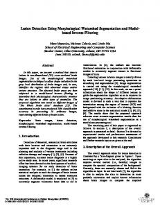
easily confused with other formations, for example, blood vessels. Therefore, we developed a semi-automated algorithm. A radiologist interactively specifies rectangular regions of interest (ROI) which are likely to contain DAI lesions. After this marking procedure each MRI section of the current stack may include none, one or several square regions (Figure 1). Then the lesions inside these specified regions are segmented fully automatically. So there is a trade-off between speed and accuracy.
A proposed algorithm for automated segmentation of DAI lesions inside specified regions is based on contouring algorithm which treats the image as a 3D topological map where a pixel’s intensity corresponds to its height. It builds isolines of an intensity function. Lesion contours are among these isolines. In order to distinguish true lesion contours from other closed contours obtained by contouring algorithm machine learning approach is exploited. A labeled training base with positive (lesions) and negative (non-lesions or lesion parts) examples of closed contours is used to train the classifier. The algorithm was evaluated with real T2*-weighted brain MRI images. Keywords: Diffuse Axonal Injury, Brain MRI, Brain Lesion Segmentation, Contour Levels.
Figure 1: ROI selected on T2*-weighted MRI.
1. INTRODUCTION Recently automated and semi-automated algorithms for medical image processing are widely used by radiologists during evaluation of a patient’s state and diagnostics of various diseases. In this paper we introduce a semi-automated method for detection of diffuse axonal injury (DAI) lesions on T2*-weighted brain MRI images. Diffuse axonal injury occurs in 50% cases of severe traumatic brain injury. Occurrence of DAI lesions in midbrain and brainstem is the most common cause of coma and subsequent disability. An accurate and convenient method for detection of DAI lesions will help a radiologist to estimate current stage of the disease and make a prognosis of a further disease flow. T2*-weighted MRI was chosen among other MRI modalities because it allows to identify DAI lesions visually rather clearly. The T2*-weighted MRI sequence is the most sensitive to the magnetic susceptibility induced by static field inhomogeneities, arising from paramagnetic blood breakdown products in DAI lesions. They look like small dark spots, usually in brain tissue, sometimes connecting each other. So some automatization can be added to the process. Fully automated algorithms for brain lesion detection usually lack accuracy and stability. True lesions can be
A proposed algorithm for DAI lesion segmentation, i.e. delineation of lesion contours, is based on contouring algorithm which is aimed to obtain closed contours which may potentially correspond to true lesion contours. In order to distinguish lesion contours from other closed contours machine learning is applied. The rest of the paper is organized as follows. A brief review of previous work is given in section 2. Contouring algorithm purpose and its application to a current problem is described in section 3. Section 4 is devoted to selection of closed contours obtained by contouring algorithm, which correspond to DAI lesions contours, via support vector machine (SVM) classifier. Implementation details and comparison with other methods is given in Section 5. Section 6 includes conclusion and discussion.
2. PREVIOUS WORK There are several works devoted to processing of MR images of brain with DAI. For the most part they deal with medical and practical aspects of the experiment rather than with image processing algorithms. Paper [8] presents a method for quantification of severity of DAI by diffusion tensor images (DTI). Several specific characteristics
are measured at multiple locations and their statistical analysis is performed for patients and healthy control subjects. Voxels with statistically significant deviation from normal values are assigned to lesions.
which is based on a quite different approach – machine learning is applied to feature vectors relative to contours, not voxels. DAI lesions usually have a specific shape so we can utilize this a priori knowledge and obtain accurate and smooth lesion contours.
In [7] two types of T2*-weighted MRI sequences are compared for detection of DAI. Lesions are defined as regions of abnormally low signal intensity.
Furthermore, in this study there is no need for 3D segmentation – each MRI section is processed separately so that a radiologist can easily control the process.
A wide variety of works on detection of brain lesions in general exist. These lesions can be caused by multiple sclerosis, aging, vascular risk factors.
3. CONTOURING ALGORITHM
One of fundamental studies in this area is [16]. The paper presents semiautomatic technique for segmentation of white matter lesions (WML) using a supervised artificial neural network classifier. Multimodal MRI data is used. The classifier is trained to assign each pixel to one of five classes – background, white matter (WM), gray matter (GM), cerebrospinal fluid (CSF) and WML – based on its intensity on different MRI modalities. A widely used method for segmentation of WML is described in [13]. It utilizes a previously proposed concept of fuzzy connected objects. An operator detects few points on dual-echo fast spinecho MR images, which correspond to WM, GM and CSF. Each of these objects is further automatically detected as a fuzzy connected set. WML’s are segmented as 3D fuzzy connected objects in the holes of these objects’ union. An important work on automated multiple sclerosis lesions detection is [11]. The method models distribution of intensities of WM, GM and CSF, and detects lesions as voxels which are not well described by these models. Markov random field is used to add contextual constraints because it is known that multiple sclerosis lesions are usually located in the vicinity of white matter. In [1] normal tissues and WMLs are segmented by thresholding on some MRI-specific values. Potential WML clusters are also analyzed for 3D shape and surrounding tissue composition.
The method proposed in this paper is based on contouring algorithm which is aimed to build smooth closed contours delineating DAI lesions. Contouring algorithm in general is used for building a contour plot – a set of level curves of different heights of a function of two variables, i.e. isolines of a function. A level curve of height h of a function f ( x, y ) is the set of all points ( x, y ) such that
f ( x, y ) = h . One or more isolines can exist for level h . Contour plots are widely used in cartography for building topographic maps, meteorology, geology and many other areas. Contouring algorithms – algorithms for building contour plots – are described in different works, for example [2]. In this work we use contouring algorithm close to the algorithm described in MATLAB online documentation [12]. In our case f ( x, y ) is a function which takes a value which equals a pixel’s intensity at the point ( x, y ) . The specificity of a current problem requires that we consider only closed contour lines because only they can delineate DAI lesions. As it is shown on Figure 2 there exists a range of contour levels on which lesions are marked perfectly with closed contour lines.
The method for automated segmentation of multiple sclerosis lesions on multimodal MRI images using fuzzy C-means (FCM) clustering is described in [4]. First, FCM is used to extract a mask for CSF and lesions which distinguishes them from other tissues. Then, FCM is reapplied to the masked image to distinguish lesions from CSF. The authors of [9] propose to use k-nearest neighbor classifier to segment WM, GM, CSF and WMLs. In [10] each voxel of brain volume is characterized by a vector of values from multiple MRI modalities – its own and of its neighbors. SVM is used for classification to lesions and nonlesions. Typical non-lesions for SVM training are selected using AdaBoost. One of the latest works [15] introduces weak labels – rectangular regions of interest specified by an expert, which contain lesions. This concept is close to one used in the present work. Lesions are segmented automatically inside these regions. First, K-means clustering is used to identify lesions cluster. Intensity distribution is calculated based on the found cluster. A confidence map is built for a whole ROI. Voxels with high probability of belonging to lesions are used as seeds for more accurate segmentation by Grow-Cut algorithm [14]. The methods mentioned above represent a wide variety of approaches to brain lesions segmentation but all of them perform voxel-wise segmentation. In this work we propose a method
Figure 2: Contour maps of image ROI.
4. SELECTION OF REQUIRED CONTOURS VIA SVM CLASSIFIER
CLOSED
After the contour lines are built, a problem which arises is how to select contours corresponding to DAI lesions contours from the whole set of closed contours. A radiologist can easily do it by sight – he uses a priori knowledge of the shape, intensity and other specific features of DAI lesions. Therefore, we can try to apply machine learning approach to this problem and train a classifier which can
distinguish lesions contours from other closed contours based on these a priori characteristics of lesions.
Classification accuracy was evaluated by 5-2 cross-validation. On 80% of images DAI lesions were segmented successfully.
4.1 Training and applying a classifier
Obtained results were compared with results of segmentation by some basic computer vision methods such as edge detection and region growing.
We used the following features to characterize each closed contour:
For edge detection Canny edge detector [5] was used. The results are not satisfactory because it gives too much clutter and furthermore it loses the contours of lesions (Figure 3b).
•
square of shape bounded by the contour;
•
perimeter;
•
mean intensity inside the contour / ROI mean intensity;
•
intensity difference inside and outside the contour;
•
elongation;
•
density.
A feature vector corresponding to a contour is used as a sample for training and testing a classifier. A training base is constructed as follows. Contour lines for a specified range of levels are built for a set of MRI images inside rectangular ROI specified by a radiologist. Only closed contours are selected among them. For each closed contour a feature vector of above mentioned features is calculated. If a contour corresponds to a DAI lesion contour it is marked as +1, otherwise it is marked as -1. Thus we obtain a labeled training set for a twoclass classifier. A classifier model is built on this set. Then, if we want to detect all DAI lesions inside a ROI on a previously unseen MRI image we should do the following: •
Build contour lines for a range of levels.
•
Select closed contours among these contour lines.
•
Calculate a feature vector for each of these contours.
•
Apply a classifier to each of these feature vectors in order to learn if a corresponding closed contour is a DAI lesion contour or not.
Algorithms based on region growing, which were also tried, start with Gaussian blurring of the original image which allows to compute local intensity minima effectively. These local minima are used as seeds for region growing because DAI lesions are regions of minimum intensity. Each local minimum can contain one pixel or a group of pixels with equal intensities which are lower than intensities of neighboring pixels. Region growing starts from these local minima – more and more pixels which satisfy special criteria are added to each region. One of such criteria is intensity threshold which can be set manually or adaptively. Another threshold used in this experiment was based on the region square. In order to filter seeds obtained from local minima third threshold for intensity value was set and finally inside and outside border mean intensities difference was also checked by threshold value. Region growing seems to be a better solution than edge detection, but in some cases it also works not correctly which results in resegmentation of odd pixels or missing some pixels which definitely belong to the lesion (Figure 3c). The proposed algorithm significantly outperforms region growing and edge detection methods. An example of its results is shown on Figure 3d.
4.2 Support vector machine (SVM) A popular SVM classifier [6] is used in this work. It constructs a hyperplane in a high-dimensional feature space, which separates the classes – a decision boundary. SVM tries to maximize the margin – the perpendicular distance between the decision boundary and the closest of the data points, which are called support vectors. The location of this boundary is determined by a subset of these support vectors. Maximizing the margin leads to better separation between classes. An important property of SVM is that the determination of the model parameters corresponds to a convex optimization problem, and so any local solution is also a global optimum [3].
(a)
(b)
(c)
(d)
5. EXPERIMENTS A proposed algorithm was implemented on C#. Implementation details and results are given below. Contour
lines
for
20
levels
in
the
intensity
min I ( x, y ) + 10, min I ( x, y ) + 70 x , y x, y
range
were built
by the contouring algorithm. The base for training and validation of SVM classifier consists of 320 lesion contours and 231 non-lesion contours obtained from 41 MRI images. A total set of 8 brain MRI volumes was used. SVM classifier with linear kernel was applied.
Figure 3: a: original ROI; b: Canny; c: region growing; d: our method.
6. CONCLUSION AND DISCUSSION A novel approach to semi-automated segmentation of DAI lesions on brain MRI images was proposed. It is based on contouring algorithm which builds level curves for a specified set of levels – these curves are isolines of an intensity function. It was shown that these level curves can catch lesion contours well. In order to select required lesion contours from other closed level contours SVM classifier was used. A proposed algorithm was evaluated on real brain MRI images provided by Children's Clinical and Research Institute Emergency Surgery and Trauma and demonstrated good results accepted by the radiologist. However, some difficulties arise in the case of DAI lesions detection in severely damaged areas where these lesions cover almost all the area of the ROI. But in this case there is a fuzzy boundary between DAI and hemorrhage. Thus detection of hemorrhage is another problem and may involve other methods for its solution. Another difficulty is that in some situations even a radiologist cannot be sure if a dark spot is a DAI lesion or a blood vessel. So one of further directions for this work is matching T2*-weighted brain MRI images with angiography images where blood vessels are well seen, in order to distinguish DAI lesions from blood vessels automatically. The future work also includes creation of a more representative MRI database of brain damaged by DAI.
7. AKNOLEDGMENTS This work was supported by federal target program "Scientific and scientific-pedagogical personnel of innovative Russia in 20092013".
8. REFERENCES [1] B. Alfano, A. Brunetti, M. Larobina et al., “Automated segmentation and measurement of global white matter lesion volume in patients with multiple sclerosis”, J. of Magnetic Resonance Imaging, vol. 12, no. 6, pp. 799—807, 2000. [2] M.J. Aramini, “The Design and Implementation of Computer Algorithms for Contour Plotting” (thesis), Stevens Institute of Technology, Hoboken, NJ 07030, (May 2, 1980). [3] C. Bishop “Pattern Recognition and Machine Learning (Information Science and Statistics)”, Springer-Verlag New York, Inc. Secaucus, NJ, USA, 2006. [4] A.O. Boudraa, S.M.R. Dehak, Y.-M. Zhu et al., “Automated Segmentation of Multiple Sclerosis Lesions in Multispectral MR Imaging Using Fuzzy Clustering”, Computers in Biology and Medicine, vol. 30, no. 1, pp. 23—40, 2000. [5] J. Canny, “A Computational Approach to Edge Detection”, IEEE Transactions on Pattern Analysis and Machine Intelligence, vol. PAMI-8, no. 6, pp. 679—698, 1986. [6] C. Cortes and V. Vapnik, “Support-Vector Networks”, Machine Learning, vol. 20, pp. 273—297, 1995. [7] E. Giugni, U. Sabatini, G.E. Hagberg et al., “Fast Detection of Diffuse Axonal Damage in Severe Traumatic Brain Injury: Comparison of Gradient-Recalled Echo and Turbo Proton Echo-Planar Spectroscopic Imaging MRI Sequences”,
American Journal of Neuroradiology, vol. 26, no. 5, pp. 1140—1148, 2005. [8] T.A.G.M. Huisman, L.H. Schwamm, P. W. Schaefer et al., “Diffusion Tensor Imaging as Potential Biomarker of White Matter Injury in Diffuse Axonal Injury”, American Journal of Neuroradiology, vol. 25, pp. 370—376, 2004. [9] C. Jongen, J. Van Der Grond, L.J. Kappelle et al. “Automated measurement of brain and white matter lesion volume in type 2 diabetes mellitus”, Diabetologia, vol 50, no, 7, pp. 1509—1516, 2007. [10] Z. Lao, D. Shen, A. Jawad, “Automated Segmentation of White Matter Lesions in 3D Brain MR Images Using Multivariate Pattern Classification”, 3rd IEEE Int. Symp. on Biomedical Imaging: Nano to Macro, pp. 307—310, 2006. [11] K. Van Leemput, F. Maes, D. Vandermeulen et al., “Automated Segmentation of Multiple Sclerosis Lesions by Model Outlier Detection”, IEEE Transactions on Medical Imaging, vol. 20, no. 8, pp. 677—688, 2001. [12] MathWorks. MATLAB Contouring Algorithm.
online
documentation.
The
http://www.mathworks.com/help/techdoc/creating_plots/f102524.html#f10-2614 [13] J.K. Udupa, L. Wei, S. Samarasekera et al., “Multiple sclerosis lesion quantification using fuzzy-connectedness principles”, IEEE Transactions on Medical Imaging, vol. 16, no. 5, pp. 598—609, 1997. [14] V. Vezhnevets, V. Konouchine, “Grow-Cut” - Interactive Multi-Label N-D Image Segmentation”, Proceedings of Graphicon 2005, pp. 150—156, 2005. [15] Y. Xie, X. Tao, “White Matter Lesion Segmentation Using Machine Learning and Weakly Labeled MR Images”, Proc. SPIE 7962, 79622G, 2011. [16] A.P. Zijdenbos, B.M. Dawant, R.A. Margolin, A.C. Palmer, “Morphometric analysis of white matter lesions on MRI images: method and validation”, IEEE Transactions on Medical Imaging, vol. 13, no. 4, pp. 716—724, 1994.
About the authors Olga Senyukova is an assistant professor at the Chair of Computer Systems Architecture, Graphics & Media Lab, Faculty of Computational Mathematics & Cybernetics, Lomonosov Moscow State University (CMC MSU). Her contact email is
[email protected] Valeriy Galanine is a student at CMC MSU. His contact email is
[email protected] Andrey Krylov is a professor, head of Laboratory of Mathematical Methods of Image Processing, CMC MSU. His contact email is
[email protected] Alexey Petraikin is a senior researcher at the Radiology Department within the Children's Clinical and Research Institute Emergency Surgery and Trauma (RD CCRIEST). His contact email is
[email protected] Tolibdjon Akhadov is a professor, head of RD CCRIEST. His contact email is
[email protected] Sergey Sidorin is a radiologist at the RD CCRIEST. His contact email is
[email protected]