Key Words: Diffuse large B-cell lymphoma; Bone marrow; Intravascular large B-cell lymphoma; Asian variant; Hemophagocytic syndrome; .... No, of Extranodal ..... Chan JKC, Jaffe ES, Ralfkiaer E. Extranodal NK/T-cell lymphoma, nasal type.
Hematopathology / DLBCL IN THE BONE MARROW
Diffuse Large B-Cell Lymphoma Initially Manifesting in the Bone Marrow Dai Kajiura, MD, Yoriko Yamashita, MD, PhD, and Naoyoshi Mori, MD, PhD Key Words: Diffuse large B-cell lymphoma; Bone marrow; Intravascular large B-cell lymphoma; Asian variant; Hemophagocytic syndrome; Immunohistochemistry DOI: 10.1309/2GW5W7KQBXF6LFAW
Abstract We histologically and immunohistochemically studied 37 cases of diffuse large B-cell lymphoma (DLBCL) initially manifesting in the bone marrow (BM). We also compared these cases with the Asian variant of intravascular large B-cell lymphoma (AIVL). Histologically, the neoplastic cells of the BM mostly had large and round nuclei and formed clusters. Immunohistochemically, all cases were positive for B-cell markers. Factor VIII staining revealed neoplastic cells within the sinusoids of BM in 8 cases; however, these cells accounted for fewer than 20% of the overall neoplastic cells. In several cases, the neoplastic cells infiltrated liver, spleen, kidneys, lungs, stomach, and adrenal glands with a mainly leukemic and infrequently intravascular pattern. Although our cases share some clinical features with AIVL, we consider DLBCL initially manifesting in the BM to be a unique entity because the neoplastic cells proliferate mainly in BM, with infrequent involvement of the sinusoids and occasional leukemic infiltration in various organs.
Secondary involvement of malignant lymphomas in bone marrow is relatively common.1 However, primary occurrence of lymphomas in bone marrow is quite rare, except in chronic lymphocytic leukemia or small cell lymphoma.2 Large cell lymphoma with primary bone marrow involvement is exceptional, although secondary involvement is sometimes observed.1,3-5 Murase et al6-8 reported the Asian variant of intravascular large B-cell lymphoma (AIVL) in which bone marrow involvement of large B lymphoma cells was frequently observed. They proposed criteria for AIVL, including clinical and laboratory criteria and histologic criteria. The former include cytopenia, hepatomegaly and/or splenomegaly, and absence of overt lymphadenopathy and tumor formation. The latter include erythrocyte hemophagocytosis, immunophenotypic evidence of neoplastic B cells, and pathologic findings of intravascular proliferation of lymphoma cells. We reviewed 37 cases of diffuse large B-cell lymphoma (DLBCL) primarily manifesting in bone marrow during the last several decades. In this article, we describe the clinical and histologic characteristics of such cases and compare them with AIVL described by Murase et al.6-8
Materials and Methods For histologic and immunohistochemical examination, tissue samples were fixed with 10% formaldehyde and embedded in paraffin. Then, 2- to 4-µm-thick sections were prepared and stained with H&E. The avidin-biotin-peroxidase complex method (Ultratech, Immunotech, Marseille, France) was used for antibody staining. Antibodies used included CD3, CD20 (L26), CD68 (KP1), CD79a (mb-1), IgG, IgA, IgM, κ, λ, and factor VIII (DAKO, Copenhagen, Denmark); 762 762
Am J Clin Pathol 2007;127:762-769 DOI: 10.1309/2GW5W7KQBXF6LFAW
© American Society for Clinical Pathology
Hematopathology / ORIGINAL ARTICLE
CD34 (Novocastra Laboratories, Newcastle upon Tyne, England); and CD5 (T cell Diagnostics, Cambridge, MA). We evaluated the degree of neoplastic cell infiltration in sinusoids of the bone marrow in 4 grades using factor VIII staining: grades 0, 1, 2, and 3 represent 0%, 0% to 10%, 10% to 20%, and more than 20% of the total neoplastic cells, respectively. We also examined hemophagocytosis using H&E-stained specimens of the bone marrow and CD68 staining, which is a marker of histiocytes. Mild, moderate, and severe degrees represent 1 to 3 cells, 4 to 6 cells, and more than 7 cells in 10 high-power fields, respectively.
Results We studied 37 cases of DLBCL of the bone marrow at initial diagnosis. Clinical characteristics were obtained in 35
cases ❚Table 1❚. The median age of the patients was 67 years, with a range from 45 to 84 years. The patients comprised 17 men and 20 women. Initial symptoms included fever (24/37 [65%]), general fatigue (14/37 [38%]), anorexia (11/37 [30%]), weight loss (5/37 [14%]), and hepatosplenomegaly (25/37 [68%]). Hemophagocytic syndrome was observed clinically in 13 cases. Laboratory data indicated high lactate dehydrogenase levels in 34 cases. Clinical stages were IVB in 23 (66%), IVA in 3 (9%), IB in 6 (17%), and IA in 3 (9%). The mean survival was 14.9 months in the 35 patients with DLBCL of the bone marrow ❚Figure 1❚. Of 35 patients, 31 underwent chemotherapy; of these, 6 were alive at last followup. About 70% of the patients in our study died within 2 years despite chemotherapy. Neoplastic cells were found in the peripheral blood in 8 cases, comprising up to 34%. International prognostic indexes were high in 26 (74%), high
❚Table 1❚ Clinical Findings for Patients With Diffuse Large B-Cell Lymphoma of Bone Marrow at Initial Examination Case No./Sex/ Age (y)
PS
LDH
Lymphoma Cells in Peripheral Blood (%)
No, of Extranodal Lesions (Organ)
HPS
Stage
1/F/72 2/F/84 3/F/56 4/F/64 5/F/76 6/F/70 7/F/68 8/F/45 9/F/70 10/F/75 11/F/77 12/F/73 13/F/54 14/F/49 15/F/79 16/F/82
2 3 3 1 3 3 4 4 2 4 3 3 3 2 4 NA
High High High High High High High High High High High High High High High High
0 1 1 NA NA NA 0 NA NA 0 0 0 0 NA NA NA
2 (BM, Lv) 4 (BM, Lv, Sp, IpM) 2 (BM, Sp) 4 (BM, Lu, Lv, Sp) 3 (BM, Lv, Sp) 4 (BM, Lv, Sp, Kd) 3 (BM, Lu, Sp) 1 (BM) 1 (BM) 2 (BM, Sp) 3 (BM, Kd, St) 6 (BM, Lv, Sp, Lu, AG, Kd) 1 (BM) 1 (BM) 4 (BM, Lv, Sp, Kd) 1 (BM)
– + – – + + + – – – – – + – + –
IVB IVA IVB IVB IVB IVB IVB IB IB IVB IVB IVB IB IB IVB IB
17/F/59 18/M/81 19/M/54 20/M/55 21/M/69 22/M/54 23/M/66 24/M/63 25/M/67 26/M/74 27/M/66 28/M/74 29/M/51 30/F/63 31/F/61 32/M/66 33/M/58 34/M/67 35/M/70
4 3 2 0 2 1 2 2 3 3 3 1 1 3 4 3 2 4 0
High High High Normal High High High High High High High High High High High High High High High
NA 19 2 NA 0 0 0 NA NA 30 0 32 0 0 3 NA NA NA 34
6 (BM, Lv, Kd, AG, Br, Rt) 2 (BM, Sp) 6 (BM, Lv, Sp, Br, Lu, Kd) 1 (BM) 3 (BM, Lv, Sp) 1 (BM) 2 (BM, Lv) 3 (BM, Lv, Sp) 3 (BM, Lv, Sp) 2 (BM, Sp) 3 (BM, Lv, Sp) 1 (BM) 3 (BM, Lv, Sp) 3 (BM, Lv, Sp) 3 (BM, Lv, Sp) 3 (BM, Lv, Sp) 2 (BM, Sp) 4 (BM, Lv, Sp, St) 1 (BM)
+ + – – – – – – + + + – + – – – – + –
IVA IVB IVB IA IVB IB IVB IVB IVB IVB IVB IA IVB IVB IVB IVA IVB IVB IA
IPI High High High High High High High High intermediate High intermediate High High High High intermediate High intermediate High Low ~ high intermediate High High High Low High Low intermediate High High High High High Low High High High High High High Low intermediate
Clinical Course, mo* (Outcome) 35 (D) 13 (D) 26 (D) 12 d (D) 1 d (D) 1 d (D) 26 (D) 13 (D) 6 (D) 14 d (D) 9 (D) 2 (D) 37 (D) 14 (D) 1 (D) 10 d (D) 5 (D) 5 (D) 16 (D) 36 (D) 10 (D) 19 (D) 7 (D) 17 (D) 15 (D) 9 (D) 2 (D) 2 (D) 7 (D) 13 (AWOD) 107 (AWOD) 46 (AWOD) 7 (AWOD) 13 (AWOD) 1 (AWD)
AG, adrenal gland; AWD, alive with disease; AWOD, alive without disease; BM, bone marrow; Br, brain; D, died; HPS, hemophagocytic syndrome; IPI, International Prognostic Index; IpM, iliopsoas muscle; Kd, kidney; LDH, lactate dehydrogenase level; Lu, lung; Lv, liver; NA, not available; PS, performance status; Rt, retina; Sp, spleen; St, stomach; +, present; –, absent. * Unless otherwise indicated.
Am J Clin Pathol 2007;127:762-769
© American Society for Clinical Pathology 763
DOI: 10.1309/2GW5W7KQBXF6LFAW
763 763
Kajiura et al / DLBCL IN THE BONE MARROW
100
Survival Rate (%)
90 80 70 60 50 40 30 20 10 0 0
20
40
60
80
100
120
Observation Period (mo)
❚Figure 1❚ Survival of 35 patients with diffuse large B-cell lymphoma of bone marrow at initial examination. About 70% of the patients died within 2 years despite chemotherapy.
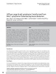
or high to low intermediate in 5 (14%), low intermediate in 2 (6%), and low in 2 (6%). The clinical and laboratory criteria for AIVL were fulfilled by 18 cases, and, of these, 5 additionally fulfilled the histologic criteria. H&E staining revealed bone marrow that was hypercellular in 12 (32%) of 37 cases, normocellular in 17 (46%), and hypocellular in 8 (22%). The neoplastic cells typically had a large and round nucleus ❚Image 1A❚, but cleaved nuclei were observed in 4 cases. Of these, large atypical lymphoid cells were present in clusters in 28 (76%) of 37 cases, whereas scattered cells were observed in 9 (24%). Immunohistochemical studies ❚Table 2❚ indicated that all cases were positive for CD20 (L26) ❚Image 1B❚. All cases but 1 were CD79a+. Immunohistochemical staining using factor VIII revealed that
A
B
C
D
❚Image 1❚ A, Bone marrow biopsy specimen. The neoplastic cells have large and round nuclei (H&E, ×400). B, CD20 staining of a bone marrow biopsy specimen. The neoplastic cells are CD20+ and formed clusters in the extrasinusoidal space (×200). C, Factor VIII staining of a bone marrow biopsy specimen. The neoplastic cells are present in the sinusoid, but neoplastic cells in larger numbers proliferate in the extrasinusoidal space (×400). D, CD5 staining of a bone marrow biopsy specimen. The neoplastic cells are CD5+ (×200). 764 764
Am J Clin Pathol 2007;127:762-769 DOI: 10.1309/2GW5W7KQBXF6LFAW
© American Society for Clinical Pathology
Hematopathology / ORIGINAL ARTICLE
neoplastic cells were present in the sinusoids in 8 cases ❚Image 1C❚; of these, 7 were grade 1, 1 was grade 2, and none were grade 3. Of the 37 cases studied immunohistochemically, 10 were CD5+ ❚Image 1D❚. Hemophagocytosis was found in 33 (89%) of 37 cases overall ❚Image 1E❚, to a severe degree in 4 (11%), a moderate degree in 13 (35%), and a slight degree in 16 (43%). All cases were CD68+, a marker for active hemophagocytosis ❚Image 1F❚. Autopsy was performed in 7 cases, but 1 (case 8) was regional autopsy including brain and kidneys, and neoplastic lesions were not observed ❚Table 3❚. In addition, the stomach was biopsied in 2 cases, the liver in 1, and the kidney in 1. Of the cases in which biopsy or autopsy was performed,
involvement of the liver was observed in 6, the spleen in 5, the lungs in 3, the kidneys in 5, the stomach in 2, and the adrenals in 1. Of the 6 cases in which liver involvement was observed, neoplastic cells were mainly found in the portal areas in 5 cases ❚Image 1G❚; in 1 case, infiltration was found predominantly in the sinusoids. Five of the affected spleens exhibited diffuse infiltration. The neoplastic cells mainly infiltrated in the red pulp, forming variously sized clusters, accompanied by congestion and occasional necrosis, with almost total effacement of the white pulp. In lungs, neoplastic cells were observed in the vessels in 2 cases and in the interstitial tissue in 1 case. Of the 2 cases in which the stomach mucosa was
E
F
G
H
E, Bone marrow biopsy specimen. Hemophagocytosis is observed in the bone marrow (arrows) (H&E, ×400). F, CD68 staining of a bone marrow biopsy specimen. Erythrophagocytosis by CD68+ histiocytes is observed (arrows) (×400). G, Autopsy specimen of liver. The neoplastic cells predominantly infiltrate in the portal area, and a part of them invade in the sinusoids (H&E, ×100). H, Autopsy specimen of kidney. The neoplastic cells are present mainly in the blood vessels and glomerulus (H&E, ×100).
Am J Clin Pathol 2007;127:762-769
© American Society for Clinical Pathology 765
DOI: 10.1309/2GW5W7KQBXF6LFAW
765 765
Kajiura et al / DLBCL IN THE BONE MARROW
❚Table 2❚ Results of Histologic and Immunohistochemical Analysis of Bone Marrow* Case No.
CD20 (L26)
1 2 3 4 5 6 7 8 9 10 11 12 13 14 15 16 17 18 19 20 21 22 23 24 25 26 27 28 29 30 31 32 33 34 35 36 37
+ 3+ + + 3+ 2+ 2+ + + + + 2+ 2+ + 2+ 2+ + 3+ 3+ + + + 2+ 2+ + 2+ 3+ 2+ 3+ 3+ 3+ 2+ 3+ 2+ 3+ 3+ +
CD79a (mb-1) + 2+ + 2+ 2+ 2+ 3+ 2+ + + + + 2+ 2+ 2+ 2+ + 3+ 3+ 3+ + + 2+ 2+ – 3+ 3+ 3+ 3+ 3+ 2+ 2+ 2+ 2+ 2+ 3+ +
CD68 (KP-1)
CD5
+ 2+ 2+ 3+ + 2+ + 2+ 2+ 3+ 2+ + 2+ 2+ 2+ 2+ 2+ + 2+ + + 3+ + + + 2+ + 2+ 2+ + 2+ 3+ 2+ 2+ + + 2+
Factor VIII (Findings of Intrasinusoidal Involvement)
– – – + – ND – – – + – – + – + + + – + – – + – – – + – – + – – – – – – – –
Grading of Intrasinusoidal Involvement
Hemophagocytosis
0 0 0 0 0 0 0 0 0 0 0 0 0 0 0 1 2 0 1 0 0 0 0 1 0 0 1 0 0 1 1 0 1 0 0 0 0
+ 2+ + + + 2+ – + – 3+ 2+ 2+ 2+ 2+ + + + + 2+ 2+ 2+ – 2+ 2+ + 3+ 3+ + + + + 2+ + 3+ – + 2+
– – – – – ND – – – – – – – – – + + – + – – – – + – – + – – + + – + – – – –
ND, not done. * Scoring or grading was as follows: for CD20, CD79a, and CD5 (overall neoplastic cells positive): 3+, >80%; 2+, 30%-80%; +, 5%-30%; –, 20%; hemophagocytosis (cells in 10 high-power fields): 3+, >7; 2+, 4-6; +, 1-3; –, none.
❚Table 3❚ Infiltration Sites and Pattern of Lymphoma Cells in Autopsy Specimens Case No. Bone Marrow 6
Liver
Spleen
Cluster formation Massive cluster formation Cluster formation
Portal area infiltration Portal area infiltration Sinusoidal infiltration
Diffuse infiltration Diffuse infiltration Diffuse infiltration
16
Cluster formation
Portal area infiltration
Diffuse infiltration
17 27
Not involved Massive cluster formation
Not involved Portal area and sinusoidal infiltration
Not involved Diffuse infiltration
7 12
Brain
Lung
Adrenal Gland
Kidney
ND
Not involved
Not involved
Interstitial infiltration
ND
Mild intravascular infiltration Intravascular infiltration
Not involved
Not involved
Intravascular infiltration
Intravascular infiltration and small amount of glomerular infiltration Interstitial infiltration and small amount of glomerular infiltration
ND
Perivascular and parenchymal infiltration ND Not involved
Interstitial infiltration
Not involved
Not involved Not involved
Not involved Not involved
Not involved Interstitial infiltration
ND, not done.
766 766
Am J Clin Pathol 2007;127:762-769 DOI: 10.1309/2GW5W7KQBXF6LFAW
© American Society for Clinical Pathology
Hematopathology / ORIGINAL ARTICLE
infiltrated, gland destruction was seen in one case and cluster formation observed in the other. In kidneys, neoplastic cells were found in the interstitial tissue in 2 cases, in the glomerular capillaries in another 2, and in both in 1 case ❚Image 1H❚. In the adrenal gland, neoplastic cells were observed in the sinusoids in 1 case.
Discussion Intravascular lymphomatosis was first described as angioendotheliomatosis proliferans systemisata by Pfleger and Tappeiner9 in 1959. Subsequently, various reports10-17 have indicated that this neoplasm originated not from endothelial cells but from lymphocytes. Some reports10,11,14,18 indicated that surface immunoglobulin was demonstrated on the neoplastic cells. Although most reports10,11,13,14,17-19 indicated a B-lymphocytic origin for the neoplastic cells, some suggested that they arose from T lymphocytes.13,20-25 This rare disease is characterized by multifocal, intravascular proliferation of large pleomorphic cells within small vessels of most organs, with a predilection for the skin and central nervous system. Clinically, patients with intravascular lymphomatosis have focal neurologic signs and a progressive decline in mental status, frequently resulting in circulatory disturbances, followed by death in a few months.15,21,22 In our study, the mean survival was 14.9 months in the 35 patients with DLBCL of the bone marrow. This result was not so different from a past report.5 Conlan et al1 reported that the presence of large cell lymphoma of bone marrow predicted a short survival. There have been reports indicating that the deficiency or absence of adhesion molecules might contribute to the inability of lymphoma cells to extravasate and invade primary tissues.16,26 Murase et al6-8 reported AIVL. According to their report, patients had high fever, anemia, thrombocytopenia, hepatosplenomegaly, hemophagocytosis, bone marrow invasion, respiratory disturbances, and disseminated intravascular coagulopathy but usually lacked lymphadenopathy, mass formation, neurologic abnormalities, and skin lesions. Histologic analysis revealed large lymphoid cells infiltrated into vessels and/or sinusoids of the liver, marrow, lung, kidney, and other organs. Hemophagocytic syndrome is frequently seen in T-/natural killer (NK)-cell lymphomas.27,28 This syndrome is due to the cytokinemia produced by neoplastic T/NK cells. However, the mechanism underlying the hemophagocytic syndrome of diffuse large cell lymphoma of the bone marrow remains totally unknown.6-8,29-34 Secondary involvement of lymphoma in the bone marrow is rarely accompanied by hemophagocytic syndrome. It is quite remarkable that the hemophagocytic syndrome is relatively specific for DLBCL
in the bone marrow at diagnosis. Western cases showing such features have been also reported.31,32,35,36 In the present study, we examined 37 cases of DLBCL of the bone marrow at initial diagnosis. Some cases were accompanied by hemophagocytic syndrome. In addition, large numbers of hemophagocytic histiocytes were observed in bone marrow specimens, as highlighted by CD68 immunostaining. The neoplastic cells in these cases typically had large and round nuclei, with a cleaved nucleus in a small number of cases. These neoplastic cells proliferated diffusely or formed clusters in the bone marrow constituent cells. Only 8 cases had a sinusoidal involvement of bone marrow, in which fewer than 20% of the overall neoplastic cells were in the sinusoids. Autopsy was performed in 7 cases. Of these, 5 cases had lymphomatous involvement of various organs, including liver (5 cases), spleen (5 cases), adrenals (1 case), lungs (3 cases), and kidneys (4 cases). Of the cases in which the neoplastic cells were found in the liver at autopsy, infiltration in the portal areas was predominant in 4, whereas sinusoidal involvement was predominant in 1 (case 12). These findings indicated that although predominant infiltration of neoplastic cells in the vessels was observed in 1 case, in other cases, infiltration of neoplastic cells in various organs was usually not limited to the blood vessels, but also extended to the tissues themselves. In this respect, the infiltration pattern seems more leukemic than intravascular. Wong et al4 reported 14 cases of large cell lymphoma with initial manifestation in the bone marrow, of which 4 exhibited a B-cell phenotype, 3 a T-cell phenotype, and 1 a non-T non-B phenotype. Most patients had “swinging” fever and peripheral blood cytopenia. Reactive hemophagocytic syndrome was a common finding, causing significant morbidity and mortality. In some patients, involvement was apparently restricted to the bone marrow. The 4 cases in their report with a B-cell phenotype were mostly consistent with our current cases. Their report and ours suggest that diffuse large cell lymphoma in the bone marrow at initial diagnosis exists and that it shows quite unusual clinical features: in most of the involved sites other than bone marrow, the infiltration pattern was leukemic rather than intravascular. There was a report of large cell lymphoma cases showing a leukemic picture at diagnosis.37 Morice et al37 reported 2 cases of DLBCL with splenic and bone marrow involvement. They indicated that although there was prominent, but not exclusive, intravascular/intrasinusoidal lymphomatous marrow infiltration, this pattern of infiltration could not be called intravascular lymphomatosis because in these cases, diffuse infiltration of the neoplastic cells in splenic red pulp and lymph nodes was observed. Therefore, they concluded that intrasinusoidal infiltration of large cell lymphoma in bone marrow might be seen distinct from intravascular lymphomatosis. Am J Clin Pathol 2007;127:762-769
© American Society for Clinical Pathology 767
DOI: 10.1309/2GW5W7KQBXF6LFAW
767 767
Kajiura et al / DLBCL IN THE BONE MARROW
This large B-cell lymphoma of the bone marrow is reported mostly in Asian countries, including Japan,4,6-8,29,38,39 with only a few reported Western cases.31,32,35,37,40 This difference in incidence is converse to that seen for chronic lymphocytic leukemia, which is relatively uncommon in Japan and other Asian countries compared with Western countries.41 This can be compared with the fact that the incidence of low-grade lymphoma in Western countries is relatively high compared with Asian countries, whereas the incidence of large cell lymphoma in Asian countries is higher than in Western countries.42-44 There have been several reports of CD5 expression in the cases of DLBCL in the bone marrow or AIVL.21,33,40,45,46 Khalidi et al45 reported CD5 antigen expression in 5 cases of intravascular lymphomatosis and suggested that these cases were related to or represented unusual histologic forms of transformation from chronic lymphocytic leukemia/small lymphocytic lymphoma or mantle cell lymphoma. In our series, 10 cases were CD5+, suggesting de novo large cell transformation of chronic lymphocytic leukemia or small cell lymphoma in the bone marrow. We consider DLBCL of bone marrow at initial diagnosis to be a distinct entity in which the neoplastic cells proliferate mainly in the bone marrow, with infrequent involvement of the sinusoids and leukemic infiltration in a variety of organs, including liver and spleen. From the Department of Pathology of Biological Response, Nagoya University Graduate School of Medicine, Nagoya, Japan. Address reprint requests to Dr Kajiura: Dept of Pathology of Biological Response, Nagoya University Graduate School of Medicine, Tsurumai-cho 65, Showa-ku, Nagoya 466-8550, Japan.
References 1. Conlan MG, Bast M, Armitage JO, et al. Bone marrow involvement by non-Hodgkin’s lymphoma: the clinical significance of morphologic discordance between the lymph node and bone marrow. J Clin Oncol. 1990;8:1163-1172. 2. Mueller-Hermelink HK, Catovsky D, Montserrat E, et al. Chronic lymphocytic leukaemia/small lymphocytic lymphoma. In: Jaffe ES, Harris NL, Stein H, et al, eds. Pathology and Genetics of Tumours of Haematopoietic and Lymphoid Tissues. Lyon, France: IARC Press; 2001:127-130. World Health Organization Classification of Tumours. 3. Arber DA, George TI. Bone marrow biopsy involvement by non-Hodgkin’s lymphoma: frequency of lymphoma types, patterns, blood involvement, and discordance with other sites in 450 specimens. Am J Surg Pathol. 2005;29:1549-1557. 4. Wong KF, Chan JK, Ng CS, et al. Large cell lymphoma with initial presentation in the bone marrow. Hematol Oncol. 1992;10:261-271. 5. Hodges GF, Lenhardt TM, Cotelingam JD. Bone marrow involvement in large-cell lymphoma: prognostic implications of discordant disease. Am J Clin Pathol. 1994;101:305-311. 6. Murase T, Nakamura S, Tashiro K, et al. Malignant histiocytosis–like B-cell lymphoma, a distinct pathologic variant of intravascular lymphomatosis: a report of five cases and review of the literature. Br J Haematol. 1997;99:656-664.
768 768
Am J Clin Pathol 2007;127:762-769 DOI: 10.1309/2GW5W7KQBXF6LFAW
7. Murase T, Nakamura S. An Asian variant of intravascular lymphomatosis: an updated review of malignant histiocytosis– like B-cell lymphoma. Leuk Lymphoma. 1999;33:459-473. 8. Murase T, Nakamura S, Kawauchi K, et al. An Asian variant of intravascular large B-cell lymphoma: clinical, pathological and cytogenetic approaches to diffuse large B-cell lymphoma associated with haemophagocytic syndrome. Br J Haematol. 2000;111:826-834. 9. Pfleger L, Tappeiner J. Zur Kenntnis der Systemisierten Endotheliomatose der cuanen Blutegefasse (Reticuloendoptheliose?). Hautarzt. 1959;10:359-363. 10. Ansell J, Bhawan J, Cohen S, et al. Histiocytic lymphoma and malignant angioendotheliomatosis: one disease or two? Cancer. 1982;50:1506-1512. 11. Bhawan J, Wolff SM, Ucci AA, et al. Malignant lymphoma and malignant angioendotheliomatosis: one disease. Cancer. 1985;55:570-576. 12. Wrotnowski U, Mills SE, Cooper PH. Malignant angioendotheliomatosis: an angiotropic lymphoma? Am J Clin Pathol. 1985;83:244-248. 13. Sheibani K, Battifora H, Winberg CD, et al. Further evidence that “malignant angioendotheliomatosis” is an angiotropic large-cell lymphoma. N Engl J Med. 1986;314:943-948. 14. Wick MR, Mills SE, Scheithauer BW, et al. Reassessment of malignant “angioendotheliomatosis”: evidence in favor of its reclassification as “intravascular lymphomatosis.” Am J Surg Pathol. 1986;10:112-123. 15. Carroll TJ Jr, Schelper RL, Goeken JA, et al. Neoplastic angioendotheliomatosis: immunopathologic and morphologic evidence for intravascular malignant lymphomatosis. Am J Clin Pathol. 1986;85:169-175. 16. Jalkanen S, Aho R, Kallajoki M, et al. Lymphocyte homing receptors and adhesion molecules in intravascular malignant lymphomatosis. Int J Cancer. 1989;44:777-782. 17. Theaker JM, Gatter KC, Esiri MM, et al. Neoplastic angioendotheliosis: further evidence supporting a lymphoid origin. Histopathology. 1986;10:1261-1270. 18. Otrakji CL, Voigt W, Amador A, et al. Malignant angioendotheliomatosis: a true lymphoma: a case of intravascular malignant lymphomatosis studied by Southern blot hybridization analysis. Hum Pathol. 1988;19:475-478. 19. Mori S, Itoyama S, Mohri N, et al. Cellular characteristics of neoplastic angioendotheliosis: an immunohistological marker study of 6 cases. Virchows Arch A Pathol Anat Histopathol. 1985;407:167-175. 20. Stroup RM, Sheibani K, Moncada A, et al. Angiotropic (intravascular) large cell lymphoma: a clinicopathologic study of seven cases with unique clinical presentations. Cancer. 1990;66:1781-1788. 21. Yegappan S, Coupland R, Arber DA, et al. Angiotropic lymphoma: an immunophenotypically and clinically heterogeneous lymphoma. Mod Pathol. 2001;14:1147-1156. 22. Gatter KC, Warnke RA. Intravascular large B-cell lymphoma. In: Jaffe ES, Harris NL, Stein H, et al, eds. Pathology and Genetics of Tumours of Haematopoietic and Lymphoid Tissues. Lyon, France: IARC Press; 2001:177-178. World Health Organization Classification of Tumours. 23. Au WY, Shek WH, Nicholls J, et al. T-cell intravascular lymphomatosis (angiotropic large cell lymphoma): association with Epstein-Barr viral infection. Histopathology. 1997;31:563-567. 24. Malicki DM, Suh YK, Fuller GN, et al. Angiotropic (intravascular) large cell lymphoma of T-cell phenotype presenting as acute appendicitis in a patient with acquired immunodeficiency syndrome. Arch Pathol Lab Med. 1999;123:335-337.
© American Society for Clinical Pathology
Hematopathology / ORIGINAL ARTICLE
25. Sepp N, Schuler G, Romani N, et al. “Intravascular lymphomatosis” (angioendotheliomatosis): evidence for a T-cell origin in two cases. Hum Pathol. 1990;21:1051-1058. 26. Ponzoni M, Arrigoni G, Gould VE, et al. Lack of CD 29 (beta1 integrin) and CD 54 (ICAM-1) adhesion molecules in intravascular lymphomatosis. Hum Pathol. 2000;31:220-226. 27. Cheung MM, Chan JK, Lau WH, et al. Primary nonHodgkin’s lymphoma of the nose and nasopharynx: clinical features, tumor immunophenotype, and treatment outcome in 113 patients. J Clin Oncol. 1998;16:70-77. 28. Chan JKC, Jaffe ES, Ralfkiaer E. Extranodal NK/T-cell lymphoma, nasal type. In: Jaffe ES, Harris NL, Stein H, et al, eds. Pathology and Genetics of Tumours of Haematopoietic and Lymphoid Tissues. Lyon, France: IARC Press; 2001:204-207. World Health Organization Classification of Tumours. 29. Wong KF, Chan JK. Reactive hemophagocytic syndrome: a clinicopathologic study of 40 patients in an Oriental population. Am J Med. 1992;93:177-180. 30. Ohno T, Miyake N, Hada S, et al. Hemophagocytic syndrome in five patients with Epstein-Barr virus negative B-cell lymphoma. Cancer. 1998;82:1963-1972. 31. Dufau JP, Le Tourneau A, Molina T, et al. Intravascular large B-cell lymphoma with bone marrow involvement at presentation and haemophagocytic syndrome: two Western cases in favour of a specific variant. Histopathology. 2000;37:509-512. 32. Allory Y, Challine D, Haioun C, et al. Bone marrow involvement in lymphomas with hemophagocytic syndrome at presentation: a clinicopathologic study of 11 patients in a Western institution. Am J Surg Pathol. 2001;25:865-874. 33. Shimazaki C, Inaba T, Shimura K, et al. B-cell lymphoma associated with haemophagocytic syndrome: a clinical, immunological and cytogenetic study. Br J Haematol. 1999;104:672-679. 34. Miyahara M, Sano M, Shibata K, et al. B-cell lymphoma–associated hemophagocytic syndrome: clinicopathological characteristics. Ann Hematol. 2000;79:378-388. 35. Tucker TJ, Bardales RH, Miranda RN. Intravascular lymphomatosis with bone marrow involvement. Arch Pathol Lab Med. 1999;123:952-956.
36. Terrier B, Aouba A, Vasiliu V, et al. Intravascular lymphoma associated with haemophagocytic syndrome: a very rare entity in western countries. Eur J Haematol. 2005;75:341-345. 37. Morice WG, Rodriguez FJ, Hoyer JD, et al. Diffuse large B-cell lymphoma with distinctive patterns of splenic and bone marrow involvement: clinicopathologic features of two cases. Mod Pathol. 2005;18:495-502. 38. Kanda M, Suzumiya J, Ohshima K, et al. Intravascular large cell lymphoma: clinicopathological, immuno-histochemical and molecular genetic studies. Leuk Lymphoma. 1999;34:569580. 39. Tokura T, Murase T, Toriyama T, et al. Asian variant of CD5+ intravascular large B-cell lymphoma with splenic infarction. Intern Med. 2003;42:105-109. 40. Estalilla OC, Koo CH, Brynes RK, et al. Intravascular large B-cell lymphoma: a report of five cases initially diagnosed by bone marrow biopsy. Am J Clin Pathol. 1999;112:248-255. 41. Kikuchi M, Mori S. Chronic lymphocytic leukemia/small lymphocytic lymphoma [in Japanese]. In: Kikuchi M, Mori S, eds. New Atlas of Malignant Lymphoma. Tokyo, Japan: Bunkodo; 2004:103-105. 42. Lymphoma Study Group of Japanese Pathologists. The World Health Organization classification of malignant lymphomas in Japan: incidence of recently recognized entities. Pathol Int. 2000;50:696-702. 43. Gatter KC, Warnke RA. Diffuse large B-cell lymphoma. In: Jaffe ES, Harris NL, Stein H, et al, eds. Pathology and Genetics of Tumours of Haematopoietic and Lymphoid Tissues. Lyon, France: IARC Press; 2001:171-174. World Health Organization Classification of Tumours. 44. Kikuchi M. Diffuse large cell lymphoma [in Japanese]. In: Kikuchi M, Mori S, eds. New Atlas of Malignant Lymphoma. Tokyo, Japan: Bunkodo; 2004:162-164. 45. Khalidi HS, Brynes RK, Browne P, et al. Intravascular large B-cell lymphoma: the CD5 antigen is expressed by a subset of cases. Mod Pathol. 1998;11:983-988. 46. Yamada N, Uchida R, Fuchida S, et al. CD5+ Epstein-Barr virus–positive intravascular large B-cell lymphoma in the uterus co-existing with huge myoma. Am J Hematol. 2005;78:221-224.
Am J Clin Pathol 2007;127:762-769
© American Society for Clinical Pathology 769
DOI: 10.1309/2GW5W7KQBXF6LFAW
769 769