Hindawi Publishing Corporation Bioinorganic Chemistry and Applications Volume 2010, Article ID 867195, 9 pages doi:10.1155/2010/867195
Research Article Diorganotin Complexes of a Thiosemicarbazone, Synthesis: Properties, X-Ray Crystal Structure, and Antiproliferative Activity of Diorganotin Complexes Joanna Wiecek,1 Dimitra Kovala-Demertzi,1 Zbigniew Ciunik,2 Maria Zervou,3 and Mavroudis A. Demertzis1 1 Sector
of Inorganic and Analytical Chemistry, Department of Chemistry, University of Ioannina, 45110 Ioannina, Greece of Chemistry, University of Wrocław, 14 F. Joliot-Curie Street, 50-383 Wrocław, Poland 3 Institute of Organic and Pharmaceutical Chemistry, National Hellenic Research Foundation, Vas. Constantinou 48, 11635 Athens, Greece 2 Faculty
Correspondence should be addressed to Dimitra Kovala-Demertzi,
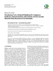
[email protected] Received 8 February 2010; Accepted 31 March 2010 Academic Editor: Spyros Perlepes Copyright © 2010 Joanna Wiecek et al. This is an open access article distributed under the Creative Commons Attribution License, which permits unrestricted use, distribution, and reproduction in any medium, provided the original work is properly cited. The synthesis and spectral characterization of novel diorganotin complexes with 3-hydroxypyridine-2-carbaldehyde thiosemicarbazone, H2 L (1), [SnMe2 (L)] (2), [SnBu2 (L)] (3), and [SnPh2 (L)] (4) are reported. The single-crystal X-ray structure of complex [SnPh2 (L)(DMSO)] (5) shows that the ligand is doubly deprotonated and is coordinated as tridentate ligand. The six coordination number is completed by two carbon atoms of phenyl groups. There are two similar monomers 5a (Sn1) and 5b (Sn51) in the asymmetric unit. The monomers 5a and 5b are linked through intermolecular hydrogen bonds of N–H–O and C–H–S type. C–H → π, intermolecular interactions, intra- and intermolecular hydrogen bonds stabilize this structure and leads to aggregation and a supramolecular assembly. The IR and NMR (1 H, 13 C and 119 Sn) spectroscopic data of the complexes are reported. The in vitro cytotoxic activity has been evaluated against the cells of three human cancer cell lines: MCF-7 (human breast cancer cell line), T-24 (bladder cancer cell line), A-549 (nonsmall cell lung carcinoma) and a mouse L-929 (a fibroblast-like cell line cloned from strain L). Compounds 1, 3, and 4 were found active against all four cell lines. Selectivity was observed for complexes 3 and 4 which were found especially active against MCF-7 and T-24 cancer cell lines.
1. Introduction Organotin(IV) compounds find wide applications as catalysts and stabilizers, and certain derivatives are used as biocides, as antifouling agents and for wood preservation. It has been observed that several diorganotin adducts show potential as antineoplastic and antituberculosis agents [1–4]. Thiosemicarbazone derivatives are of considerable interest due to their antibacterial, antimalarial, antiviral, and antitumor activitiy [5, 6]. In our laboratory, the chemistry of thiosemicarbazones has been an extremely active area of research, primarily because of the beneficial biological (namely, antiviral and antitumor) activities of their transition-metal complexes [7–9]. 3-Hydroxypyridine-2carbaldehyde thiosemicarbazone (1) is a member of the socalled α-(N)-heterocyclic carbaldehyde thiosemicarbazones
(HCTs), which are the most potent known inhibitors of ribonucleoside diphosphate reductase. Compounds (1) and 5-hydroxypyridine-2-carbaldehyde thiosemicarbazone have shown high anticancer activity in animal models but were found to be readily glucuronidated and rapidly excreted [10]. The multiple dissociation constants of the ligand H2 L (1) and the crystal structure of he complex of Pd(II) with 1, [Pd(HL)Cl] have been studied by us [11]. This work is an extension of previously studied complexes of thiosemicarbazones with palladium(II), platinum(II), zinc(II), and organotin(IV) with potentially interesting biological activities [12–14]. The present paper includes the interaction of SnPh2 O (where R is methyl, butyl, and phenyl groups) with 3Hydroxypyridine-2-carbaldehyde thiosemicarbazone (H2 L) and the crystal structure of the complex [SnPh2 (L)(DMSO)]
2 (5). IR and NMR spectroscopic data are reported. The results of the cytotoxic activity of diorganotin complexes have been evaluated for antiproliferative activity in vitro against human cancer cell lines: MCF-7, T-24, A-549, and a mouse L-929.
2. Experimental 2.1. General and Instrumental. All reagents were commercially available (Aldrich or Merck) and used as supplied. Solvents were purified and dried according to standard procedures. Melting points (m.p.) were determined in open capillaries and are uncorrected. IR and far-IR spectra were recorded on a Perkin Elmer Spectrum GX Fourier transform spectrophotometer using KBr pellets (4000–400 cm−1 ) and nujol mulls dispersed between polyethylene disks (400– 40 cm−1 ). The free ligand was dissolved in (CD3 )2 SO and 1 H, 1 H–1 H COSY and 13 C spectra were acquired on a BRUKER 300 MHz spectrometer. Compounds 2–4 were dissolved in CDCl3 and were spectroscopically characterized by the use of 1 D and 2 D NMR spectroscopy on a Varian 600 MHz spectrometer. Experimental data were processed using VNMR and WinNMR routines. Chemical shifts (δ) are reported in ppm while spectra were referenced by the standard experimental setup. 119 Sn NMR spectra were acquired on the Varian 600 MHz and tin spectra were referenced by the use of external solution of Me4 Sn in C6 D6 . The splitting of proton resonances in the reported 1 H-NMR spectra is defined as s = singlet, d = doublet, t = triplet, and m = multiplet. The chemical shifts are reported in ppm for 1 H and 13 C NMR. Elemental analyses were carried out by the microanalytical service of the University of Ioannina, Greece. 2.2. Synthesis of the Ligand and the Complexes 3-Hydroxypyridine-2-Carbaldehyde Thiosemicarbazone (1). The ligand was synthesized according to a published procedure [11]. The white powder was recrystallized from cold ethanol and was dried in vacuo over silica gel. Yield: 75%. Bright-yellow powder. M.p. 209◦ C. IR (cm−1 ): 3555 s, 3451 m v(OH); 3291 m, 3194 m, v(NH2 , NH); 1639 s, v(C=N); 1229 s, v(C–O); 1098 s, v(NN); 827 s, v(C=S). 1 HNMR: 11.50 (s, OH); 9.62 (s, H–N(3); 8.12 (d, H–C(1), 3 J = 4.2 Hz); 7.26 (dd, H–C(2) 3 J = 8.5, 4 J = 4.2 Hz); 7.30 (dd, H–C(3) 3 J = 8.4, 4 J = 1.4 Hz); 8.34 (s, H–C(6)); 8.00, 8.24 (br. s, NH2). 13 C-NMR: 178.0 C(7); 153.1 C(4); 144.4 C(6); 141.1 C(1); 137.6 C(5); 125.3 C(2); 124.2 C(3). Anal. calc. for C7 H8 N4 OS (196.04): C, 42.8; H, 4.1; N, 28.6; S, 16.3; found: C, 42.6; H, 3.9; N, 28.2; S, 16.0%. SnMe2 (L) (2). Dimethyltin(IV) oxide (0.033 g, 0.2 mmole) and 3-hydroxypyridine-2-carbaldehyde Thiosemicarbazone (0.0392 g, 0.2 mmole) in benzene (20 mL) were stirred and were refluxed for 12 hours under azeotropic removal of water (Dean-Stark trap). The resulting clear solution was rotary evaporated under vacuum to a small volume (2 mL), chilled and triturated with diethyl ether to give a white solid. The powder was recrystallized from distilled diethyl ether and dried in vacuo over silica gel to give yellow solid; mp.
Bioinorganic Chemistry and Applications 228–230◦ C, Yield 35%. IR (cm−1 ): 3296 m, v(NH2 ); 1580 s, v(C=N); 1176 s, v(C–O); 1111 s, v(NN); 804 w, v(C=S); 583 m, 564 v(Sn–C); 431 m, v(Sn–N); 231 m, v(Sn–O); 376s, ν(Sn–S). 1 H-NMR: 8.09 (dd, H–C(1), 3 J = 4.2,4 J = 1.4 Hz); 7.18 (dd, H–C(2) 3 J = 8.5, 4 J=4.2 Hz); 7.08 (d, H–C(3) 3 J = 8.5,4 J = 1.4 Hz); 8.76 (s, H–C(6)); 5.09 (br. s, NH2 ); 0.89 (s, CH3 , 2 J (117/119 Sn–1 H) = 35.7 Hz). 13 C-NMR: 168.8 C(7); 163.4 C(4); 161.5 C(6); 140.2 C(1); 135.4 C(5); 127.6 C(2); 128.9 C(3); 6.22 (CH3 , J (117/119 Sn–13 C) = 309 Hz). 119 Sn NMR: δ = −94.5. Anal. calc. for C9 H12 N4 OSSn (343.0): C, 31.5; H, 3.5; N, 16.3; S, 9.3; found: C, 31.5; H, 3.7; N, 16.0;, S, 9.1 %. SnBu2 (L) (3). Dibutylltin(IV) oxide (0.0498 g, 0.2 mmole) and 3-hydroxypyridine-2-carbaldehyde Thiosemicarbazone (0.0392 g, 0.2 mmole) in benzene (20 mL) were stirred and were refluxed for 12 hours under azeotropic removal of water (Dean-Stark trap). The resulting clear solution was rotary evaporated under vacuum to a small volume (2 mL), chilled and triturated with diethyl ether to give a white solid. The powder was recrystallized from distilled diethyl ether and dried in vacuo over silica gel to give yellow solid; mp. 126–128◦ C, Yield 41%. IR (cm−1 ): 3292 m, v(NH2 ); 1577 s, v(C=N); 1173 s, ν(C–O); 1114 s, v(NN); 805 w, ν(C=S); 578 ms, 560 sh ν(Sn–C); 418 m, ν(Sn–N); 247 m, ν(Sn–O); 394 ms, ν(Sn–S). 1 H-NMR: 8.05 (dd, H–C(1), 3 J = 4.2, 4 J = 1.3 Hz); 7.16 (dd, H–C(2) 3 J = 8.5, 4 J = 4.2 Hz); 7.07 (dd, HC(3) 3 J = 8.5, 4 J = 1.3 Hz); 8.82 (s, H-C(6)); 5.22 (br. s, NH2 ); 0.87 (t, 7.3 Hz, Hδ); 1.32 (m, Hγ); 1.54, 1.64 (Hα, β). 13 CNMR: 167.7 C(7); 162.5 C(4); 160.0 C(6); 138.7 C(1); 134.5 C(5); 126.8 C(2); 128.0 C(3); 26.9 (Cα, J (117/119 Sn−13 C) = 265 Hz); 28.2 (Cβ); 27.3 (Cγ); 14.6 (Cδ). 119 Sn NMR: δ = −194.4. Anal. calc. for C16 H18 N4 OSSn (537.2): C, 42.2; H, 5.7; N, 13.1; S, 7.5; found: C, 42.0; H, 5.9; N, 13.2; S, 7.4 %. SnPh2 (L) (4). Diphenylltin(IV) oxide (0.0578 g, 0.2 mmole) and 3-hydroxypyridine-2-carbaldehyde thiosemicarbazone (0.0392 g, 0.2 mmole) in benzene (20 mL) were stirred and were refluxed for 12 hours under azeotropic removal of water (Dean-Stark trap). The resulting clear solution was rotary evaporated under vacuum to a small volume (2 mL), chilled and triturated with diethyl ether to give a white solid. The powder was recrystallized from distilled diethyl ether and dried in vacuo over silica gel to give yellow solid : mp. 186–188◦ C, Yield 34%. IR (cm−1 ): 3269 m, ν(NH2 ); 1589 s, ν(C=N); 1185 s, v(C–O); 1118 s, v(NN); 808 m, v(C=S); 322 ms, 303 sh ν(Sn–C); 419 m, ν(Sn–N); 248 m, ν(Sn–O); 370 ms, ν(Sn–S). 1 H-NMR: 8.02(d, H–C(1), 3 J = 4.2 Hz); 7.18(dd, H-C(2), 3 J = 8.5, 4 J = 4.2 Hz); 7.29 (d, H-C(3), 3 J = 8.5 Hz); 8.88 (s, H–C(6)); 5.37 (br. s, NH ); 7.82 (d, 2 7.7 Hz, Ho-Ph); 7.34–7.30 (m, Hm, p-Ph). 13 C-NMR: 167.4 C(7); 164.0 C(4); 161.5 C(6); 140.2 C(1); 135.4 C(5); 127.8 C(2); 129.3 C(3); 142.1 (Sn-Cph, J (117/119 Sn–13 C) = 424 Hz); 135.8 (Co-Ph, 2 J (117/119 Sn–13 C) = 28 Hz); 128.8 (Cm-Ph, 3 J (117/119 Sn–13 C) = 37 Hz); 130.2 (Cp-Ph, 4 J (117/119 Sn– 13 C) = 8.7 Hz); 119 Sn NMR: δ = −227.2. Anal. calc. for C19 H16 N4 OSSn (663.1): C, 48.9; H, 3.5; N, 12.0; S, 6.9; found: C, 48.6; H, 3.5; N, 10.7; S, 6.8%.
Bioinorganic Chemistry and Applications Table 1: X-ray crystal data and structure refinement. 5 Empirical formula C21 H22 N4 O2 S2 Sn Formula weight 545.24 Temperature/ (K) 100 (2) Wavelength/ (A) 0.71073 Crystal system Triclinic Space group P-1 ˚ a (A) 9.4663 (4) ˚ b (A) 14.7350 (7) ˚ c (A) 16.6374 (7) ◦ 94.871 (4) α( ) 96.434 (4) β (◦ ) 90.793 (4) γ (◦ ) 2297.1 (2) Volume (A3 ) Z 4 1.577 Dc (Mg/m3 ) 1.319 Absorption coefficient (mm−1 ) F(000) 1096 Crystal size (mm) 0.32 × 0.28 × 0.16 Diffractometer Kuma KM4CCD 3.14–36.65 Theta range for data collection (◦ ) −15 → 15, −24 → 20, −27 → 27 Ranges of h, k, l Reflections collected 35235 Independent reflections (Rint) 18670 (0.0381) 81.9% Completeness to 2θ = 36.65 Data/parameters 18670/541 0.920 Goodness-of-fit (F 2 ) Final R1/wR2 indices [I>2σ(I)] 0.0336/0.0768 2.090/−1.098 Largest diff. peak/hole (e/A˚ 3 )
2.3. X-Ray Crystallography. Crystals of complex 5, suitable for X-ray analysis, were obtained by slow crystallization of 4 from a mixture of solvents C6 H6 /toluene/DMSO/CH3 CN. Crystal data 5 are given in Table 1, together with refinement details. All measurements of crystals were performed in low temperature using an Oxford Cryosystem device on a Kuma KM4CCD κ-axis diffractometer with graphitemonochromated Mo Ka radiation. The data were corrected for Lorentz and polarization effects. No absorption correction was applied. Data reduction and analysis were carried out with the Oxford Diffraction (Poland) Sp. z o.o (formerly Kuma Diffraction Wroclaw, Poland) programs. Crystal structure was solved by direct methods (program SHELXS97) and refined by the full-matrix least-squares method on all F 2 data using the SHELXL97 [15] programs. Nonhydrogen atoms were refined with anisotropic displacement parameters; hydrogen atoms were included from geometry of molecules and Δρ maps. During the refinement process they treated as riding atoms. Molecular graphics were performed from PLATON2003 [16, 17]. Crystallographic data, that is, atomic coordinates, thermal parameters, bond lengths, and bond angles (CCDC number 634270 for 5), have been deposited with the
3 Cambridge Crystallographic Data Centre. Copies of available material can be obtained, free of charge, on application to CCDC, 12 Union Road, Cambridge CB2 1EZ, UK, (fax: +44-1223-336033 or e-mail:
[email protected] or http://www.ccdc.cam.ac.uk). 2.4. Antiproliferative Assay In Vitro. The results of cytotoxic activity in vitro are expressed as IC50 -the concentration of compound (in μM) that inhibits a proliferation rate of the tumor cells by 50% as compared to control untreated cells. The compounds 1–4 were tested for their antiproliferative activity in vitro against the cells of four human cancer cell lines: against the cells of three human cancer cell lines: MCF7 (human breast cancer cell line), T-24 (bladder cancer cell line), A-549 (nonsmall cell lung carcinoma), and a mouse fibroblast L-929 cell line. Compounds. Test solutions of the compounds tested (1 mg/mL) were prepared by dissolving the substance in 100 μL of DMSO completed with 900 μL of tissue culture medium. Afterwards, the tested compounds were diluted in culture medium to reach the final concentrations of 100, 50, 10, 1 and 0.1 ng/μL. The solvent (DMSO) in the highest concentration used in test did not reveal any cytotoxic activity. Cells. The cell lines are maintained in the Cell Culture Collection of the University of Ioannina. Twenty-four hours before addition of the tested agents, the cells were plated in 96-well plates at a density of 104 cells per well. The MCF-7 cells were cultured in the DMEM (Modified Eagle’s Medium) medium supplemented with 1% antibiotic and 10% fetal calf serum. L-929 cells were grown in Hepes-buffered RPMI 1640 medium supplemented with 10% fetal calf serum, penicillin (50 U/mL), and streptomycin (50 mg/mL). A-549 cells were grown in F-12K Ham’s medium supplemented with 1% glutamine, 1% antibiotic/antimycotic, 2% NaHCO3 , and 10% fetal calf serum. The cell cultures were maintained at 37◦ C in a humid atmosphere saturated with 5% CO2 . Cell number was counted by the Trypan blue dye exclusion method. MCF-7, L-929, and A-549 cells were determined by the sulforhodamine B assay [18], while T-24 cells by the MTT assay [19]. The in vitro tests were performed as described previously [20].
3. Results and Discussion 3.1. Spectroscopy 3.1.1. Infrared Spectroscopy. The bands at 3555 and 3451 cm−1 are assigned to v(OH) mode while the mediumstrong intensity bands at around 3291 and 3194 cm−1 in the spectra of H2 L are assigned to v(NH2 ) and v(NH), respectively. The significant changes in the ligand bands upon complexing are the decrease in ν(C=N) and increase in ν(N–N) and the absence of the large systematic shifts of νas (NH2 ) to lower frequencies. These data indicate coordination through the nitrogen of the azomethine group and no interaction between the terminal amino nitrogen and the metal ion. The ν(CS) band at 827 cm−1 is less intense
4 in the complexes 2–4 and is shifted to a lower frequency, suggesting coordination of the metal through sulfur. Coordination of the thiolato S-atom was further indicated by a decrease in the energy of the thioamide band as well as by the presence of a band at ca. 370 cm−1 assignable to ν(Sn–S). An IR band at 1229 cm−1 for 1 was assigned to ν(C– O). This band was found to be shifted to 1185–1173 cm−1 , in the spectra of the complexes 2–4. Coordination of the phenolic-O atom was further indicated by the presence of a band at ca. 250–230 cm−1 assignable to ν(Sn–O). From the metal-ligand stretching vibrations, which are below 600 cm−1 , it is possible to assign the bands characteristic for ν(SnC). Also, the bands at 394–370 cm−1 and 431–418 cm−1 are assigned to ν(SnS) and ν(SnN), respectively, and the bands at 250–230 cm−1 are assigned at ν(SnO) stretching vibrations [20, 21]. 3.1.2. NMR Spectra. 1 H and 13 C resonances of the ligand H2 L as well as of the complexes 2–4 bearing di-methyl, nbutyl, and phenyl groups attached to the central tin atom were unambiguously assigned by the use of 2D 1 H - 1 H gCOSY, 1 H, 13 C-HSQC, and 1 H, 13 C-HMBC experiments. In the 1 H NMR spectra of (H2 L) (1) the broad singlet at δ 11.60 was attributed to OH group in accordance with [22] and the broad signal at δ 9.75 ppm was assigned to NH group. These two groups apparently participate in Hbonding with the nucleophilic solvent molecules (DMSO) or with other ligand molecules. These two signals are abolished upon interaction with the metal, indicating deprotonation of these groups and possible coordination to the tin(IV) atom at 2–4. The absence of peaks corresponding to the imino proton NH and OH proton in the spectrum of 2– 4 indicates that the nitrogen and oxygen are present in the deprotonated form and the ligand is dideprotonated. A sharp resonance peak appearing at ca. 5 ppm in all complexes is attributed to the NH2 group. This is also corfirmed by integration of the 1 H spectral profile while additionally the use of CDCl3 eliminates the formation of H-bonding or complexation with the participation of solvent molecules as was the case with the ligand alone. In the 1 H-NMR spectra of the complexes 2–4, the formyl H-atom H–C(6) was shifted upon coordination, which indicated variations in the electron density at position 6. This signal was shifted downfield, in accordance with a decreased electron density at site C6 in the complexes. The C=S resonance of the thiosemicarbazone moiety in the free ligand resonated at 178.0 ppm. All complexes showed an upfield shift of C7 peak in the order of ∼10 ppm compared with the free ligand, indicating the complexation of tin(IV) to the sulphur atom which apparently is related with an increased electron density at this site on complexation, due probably to π-back bonding for thiolato sulphur [21]. All the complexes exhibit downfield shifts of the C3, C4, and C6. This deshielding, in accordance with decreased electron density upon complexation, is indicative of Sn–O and Sn– N (azomethine nitrogen) bonds. These data indicate that the complexes are formed by ligand deprotonation followed by metallation, a structural motive that seems to be stable both
Bioinorganic Chemistry and Applications in the solid state and in CDCl3 solution. 119 Sn chemical shifts of compounds 2–4 were found between −94.4 and −227.2 in accordance with five-coordinate tin center [23]. 3.2. Molecular Structure. Crystals of complex 5, suitable for X-ray analysis, were obtained by slow crystallization of 4 from a mixture of solvents C6 H6 /toluene/DMSO/CH3 CN. The crystal structure is shown in Figure 1. Crystal data are given in Table 1, together with refinement details. Bond lengths and angles are given in Table 2. There are two similar monomers 5a (Sn1) and 5b (Sn51) in the asymmetric unit. The double deprotonated ligand is coordinated as tridentate ligand via the phenolic oxygen O(1), the azomethine nitrogen N(3), and thiolato sulfur S(1) atoms. The molecule of DMSO is coordinated to the tin through oxygen O(2) atom. The six coordination number is completed by two carbon atoms of phenyl groups. The organic molecule acts as anionic tridentate with the ONS donors placed in the same side. The dianionic, tridentate ligand has a ZEZ configuration for the oxygen, nitrogen, and sulfur donor centers. The coordinated ligand consists of three rings, one heterocyclic and two chelates, SnSNNC and SnONCC, I and II, respectively. For monomers 5a and 5b the dihedral angles between the planes of the rings I and II are 14.47(6) and 12.59(6)◦ , respectively and between the ring II and the pyridyl ring are 7.4(5) and 8.1(2)◦ , respectively, indicating that the ligand as a whole in the two monomers deviates from planarity. The C–S bond lengths 1.747(2), 1.749(2) A˚ for 5a and ˚ but 5b, respectively, are shorter than a single bond (1.81 A), ˚ suggesting partial single longer than a double bond (1.62 A), ˚ is bond character. The C(14)–N(3) bond length, 1.301(2) A, ˚ The S–C bond distances close to a double bond (1.28 A). are consistent with increased single-bond character while both thioamide C–N distances indicate increased double bond character. The negative charge of the deprotonated ligand is delocalized over the thiosemicarbazonato moiety. This is indicative of the coordinated thiosemicarbazone’s greater conjugation and more delocalized electron density. The Sn–N(3) bond distance is longer than the sum of the ˚ which indicates strong bond, while covalent radii (2.15 A), ˚ though much the bond distance Sn–S, 2.5141(5), 2.519(5) A, ˚ shorter than the sum of the van der Waals radii (4.0 A), indicates a weak bond [21]. The C–Sn C bond angle is equal to 105.87(7), 104.58(7)◦ , and the bond angles C(7)–Sn–N(3) and C(1) –Sn–O(2) are 160.47(6), 165.59(6) and 162.80(6), 166.89(6)◦ for 5a and 5b, respectively. The two monomers 5a and 5b are linked through two intermolecular hydrogen bonds of N(51)–H(51B)–O(1) and of C(8)–H(8)–S(51) type (The N–O distance is 3.013(2) A˚ ˚ The monomers of and the C–S distance is 3.673(2) A. 5a form hydrogen-bonded dimers linked by two N(1)– H(1A)–N(4) hydrogen bonds involving the amino N(1)– H(1A) hydrogen atom and the pyridyl N(4) nitrogen. The monomers of 5a are also linked by another C(14)–H(14)– N(2) hydrogen bond involving the formyl C(14)–H(14) hydrogen and the adjacent imino nitrogen N(2) and vice versa of centro-symmetrically related pairs of molecules. The observed hydrogen bonding patterns are of the DA=AD
Bioinorganic Chemistry and Applications
C54
5
C20
C55
C53
N1
C21
S2 C11
C56 O2 C66 N54 C52 C51 C65
C67
C13 C12
S1
C10
C64
C68 C69
C8 C63
O51
Sn51
N2
C9
N53 N52 N51
C7 N3
S51
Sn1 C14
O1 C15
C62 C57 O52
C1
C58
C2
C19
C61 C60
C59 S52
C70
N4
C18
C3
C6 C5
C17
C71
C4
C16
(a)
(b)
Figure 1: Molecular structure of the diorganotin complex 5.
˚ and angles (◦ ) for complex 5. Table 2: Bond lengths (A) 5a Sn(1)–S(1) Sn(1)–O(1) Sn(1)–O(2) Sn(1)–N(3) Sn(1)–C(1) Sn(1)–C(7) S(1)–C(13) S(2)–O(2) S(2)–C(20) S(2)–C(21) O(1)–C(19) N(2)–N(3) S(1)–Sn(1)–O(1) S(1)–Sn(1)–O(2) S(1)–Sn(1)–N(3) S(1)–Sn(1)–C(1) S(1)–Sn(1)–C(7) O(1)–Sn(1)–O(2) O(1)–Sn(1)–N(3) O(1)–Sn(1)–C(1) O(1)–Sn(1)–C(7) O(2)–Sn(1)–N(3) O(2)–Sn(1)–C(1) O(2)–Sn(1)–C(7) N(3)–Sn(1)–C(1) N(3)–Sn(1)–C(7) C(1)–Sn(1)–C(7)
2.5141(5) 2.087(2) 2.337(2) 2.251(2) 2.164(2) 2.149(2) 1.747(2) 1.528(2) 1.791(3) 1.789(2) 1.326(2) 1.380(2) 156.25(4) 84.87(3) 77.37(4) 101.71(5) 95.51(5) 76.08(5) 84.11(5) 94.05(6) 97.14(6) 75.34(5) 165.59(6) 86.01(6) 93.44(6) 160.47(6) 105.87(7)
5b Sn(51)–S(51) Sn(51)–O(51) Sn(51)–O(52) Sn(51)–N(53) Sn(51)–C(51) Sn(51)–C(57) S(51)–C(63) S(52)–O(52) S(52)–C(70) S(52)–C(71) O(51)–C(69) N(52)–N(53) S(51)–Sn(51)–O(51) S(51)–Sn(51)–O(52) S(51)–Sn(51)–N(53) S(51)–Sn(51)–C(51) S(51)–Sn(51)–C(57) O(51)–Sn(51)–O(52) O(51)–Sn(51)–N(53) O(51)–Sn(51)–C(51) O(51)–Sn(51)–C(57) O(52)–Sn(51)–N(53) O(52)–Sn(51)–C(51) O(52)–Sn(51)–C(57) N(53)–Sn(51)–C(51) N(53)–Sn(51)–C(57) C(51)–Sn(51)–C(57)
2.5190(5) 2.088(2) 2.345(2) 2.262(2) 2.157(2) 2.151(2) 1.749(2) 1.528(2) 1.777(3) 1.787(3) 1.320(2) 1.375(2) 155.68(4) 83.23(3) 77.71(4) 100.07(5) 100.30(5) 77.25(5) 83.46(5) 95.98(6) 93.14(6) 75.65(5) 166.89(6) 87.15(6) 92.55(7) 162.80(6) 104.58(7)
6
Bioinorganic Chemistry and Applications
Figure 2: Arrangement of the intermolecular hydrogen bonds in 5. Table 3: C–H → π interactions and intermolecular hydrogen bonds for 5.
D
C–H(I) → Cg(J)a C(11)–H(11) [1] → Cg(4)(i) C(60)–H(60) [2] → Cg(5)(ii) H Ab (iii) N(1)–H(1A)· · · N(4) N(1)–H(1B)· · · O(51)(iv) N(1)–H(1B)· · · O(52)(iv) N(51)–H(51A)· · · N(54)(v) N(51)–H(51B)· · · O(1) C(8)–H(8)· · · S(51) C(14)–H(14)· · · N(2)(iii) C(58)–H(58)· · · S(51) C(62)–H(62)· · · O(51) C(64)–H(64)· · · N(52)(v)
H–Cg 2.71 2.66 H ··· A 2.13 2.28 2.55 2.16 2.24 2.84 2.50 2.85 2.55 2.58
C–Cg 3.5394 3.4118 D ··· A 3.005(2) 3.065(2) 3.124(2) 2.993(3) 3.013(2) 3.673(2) 3.442(2) 3.564(2) 3.132(2) 3.483(2)
∠C–H–Cg 159 135 ∠D–H · · · A 165 150 124 168 152 143 175 127 118 174
a Where Cg(4) and Cg(5) are referred to the rings C(1)–C(6) and C(7)–C(12); b Cg–Cg is the distance between ring centroids; symmetry transformations, (i) 1 − x, − y, −z; (ii) 1−x, 1− y, −z; (iii) 1−x, − y,1−z; (iv) x, −1 + y, z; (v) 2 − x, 1 − y, 1 − z.
type. Also, the monomers of 5b form hydrogen-bonded dimers and are linked by two hydrogen bonds, the N(51)– H(51A)–N(54) and C(64)–H(64)–N(52) (Figure 2). C–H → π intermolecular interactions intra- and intermolecular hydrogen bonds [24] stabilize this structure and lead to a supramolecular assembly, and Table 3 and Figure 3. 3.3. Pharmacology. Antiproliferative Activity In Vitro. Complexes of N4-ethyl 2-acetylpyridine thiosemicarbazone with platinum(II) or palladium(II) were tested in a panel of human tumor cell lines of different origins (breast, colon,
and ovary cancers) and cis-platin-refractory/resistant cell lines and were found to exhibit very remarkable growth inhibitory activities with mean IC50 values of 0.9–0.5 nM and support the hypothesis that both [Pt(Ac4Et)2 ] and [Pd(Ac4Et)2 ] complexes can be characterized by cellular pharmacological properties distinctly different from those of cis-platin [8]. The complexes [ZnCl2 (Fo4Npypipe)] and [ZnCl2 (Ac4Npypipe)] where Fo4Npypipe and Ac4Npypipe are the monoion of 2-formyl pyridine N(4)-1-(2-pyridyl)piperazinyl thiosemicarbazone and 2-acetyl pyridine N(4)1-(2-pyridyl)-piperazinyl thiosemicarbazone have been evaluated in vitro against MCF-7, T-24, A-549, and L-929 cell
Bioinorganic Chemistry and Applications
7
Figure 3: A view of the extended network of 5 along the b axis.
T-24
50
MCF7
4
40 IC50
3
2
[SnBu2 (L)](3) [SnPh2 (L)](4) (a)
cis-platin
Ph2 SnO
cis-platin
H2 L (1)
Bu2 SnO
0
Me2 SnO
0 [SnPh2 (L)](4)
10
[SnBu2 (L)](3)
1
H2 L (1)
20
[SnMe2 (L)](2)
IC50
30
(b)
Figure 4: The antiproliferative activity in vitro expressed as IC50 ± SD (μM) against (a) T-24 and (b) MCF-7 cancer cell lines.
8
Bioinorganic Chemistry and Applications
Table 4: The antiproliferative activity in vitro of 1–4, expressed as as IC50 ± SD (μM) against MCF-7, T-24, A-549, and L-929 cancer cell lines. H2 L (1) [SnMe2 (L)] (2) [SnBu2 (L)] (3) [SnPh2 (L)] (4) Me2 SnO Bu2 SnO Ph2 SnO cisplatin
L929 2.5 ± 0.03 7.29 ± 0.04 1.05 ± 0.02 1.37 ± 0.03


![64Cu-Azabicyclo[3.2.2]Nonane Thiosemicarbazone Complexes ...](https://m.moam.info/img/260x300/64cu-azabicyclo322nonane-thiosemicarbazone-complex_5a399ccf1723dd00995c24e5.jpg)